. 2009 Mar;33(3):499-508.
doi: 10.1016/j.nbd.2008.12.004. Epub 2008 Dec 25.

Absence epilepsy in apathetic, a spontaneous mutant mouse lacking the h channel subunit, HCN2

Affiliations

Absence epilepsy in apathetic, a spontaneous mutant mouse lacking the h channel subunit, HCN2

Wendy K Chung et al. Neurobiol Dis. 2009 Mar.

Abstract

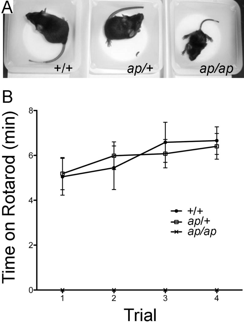
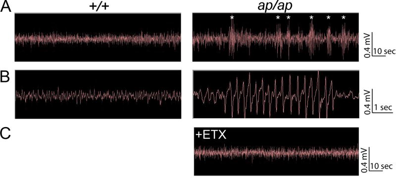
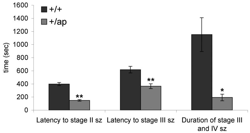
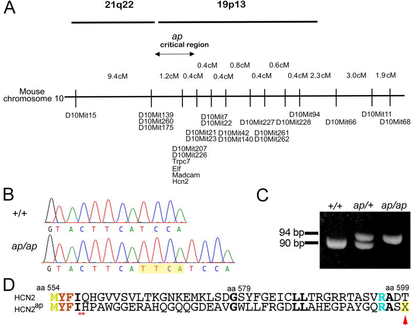
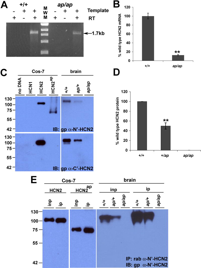
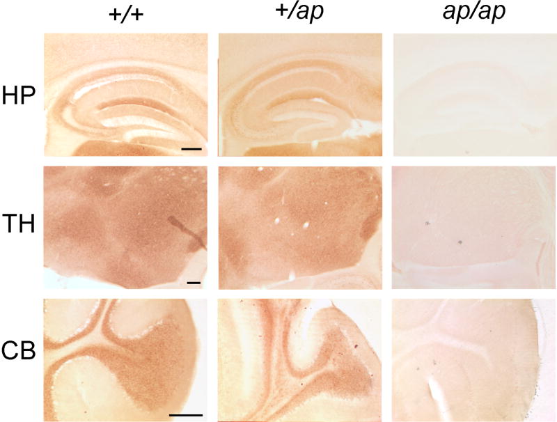
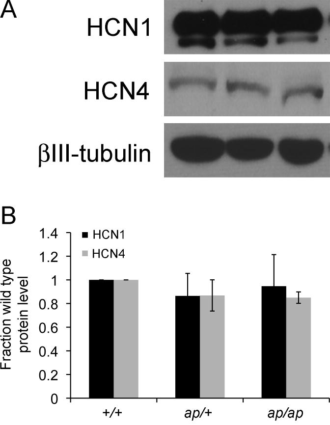
Analysis of naturally occurring mutations that cause seizures in rodents has advanced understanding of the molecular mechanisms underlying epilepsy. Abnormalities of I(h) and h channel expression have been found in many animal models of absence epilepsy. We characterized a novel spontaneous mutant mouse, apathetic (ap/ap), and identified the ap mutation as a 4 base pair insertion within the coding region of Hcn2, the gene encoding the h channel subunit 2 (HCN2). We demonstrated that Hcn2(ap) mRNA is reduced by 90% compared to wild type, and the predicted truncated HCN2(ap) protein is absent from the brain tissue of mice carrying the ap allele. ap/ap mice exhibited ataxia, generalized spike-wave absence seizures, and rare generalized tonic-clonic seizures. ap/+ mice had a normal gait, occasional absence seizures and an increased severity of chemoconvulsant-induced seizures. These findings help elucidate basic mechanisms of absence epilepsy and suggest HCN2 may be a target for therapeutic intervention.


Conflict of interest statement

Disclosure of Conflicts of Interest None of the authors has any conflict of interest to disclose.

Figures

Figure 1
Figure 1
Apathetic mice have smaller body size and an ataxic gait. A, photograph of postnatal day 25 wild type (+/+), heterozygous (ap/+) and apathetic (ap/ap) mice reveals smaller body size (also note the wide-based stance of the ap/ap mouse. B, Rotarod test. The averages ± S.E.M for time spent on the rotarod across four test trials for +/+ (filled circles), ap/+ (open squares) and ap/ap (x shape) mice. n=6 each male and female +/+, n=10 each male and female ap/+ and n=5 each ap/ap male and female. Note the ap/ap mice are totally unable to walk on rotarod, whereas +/+ and ap/+ mice perform equally well.
Figure 2
Figure 2
Epidural electrocephalographic (EEG) recordings from +/+ and ap/ap mice. A, Continuous two minute EEG recording in +/+ littermate (left panel) and ap/ap (right panel) mice. Asterisks (*) indicate typical spike-wave discharges observed in the ap/ap mice. B, Expanded time scale of EEG activity in +/+ (left trace) and ap/ap (right panel) mice demonstrates representative (3-7 Hz, 1-4 sec) spike-wave discharge recorded during behavioral arrest in an ap/ap mouse (right panel). C, Ethosuximide (ETX, 200 mg/kg, i.p.) injection in an ap/ap mouse eliminates behavioral arrests and spike-wave seizures detected by EEG.
Figure 3
Figure 3
Seizure severity of ap/+ mice is higher than +/+ mice. Generalized seizures were induced by injecting +/+ (n=15) and ap/+ (n=17) mice with 4-aminopyridine (4-AP; 10mg/kg). Latency to stage II and III seizure as well as duration of stage III seizure are presented. Both latency and duration of seizures were significantly shorter in +/ap mice compared to +/+ mice (*P<0.01. ** P<0.001)
Figure 4
Figure 4
Apathetic mice harbor a four base pair insertion in the Hcn2 gene. A, Genetic map of mouse chromosome 10 at the apathetic locus with the homologous human chromosomes. Map based upon 946 meioses. Maximum, non-recombinant interval containing ap indicated. B, Schematic diagram of the 4 base pair insertion (TTCA) in Hcn2 in ap/ap mice, localized to the coding region of the cyclic nucleotide binding domain of HCN2 protein. C, Electropherograms of wild type and apathetic Hcn2 gene shows insertion of 4 base pairs, TTCA in Hcn2ap gene. D, PCR amplification of the Hcn2 gene yields a 90 and 94 base pair amplicon for wild type (+) and apathetic (Hcn2ap) alleles, respectively. E, A Predicted translation products of wild type and apathetic Hcn2 alleles show the frameshift and premature stop codon (red arrow) in predicted Hcn2ap.
Figure 5
Figure 5
Hcn2ap is transcribed, but apathetic HCN2 protein is not detected. A, RT-PCR of full length of Hcn2 mRNA shows that Hcn2 mRNA is present in both wild type (+/+) and apathetic (ap/ap) mice (arrow). B, Quantitative RT-PCR shows that mRNA of Hcn2 is decreased by ~90% in ap/ap mice as compared to +/+ controls. C, Protein extracts from Cos-7 cells overexpressing wild type or apathetic HCN2 (HCN2ap), and protein extracts from +/+, ap/+, and ap/ap mouse brains were generated and separated by SDS-PAGE. Blots were labeled with gp α-HCN2 (C-terminal epitope) or gp α-N’-HCN2 (N-terminal epitope) antibodies. Gp α-HCN2 detects wild type HCN2, but not HCN2ap protein, whereas α-N’-HCN2 detects both proteins in overexpressed heterologous cells. α-N’-HCN2 does not detect HCN2ap in ap/+ or ap/ap brain, suggesting absence of HCN2ap protein. D, Protein expression of HCN2 was decreased ~50% in the brain of ap/+ compared to +/+ mice (n=4, ** P<0.01). E, HCN2ap protein was not detected even after concentrating protein extracts by immunoprecipitation. Protein extracts prepared from Cos-7 cells overexpressing wild type or HCN2ap, as well as +/+, ap/+, and ap/ap mouse brains were immunoprecipitated with rab α-HCN2 antibody and probed with gp α-N’-HCN2. Whereas both wild type and HCN2ap proteins in Cos-7 cells were concentrated and showed increased band intensity, no bands were detected from brain extracts of ap/ap mice. inp= input extract, ip= immunoprecipitated extract.
Figure 6
Figure 6
Distribution of HCN2 protein in +/+, ap/+ and ap/ap mouse brain. 50 μm-think parasagittal brain sections were generated and immunohistochemistry was performed using gp α-N’-HCN2 antibody, visualized with DAB staining. HCN2 is concentrated in the stratum lacunosum-moleculare layer of hippocampal area CA1 (HP, top panel), thalamic nuclei (TH, middle panel), and granule cell layer in the cerebellum (CB, bottom panel). Note that distribution patterns of HCN2 are not significantly different between +/+ and ap/+ brains. No immunoreactivity of HCN2 was detected in ap/ap brain.
Figure 7
Figure 7
HCN1 and HCN4 expression in +/+, +/ap, and ap/ap brain. A. Brain extracts were generated and proteins separated by SDS-PAGE. Blots were probed with gp α-HCN1 antibody, gp α-HCN4 antibody, or ms α-βIII-tubulin antibody. B. Density of HCN1 and HCN4 bands were normalized to the density of βIII-tubulin to control for loading differences. No significant change in the protein expression levels of HCN1 or HCN4 was detected when comparing +/ap or ap/ap to +/+ mice (n=3, P=.75 and .14 for HCN1 and HCN4, respectively). Data shown are mean ± S.E.M.

Similar articles

Cited by

References

    1. Abbas SY, Ying SW, Goldstein PA. Compartmental distribution of hyperpolarization-activated cyclic-nucleotide-gated channel 2 and hyperpolarization-activated cyclic-nucleotide-gated channel 4 in thalamic reticular and thalamocortical relay neurons. Neuroscience. 2006;141:1811–1825. - PubMed
    1. Bender RA, Soleymani SV, Brewster AL, Nguyen ST, Beck H, Mathern GW, Baram TZ. Enhanced expression of a specific hyperpolarization-activated cyclic nucleotide-gated cation channel (HCN) in surviving dentate gyrus granule cells of human and experimental epileptic hippocampus. J Neurosci. 2003;23:6826–6836. - PMC - PubMed
    1. Blumenfeld H, Klein JP, Schridde U, Vestal M, Rice T, Khera DS, Bashyal C, Giblin K, Paul-Laughinghouse C, Wang F, Phadke A, Mission J, Agarwal RK, Englot DJ, Motelow J, Nersesyan H, Waxman SG, Levin AR. Early treatment suppresses the development of spike-wave epilepsy in a rat model. Epilepsia. 2008;49:400–409. - PMC - PubMed
    1. Browne TR. Ethosuximide (Zarontin) and other succinimides. In: Browne TR, Feldman RG, editors. Epilepsy: Diagnosis and Management. Little Brown & Co; Boston, Mass: 1983. pp. 215–224.
    1. Budde T, Caputi L, Kanyshkova T, Staak R, Abrahamczik C, Munsch T, Pape HC. Impaired regulation of thalamic pacemaker channels through an imbalance of subunit expression in absence epilepsy. J Neurosci. 2005;25:9871–9882. - PMC - PubMed

Publication types

MeSH terms