Review
. 2009 May;19(5):453-61.
doi: 10.1093/glycob/cwp004. Epub 2009 Jan 15.

Roles of gastric mucin-type O-glycans in the pathogenesis of Helicobacter pylori infection

Affiliations
Review

Roles of gastric mucin-type O-glycans in the pathogenesis of Helicobacter pylori infection

Motohiro Kobayashi et al. Glycobiology. 2009 May.

Abstract

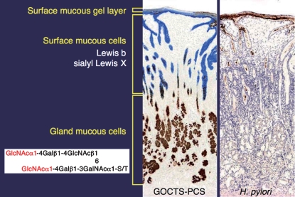
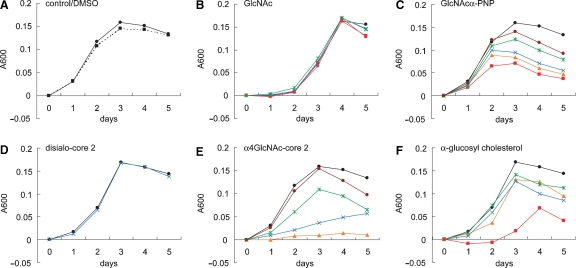
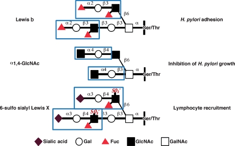
Helicobacter pylori is a Gram-negative bacterium that infects over 50% of the world's population. This organism causes various gastric diseases such as chronic gastritis, peptic ulcer, and gastric cancer. H. pylori possesses lipopolysaccharides that share structural similarity to Lewis blood group antigens in gastric mucosa. Such antigenic mimicry could result in immune tolerance against antigens of this pathogen. On the other hand, H. pylori colonizes gastric mucosa by utilizing adhesins that bind Lewis blood group antigen-related carbohydrates expressed on gastric epithelial cells. After colonization, H. pylori induces acute inflammatory responses mainly by neutrophils. This acute phase is gradually replaced by a chronic inflammatory response. In chronic gastritis, lymphocytes infiltrate the lamina propria, and such infiltration is facilitated by the interaction between L-selectin on lymphocytes and peripheral lymph node addressin (PNAd), which contains 6-sulfo sialyl Lewis X-capped O-glycans, on high endothelial venule (HEV)-like vessels. H. pylori barely colonizes gland mucous cell-derived mucin where alpha1,4-GlcNAc-capped O-glycans exist. In vitro experiments show that alpha1,4-GlcNAc-capped O-glycans function as a natural antibiotic to inhibit H. pylori growth. These findings show that distinct sets of carbohydrates expressed in the stomach are closely associated with pathogenesis and prevention of H. pylori-related diseases, providing therapeutic potentialities based on specific carbohydrate modulation.


Figures

Fig. 1
Fig. 1
Photomicrograph of gastric mucosa with (upper panel) or without (lower panel) intestinal metaplasia. (A) Complete-type intestinal metaplasia of gastric epithelial cells observed in chronically inflamed gastric mucosa. Gastric epithelial cells are replaced with absorptive cells, mucin-secreting goblet cells (shown by arrows), and Paneth cells having bright eosinophilic granules (shown by arrowheads). (B) Almost normal gastric epithelial cells in the pyloric gland area without intestinal metaplasia. These were stained by hematoxylin and eosin. Bar = 100 μm.
Fig. 2
Fig. 2
Disappearance of HEV-like vessels in the gastric mucosa after the eradication of H. pylori. Gastric mucosa infected with H. pylori was examined before and 2 months after treatment to eradicate H. pylori. (A) Before treatment, HEV-like vessels detected by MECA-79 and HECA-452 antibodies were abundant, and large numbers of mononuclear cells (lymphocytes) were present around these vessels. (B) After the eradication of H. pylori, HEV-like vessels were no longer present and very few mononuclear cells were present. CD34 was used for a marker of vascular endothelial cells. HE, hematoxylin and eosin, Bar = 100 μm. Adapted with permission from Kobayashi et al. (2004).
Fig. 3
Fig. 3
Two distinct mucins present in gastric mucosa. Surface mucous cell-derived mucins containing Lewis b and sialyl Lewis X (in blue), and gland mucous cell-derived mucins containing α1.4-GlcNAc-capped O-glycans (in brown) can be distinguished by galactose oxidase-cold thionin Schiff-paradoxical concanavalin A staining (GOCTS-PCS) (left panel). H. pylori in brown in the right panel is almost exclusively present in the surface mucous-cell derived mucin.
Fig. 4
Fig. 4
Inhibition of H. pylori growth by synthetic oligosaccharides and monosaccharides. H. pylori was cultured for 5 days in Mueller–Hinton broth supplemented with 5.5% horse serum containing various amounts of synthetic oligosaccharides and monosaccharides. Bacterial growth was measured at O.D. 600 nm, and the absorbance for control experiments at time 0 was subtracted from absorbance at later time points. Oligosaccharide and monosaccharide concentrations are 1 mM (red), 0.75 mM (orange), 0.5 mM (blue), 0.25 mM (green), 0.125 mM (brown), and control (closed circle). Two mM GlcNAc were also added in B (magenta). Oligosaccharides and monosaccharides were initially dissolved in dimethyl sulfoxide (DMSO), and the final DMSO concentration in the culture medium was 1%. The growth curve in the absence of DMSO is shown as a dotted line (A) Adapted from Lee et al. (2008).
Fig. 5
Fig. 5
Carbohydrates critical for H. pylori infection and pathogenesis. Distinct sets of carbohydrates on gastric mucosa play critical roles in H. pylori adhesion, inhibition of H. pylori growth, and recruitment of lymphocytes and facilitation of inflammatory response following H. pylori infection. BabA adhesin on H. pylori binds to Lewis b blood group antigen, thus facilitating H. pylori colonization (Ilver et al. 1998). This colonization is counteracted by α1,4-GlcNAc-capped O-glycans present in the deeper portion of gastric mucosa (Kawakubo et al. 2004). Once H. pylori infects the stomach a series of inflammatory responses is initiated, and this response is facilitated by the de novo expression of 6-sulfo sialyl Lewis X on HEV-like vessels that recruit lymphocytes to inflammatory sites (Kobayashi et al. 2004).

Similar articles

Cited by

References

    1. Alm RA, Ling LS, Moir DT, King BL, Brown ED, Doig PC, Smith DR, Noonan B, Guild BC, deJonge BL, et al. Genomic-sequence comparison of two unrelated isolates of the human gastric pathogen Helicobacter pylori. Nature. 1999;397:176–180. - PubMed
    1. Appelmelk BJ, Monteiro MA, Martin SL, Moran AP, Vandenbroucke- Grauls CM. Why Helicobacter pylori has Lewis antigens? Trends Microbiol. 2000;8:565–570. - PubMed
    1. Asahi M, Azuma T, Ito S, Ito Y, Suto H, Nagai Y, Tsubokawa M, Tohyama Y, Maeda S, Omata M, et al. Helicobacter pylori CagA protein can be tyrosine phosphorylated in gastric epithelial cells. J Exp Med. 2000;191:593–602. - PMC - PubMed
    1. Aspholm-Hurtig M, Dailide G, Lahmann M, Kalia A, Ilver D, Roche N, Vikstrom S, Sjostrom R, Linden S, Backstrom A, et al. Functional adaptation of BabA, the H. pylori ABO blood group antigen binding adhesin. Science. 2004;305:519–522. - PubMed
    1. Catrenich CE, Chestnut MH. Character and origin of vacuoles induced in mammalian cells by the cytotoxin of Helicobacter pylori. J Med Microbiol. 1992;37:389–395. - PubMed

Publication types

MeSH terms