

Mechanisms of vascular smooth muscle NADPH oxidase 1 (Nox1) contribution to injury-induced neointimal formation
- PMID: 19150879
- PMCID: PMC2734189
- DOI: 10.1161/ATVBAHA.108.181925
Mechanisms of vascular smooth muscle NADPH oxidase 1 (Nox1) contribution to injury-induced neointimal formation
Abstract
Objective: Vascular NADPH oxidases (Noxes) have been implicated in cardiovascular diseases; however, the importance of individual Nox homologues remains unclear. Here, the role of the vascular smooth muscle cell (VSMC) Nox1 in neointima formation was studied using genetically modified animal models.
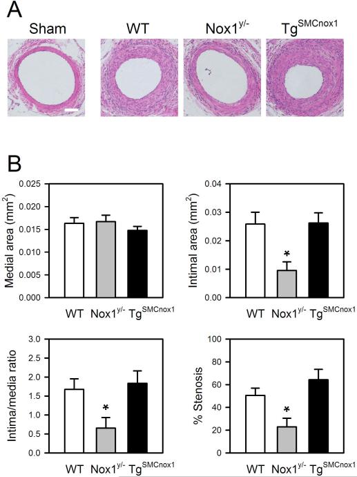
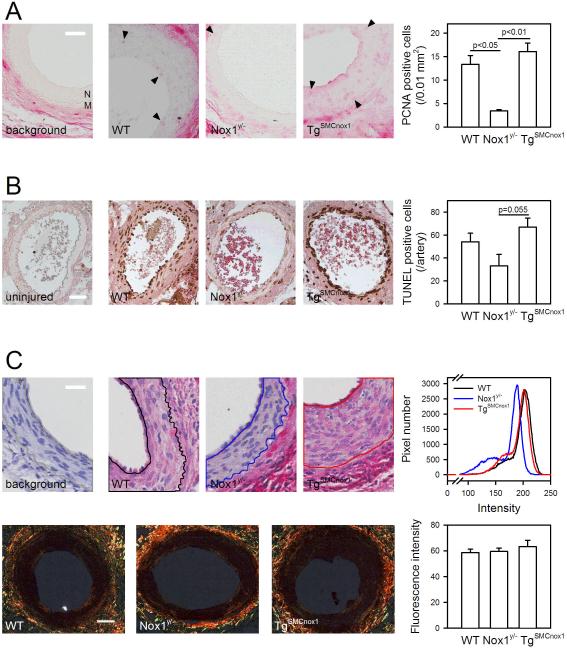
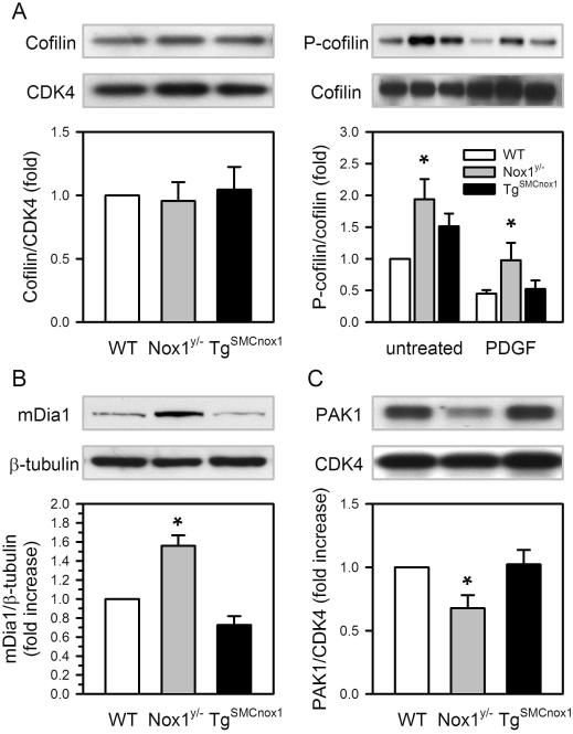
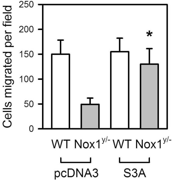
Methods and results: Wire injury-induced neointima formation in the femoral artery, along with proliferation and apoptosis, was reduced in Nox1(y/-) mice, but there was little difference in Tg(SMCnox1) mice compared with wild-type (WT) mice. Proliferation and migration were reduced in cultured Nox1(y/-) VSMCs and increased in Tg(SMCnox1) cells. Tg(SMCnox1) cells exhibited increased fibronectin secretion, but neither collagen I production nor cell adhesion was affected by alteration of Nox1. Using antibody microarray and Western blotting analysis, increased cofilin phosphorylation and mDia1 expression and decreased PAK1 expression were detected in Nox1(y/-) cells. Overexpression of S3A, a constitutively active cofilin mutant, partially recovered reduced migration of Nox1(y/-) cells, suggesting that reduction in cofilin activity contributes to impaired migration of Nox1(y/-) VSMCs.
Conclusions: These results indicate that Nox1 plays a critical role in neointima formation by mediating VSMC migration, proliferation, and extracellular matrix production, and that cofilin is a major effector of Nox1-mediated migration. Inhibition of Nox1 may be an efficient strategy to suppress neointimal formation.
Figures


Similar articles
-
Soluble Flt-1 gene transfer ameliorates neointima formation after wire injury in flt-1 tyrosine kinase-deficient mice.Arterioscler Thromb Vasc Biol. 2009 Apr;29(4):458-64. doi: 10.1161/ATVBAHA.109.183772. Epub 2009 Jan 22. Arterioscler Thromb Vasc Biol. 2009. PMID: 19164801
-
Nox1/PAK1 is required for angiotensin II-induced vascular inflammation and abdominal aortic aneurysm formation.Redox Biol. 2025 Feb;79:103477. doi: 10.1016/j.redox.2024.103477. Epub 2024 Dec 19. Redox Biol. 2025. PMID: 39721498 Free PMC article.
-
Formin mDia1 mediates vascular remodeling via integration of oxidative and signal transduction pathways.Circ Res. 2012 May 11;110(10):1279-93. doi: 10.1161/CIRCRESAHA.111.262519. Epub 2012 Apr 17. Circ Res. 2012. PMID: 22511750 Free PMC article.
-
Deficiency of NOX1/nicotinamide adenine dinucleotide phosphate, reduced form oxidase leads to pulmonary vascular remodeling.Arterioscler Thromb Vasc Biol. 2014 Jan;34(1):110-9. doi: 10.1161/ATVBAHA.113.302107. Epub 2013 Nov 14. Arterioscler Thromb Vasc Biol. 2014. PMID: 24233492
-
Zinc finger protein 191 deficiency attenuates vascular smooth muscle cell proliferation, migration, and intimal hyperplasia after endovascular arterial injury.J Vasc Surg. 2014 Feb;59(2):500-9. doi: 10.1016/j.jvs.2013.03.049. Epub 2013 Jun 4. J Vasc Surg. 2014. PMID: 23755975
Cited by
-
Vascular smooth muscle cell phenotypic switching in atherosclerosis.Heliyon. 2024 Sep 10;10(18):e37727. doi: 10.1016/j.heliyon.2024.e37727. eCollection 2024 Sep 30. Heliyon. 2024. PMID: 39309965 Free PMC article. Review.
-
Ambient Particulate Matter Induces Vascular Smooth Muscle Cell Phenotypic Changes via NOX1/ROS/NF-κB Dependent and Independent Pathways: Protective Effects of Polyphenols.Antioxidants (Basel). 2021 May 14;10(5):782. doi: 10.3390/antiox10050782. Antioxidants (Basel). 2021. PMID: 34069133 Free PMC article.
-
Redox signaling in cardiovascular pathophysiology: A focus on hydrogen peroxide and vascular smooth muscle cells.Redox Biol. 2016 Oct;9:244-253. doi: 10.1016/j.redox.2016.08.015. Epub 2016 Aug 26. Redox Biol. 2016. PMID: 27591403 Free PMC article. Review.
-
Coronary endothelial function and vascular smooth muscle proliferation are programmed by early-gestation dexamethasone exposure in sheep.Am J Physiol Regul Integr Comp Physiol. 2010 Jun;298(6):R1607-14. doi: 10.1152/ajpregu.00824.2009. Epub 2010 Mar 24. Am J Physiol Regul Integr Comp Physiol. 2010. PMID: 20335378 Free PMC article.
-
Nox isoforms in vascular pathophysiology: insights from transgenic and knockout mouse models.Redox Rep. 2010;15(2):50-63. doi: 10.1179/174329210X12650506623401. Redox Rep. 2010. PMID: 20500986 Free PMC article. Review.
References
-
- Zargham R. Preventing restenosis after angioplasty: a multistage approach. Clin Sci (Lond) 2008;114:257–264. - PubMed
-
- Ferns GA, Raines EW, Sprugel KH, Motani AS, Reidy MA, Ross R. Inhibition of neointimal smooth muscle accumulation after angioplasty by an antibody to PDGF. Science. 1991;253:1129–1132. - PubMed
-
- Lassègue B, Sorescu D, Szöcs K, Yin Q, Akers M, Zhang Y, Grant SL, Lambeth JD, Griendling KK. Novel gp91phox homologues in vascular smooth muscle cells: nox1 mediates angiotensin II-induced superoxide formation and redox-sensitive signaling pathways. Circ. Res. 2001;88:888–894. - PubMed
-
- Touyz RM, Chen X, Tabet F, Yao G, He G, Quinn MT, Pagano PJ, Schiffrin EL. Expression of a functionally active gp91phox-containing neutrophil-type NAD(P)H oxidase in smooth muscle cells from human resistance arteries: regulation by angiotensin II. Circ Res. 2002;90:1205–1213. - PubMed
Publication types
MeSH terms
Substances
Grants and funding
LinkOut - more resources
Full Text Sources
Molecular Biology Databases
Research Materials
Miscellaneous
