. 2009 Mar 27;284(13):8747-59.
doi: 10.1074/jbc.M807590200. Epub 2009 Jan 16.

PML3 Orchestrates the Nuclear Dynamics and Function of TIP60

Affiliations

PML3 Orchestrates the Nuclear Dynamics and Function of TIP60

Quan Wu et al. J Biol Chem. .

Abstract

The promyelocytic leukemia (PML) protein is a major component to govern the PML nuclear body (NB) assembly and function. Although it is well defined that PML NB is a site recruiting sumoylated proteins, the mechanism by which PML protein regulates the process remains unclear. Here we show that PML3, a specific PML isoform, interacts with and recruits TIP60 to PML NBs. Our biochemical characterization demonstrates that PML3 physically interacts with TIP60 via its N-terminal 364 amino acids. Importantly, this portion of TIP60 is sufficient to target to the PML NBs, suggesting that PML3-TIP60 interaction is sufficient for targeting TIP60 to the NBs. The PML3-TIP60 interaction is specific, since the region of TIP60 binding is not conserved in other PML isoforms. The physical interaction between PML3 and TIP60 protects TIP60 from Mdm2-mediated degradation, suggesting that PML3 competes with MDM2 for binding to TIP60. Fluorescence recovery after photobleaching analysis indicates that the PML3-TIP60 interaction modulates the nuclear body distribution and mobility of TIP60. Conversely, the distribution and mobility of TIP60 are perturbed in PML3-deficient cells, accompanied by aberrations in DNA damage-repairing response. Thus, PML3 orchestrates the distribution, dynamics, and function of TIP60. Our findings suggest a novel regulatory mechanism by which the PML3 and TIP60 tumor suppressors cooperate to ensure genomic stability.


Figures
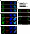
FIGURE 1.
FIGURE 1.
PML3 recruits TIP60 to PML NBs. A, TIP60 and nonsumoylatable TIP60dm diffuse throughout the nucleus. U2OS cells were stained using TIP60 antibody (a), or HeLa cells were transfected to express GFP-TIP60 (b) or GFP-TIP60dm (c). Thirty-six hours post-transfection, cells were fixed, permeabilized, and stained with PML antibody to indicate PML NBs and FLAG-PML3. Bar, 10 μm. B, PML3 facilitates TIP60 and nonsumoylatable TIP60dm to PML NBs. U2OS cells treated with UV (100 J/m2) were stained using TIP60 antibody (a), or HeLa cells were transfected to co-express FLAG-PML3 and GFP-TIP60 (b) or GFP-TIP60dm (c). Thirty-six hours post-transfection, cells were fixed, permeabilized, and stained with PML3 antibody or FLAG antibody to indicate FLAG-PML3. Bar, 10 μm. DAPI, 4′,6-diamidino-2-phenylindole. C, PML3 recruits TIP60 into PML NBs. U2OS cells grown on coverslips were transfected with Flag-PML3. Thirty-six hours post-transfection, cells were in normal condition (a) or exposed to UV (100 J/m2)(b), then cells were fixed, permeabilized and stained with TIP60 antibody and Flag antibody to indicate TIP60 and Flag-PML3. Bar: 10 μm. D, specific knockdown expression of PML3 by siRNA. U2OS cells were transfected with the PML3 siRNA oligonucleotide (150 μM) for different intervals (0, 24, 36 and 48 hours) and subjected to SDS-PAGE and immunoblotting. Top panel: PML3; middle panel: PML4; lower panel: tubulin. E, depletion of PML3 abrogates TIP60 targeting to PML NBs. U2OS cells were transfected with/without PML3 siRNA oligonucleotide (150 nM) (b, d and a, c). Forty-eight hours after transfection, cells were in normal condition or treated with UV-irradiation (100 J/m2)(a, b and c, d), and then fixed, permeabilized and stained with TIP60 and PML3 antibody to indicate TIP60 and PML3. Bar: 10 μm.
FIGURE 2.
FIGURE 2.
Direct interaction between PML3 and TIP60 is independent of their sumoylation. A, PML3 interacts with TIP60 in vivo. Whole cell extracts from normal U2OS cells and irradiated U2OS cells (UV, 100 J/m2) were subjected to co-immunoprecipitation (IP) and Western blotting with anti-TIP60 and PML3 antibody (Ab) as above. Lysates loaded onto the gel were 5% of those used for co-immunoprecipitation in U2OS cells. Cell extracts were run on a 10% SDS-polyacrylamide gel. Lanes 1, normal cells; lanes 2, cells treated with UV (100 J/m2) incubated with PML3 antibody; lanes 3, cells treated with UV (100 J/m2) incubated with control IgG. B, TIP60 interacts with PML3 in vivo. Whole-cell extracts from normal U2OS cells and U2OS cells irradiated with 100 J/m2 UV were subjected to co-immunoprecipitation (IP) and western blotting with anti-TIP60 and PML3 antibodies (Ab) as above. Lysates loaded onto the gel were 5% of those used for co-immunoprecipitation in U2O5 cells. Cell extracts were run on a 10% 5OS-PAGE gel. Lane 1, normal cell; lane 2, cells treated with UV (100 J/m2) were incubated with TIP60 antibody; lane 3, cells treated with UV (100 J/m2) were incubated with control IgG. C, PML3 interacts with TIP60 in vivo. 293T cells were transfected to co-express FLAG-PML3 and the indicated GFP-tagged TIP60 and GFP-TIP60dm constructs. Thirty-six hours after transfection, cells were collected for a co-immunoprecipitation assay with FLAG M2 beads. Immunoprecipitates were identified by Western blotting with GFP and FLAG antibodies. Lane 1, GFP + FLAG-PML3; lane 2, GFP-TIP60 + FLAG-PML3; lane 3, GFP-TIP60dm + FLAG-PML3. D, TIP60 interacts with PML3 in vivo. 293T cells were transfected to co-express FLAG-TIP60 or TIP60dm and the indicated GFP-tagged PML3 construct. Thirty-six hours after transfection, cells were collected for the co-immunoprecipitation assay with FLAG M2 beads. Immunoprecipitates were identified by Western blotting with GFP and FLAG antibodies. Lane 1, GFP + FLAG-TIP60; lane 2, GFP + FLAG-TIP60dm; lane 3, GFP-PML3 + FLAG-TIP60; lane 4, GFP-PML3 + FLAG-TIP60dm. E, schematic diagram of PML3 SUMO modification sites. F, nonsumoylatable PML33R disrupts PML NB localization of TIP60. HeLa cells were transfected to co-express the indicated constructs. Thirty-six hours after transfection, cells were fixed, permeabilized, and stained with FLAG antibody to the indicated FLAG-PML3 and FLAG-PML33R. Bar, 10 μm. G, PML33R interacts with TIP60 in vivo. 293T cells were transfected to co-express FLAG-PML3 or FLAG-PML33R and the indicated GFP-tagged TIP60 construct. Thirty-six hours after transfection, cells were collected for a co-immunoprecipitation assay with FLAG M2 beads. Immunoprecipitates were identified by Western blotting with GFP and FLAG antibodies. Lane 1, GFP + FLAG-PML3; lane 2, GFP + FLAG-PML33R; lane 3, GFP-TIP60 + FLAG-PML3; lane 4, GFP-TIP60 + FLAG-PML33R. H, PML3 interacts with TIP60 in vitro. GST-tagged recombinant PML3 protein purified on glutathione-agarose beads was used as an affinity matrix to absorb purified MBP-TIP60 and MBP-TIP60dm proteins from bacteria. GST-PML3 and MBP fusion proteins were used as loading controls. Pull-downs were identified by Western blotting with MBP antibody. Lane 1, MBP; lane 2, MBP + GST-PML3; lane 3, MBP-TIP60; lane 4, MBP-TIP60 + GST; lane 5, MBP-TIP60 + GST-PML3; lane 6, MBP-TIP60dm; lane 4, MBP-TIP60dm + GST; lane 8, MBP-TIP60dm + GST-PML3. DAPI, 4′,6-diamidino-2-phenylindole.
FIGURE 3.
FIGURE 3.
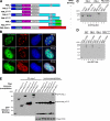
Dissection of TIP60 region required for PML3 binding and targeting of PML NBs. A, schematic diagram of TIP60 functional domains and related constructs. B, dissection of TIP60 fractions required for PML NBs targeting. HeLa cells were transfected to co-express FLAG-PML3 and the indicated GFP-tagged TIP60 deletion constructs. Thirty-six hours after transfection, cells were fixed, permeabilized, and stained with FLAG antibody to indicate FLAG-PML3. Bar, 10 μm. C, mapping regions of TIP60 responsible for PML3 binding. 293T cells were transfected to co-express FLAG-PML3 and the indicated GFP-tagged TIP60 deletion constructs. Thirty-six hours after transfection, cells were collected for a co-immunoprecipitation assay with FLAG antibody. Immunoprecipitates were identified by Western blotting with GFP antibody. Asterisks indicate the fractions of TIP60 deletions required for PML3 binding in vivo. Lanes 1, GFP; lanes 2, GFP-TIP60-(1-211); lanes 3, GFP-TIP60-(1-364); lanes 4, GFP-TIP60-(212-364); lanes 5, GFP-TIP60-(212-511); lanes 6, GFP-TIP60-(365-511). WT, wild type; DAPI, 4′,6-diamidino-2-phenylindole.
FIGURE 4.
FIGURE 4.
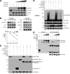
TIP60 specifies PML3 association by binding to PML3 C terminus. A, schematic diagram of PML3, PML4, PML-RARα, and various deletions. B, overexpression of PML3 isoform recruits PML NBs targeting of TIP60. HeLa cells were transfected to co-express FLAG-PML3, FLAG-PML4, FLAG-PML-RARα and the indicated GFP-tagged TIP60. Thirty-six hours after transfection, cells were fixed, permeabilized, and stained with FLAG antibody to indicate exogenous PML3, PML4, and PML-RARα. Bar, 10 μm. C, TIP60 specifically associates with PML3.293T cells were transfected to express FLAG-PML3, FLAG-PML4, and FLAG-RARα constructs. Thirty-six hours after transfection, cells were collected and lysed in 300 mm NaCl-containing lysis buffer (300 mm NaCl, 50 mm Tris-HCl, pH 7.5, 0.5% Nonidet P-40, 10% glycerol) for an in vitro binding assay. The lysates were then diluted twice and incubated with 4 μg of MBP-TIP60 or MBP alone. Pull-downs were identified by Western blotting with FLAG antibody. D, TIP60 specifically associates with PML3 in vitro. MBP fusion protein-bound amylose resin (Biolab) was used as an affinity matrix to isolate proteins interacting with TIP60 by purified MBP-tagged full-length that was expressed in bacteria. Briefly, the MBP-TIP60 fusion protein-bound amylose resin was incubated with purified proteins (GST-PML3, GST-PML4, and GST-PML-(1-394)) for 4 h at 4 °C. After the incubation, the resin was extensively washed with 0.25% Triton X-100 PBS and boiled in SDS-PAGE sample buffer, followed by fractionation of bound proteins on a 10% SDS-polyacrylamide gel. Proteins were then transferred onto a nitrocellulose membrane for Western blotting using GST antibody. E, TIP60 specifically interacts with PML3 C terminus. 293T cells were transfected to co-express FLAG-TIP60 and indicated GFP-tagged PML constructs. Thirty-six hours after transfection, cells were collected for a co-immunoprecipitation assay with FLAG antibody. Immunoprecipitates were identified by Western blotting with GFP antibody. Asterisks indicate TIP60 specifically associated with the C terminus of PML3 and PML3 full-length in vivo. Lanes 1, GFP; lanes 2, GFP-PML3-(1-394); lanes 3, GFP-PML3-(228-394); lanes 4, GFP-PML3-(1-570); lanes 5, GFP-PML4-(480-641); lanes 6, GFP-PML3-(480-633); lanes 7, GFP-PML3. DAPI, 4′,6-diamidino-2-phenylindole.
FIGURE 5.
FIGURE 5.
PML3 modulates TIP60 stability by impairing Mdm2 binding to TIP60. A, PML3 protects TIP60 stability in the presence of Mdm2. U2OS cells were transfected to express GFP-tagged TIP60 construct alone (0.5 μg; lane 1), together with HA-Mdm2 (1 μg; lane 2) or together with HA-Mdm2 and increasing amounts of PML3 (0.25 μg (lane 3), 1 μg (lane 4), 2 μg (lane 5), and 4 μg (lane 6)). Thirty-six hours after transfection, cells were harvested and subjected to SDS-PAGE for Western blotting detection. Transfection efficiency was monitored by expressing a constant amount of GFP expression plasmid (0.25 μg) in each sample. B, PML3 delays the degradation of TIP60. H1299 cells were transfected with GFP-TIP60 (1 μg) and GFP vector (0.25 μg) in the presence or absence of FLAG-PML3 (3 μg) and treated with CHX for the indicated durations. Whole cell extracts were then subjected to Western blotting using anti-tubulin, anti-FLAG, and anti-GFP antibodies. TIP60 densitometric signals were normalized to tubulin as a loading control. A 100% value was arbitrarily assigned to the signal obtained at zero time of cycloheximide treatment. Results are the means ± S.D. of three independent experiments. C, PML3 specifies TIP60 stability protection in the presence of Mdm2. U2OS cells were transfected to express GFP-TIP60 (lane 1), together with HA-Mdm2 (lane 2) or together with HA-Mdm2 and the indicated FLAG-tagged constructs (lanes 3-7). Thirty-six hours after transfection, cells were harvested and subjected to SDS-PAGE for Western blotting detection. D, PML3 specifies TIP60 stability protection by blocking Mdm2-mediated ubiquitious degradation. U2OS cells were transfected to express HA-TIP60 (lane 1), together with GFP-Mdm2 (lane 2) or together with GFP-Mdm2 and the indicated FLAG-tagged constructs plus INF-α (2 kilounits/ml) treatment (lanes 3-7). Thirty-six hours after transfection, cells were treated with MG132 (20 μm) for 6 h to block Mdm2-mediated TIP60 degradation and, as indicated, treated with INF-α (2 kilounits/ml) for 24 h. After MG132 treatment, cells were harvested and subjected to SDS-PAGE for Western blotting with HA antibody to detect TIP60 and ubiquitious TIP60 shift bands. Long exposure indicated ubiquitious TIP60 shift bands, and short exposure indicated TIP60 stability. E, PML3 competes with Mdm2 to bind to TIP60 in vivo. 293T cells were transfected to express the indicated FLAG-TIP60, together with GFP-Mdm2, or to express GFP-PML3 alone. Thirty-six hours after transfection, cells were harvested for a co-immunoprecipitation assay with FLAG M2 beads. Then using co-immunoprecipitation, immunoprecipitated beads were incubated with increasing amounts of cell lysates of GFP-PML3. Immunoprecipitates were identified by Western blotting with GFP antibody.
FIGURE 6.
FIGURE 6.
PML3 governs the dynamics of TIP60 in PML NBs. A, dynamic association of TIP60 with PML-NBs. HeLa cells were transiently transfected to express RFP-PML3, GFP-TIP60, GFP-TIP60 plus RFP-PML3, GFP-TIP60-(1-364), GFP-TIP60-(1-364) plus RFP-PML3, GFP-TIP60-(1-364), and GFP-TIP60-(1-364) plus RFP-PML3. RFP and GFP fluorescence was monitored in the aforementioned experimentation live cells. After a region of interest was selected, corresponding to nuclear dots (a, d, and f) or areas in nucleoplasm (b, c, e, g, and h), photobleaching was initiated, and the fluorescence recovery was quantified. The images were collected every 4 s. Representative images were selected and presented from given time points. B, quantification fluorescence recovery t½ for the bleached areas. The results are presented as mean values and S.E. of the percentages of the postbleach fluorescence intensities at the bleached areas relative to the prebleach intensity from 10 independent experiments.

Similar articles

Cited by

References

    1. Kuo, M. H., and Allis, C. D. (1998) BioEssays 20 615-626 - PubMed
    1. Struhl, K. (1998) Genes Dev. 12 599-606 - PubMed
    1. Sapountzi, V., Logan, I. R., and Robson, C. N. (2006) Int. J. Biochem. Cell Biol. 28 1496-1509 - PubMed
    1. Wu, H., Chen, Y., Liang, J. Shi, B., Wu, G., Zhang, Y., Wang, D., Li, R., Yi, X., Zhang, H., Sun, L., and Shang, Y. (2005) Nature 438 981-987 - PubMed
    1. Ikura, T., Ogryzko, V. V., Grigoriev, M., Groisman, R., Wang, J., Horikoshi, M., Scully, R., Qin, J., and Nakatani, Y. (2000) Cell 102 463-473 - PubMed

Publication types

MeSH terms