. 2009 Mar 27;284(13):8486-94.
doi: 10.1074/jbc.M808476200. Epub 2009 Jan 15.

Structure-guided Mutational Analysis of the Nucleotidyltransferase Domain of Escherichia coli DNA Ligase (LigA)

Affiliations

Structure-guided Mutational Analysis of the Nucleotidyltransferase Domain of Escherichia coli DNA Ligase (LigA)

Li Kai Wang et al. J Biol Chem. .

Abstract

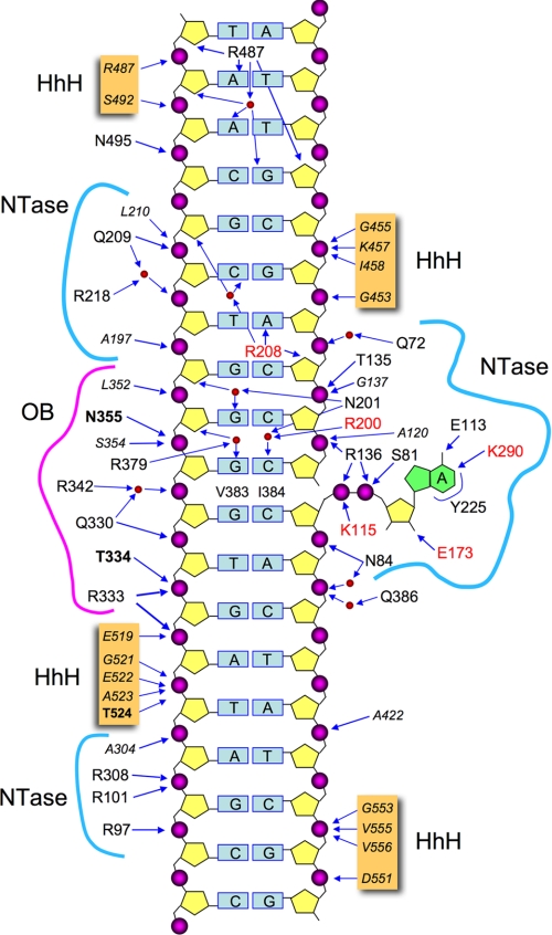
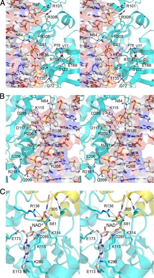
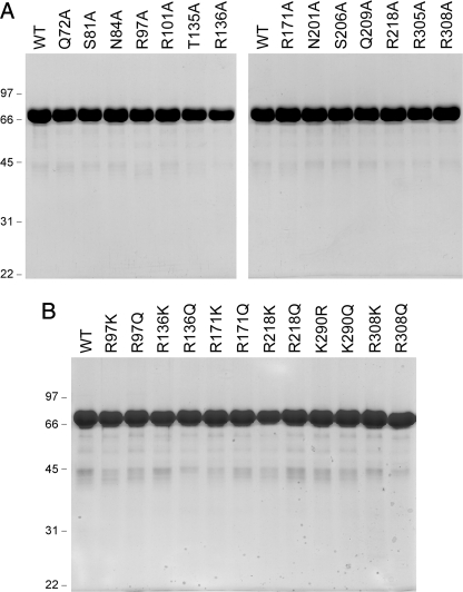
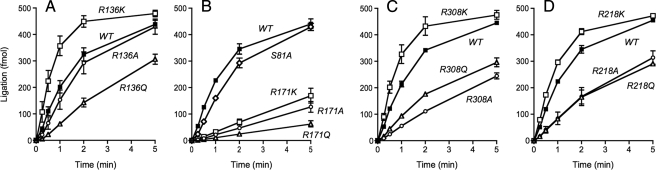
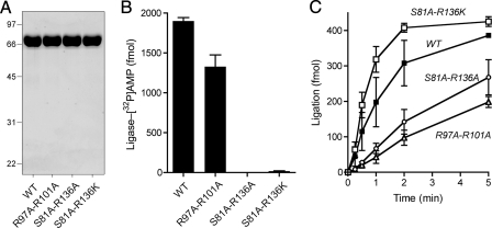
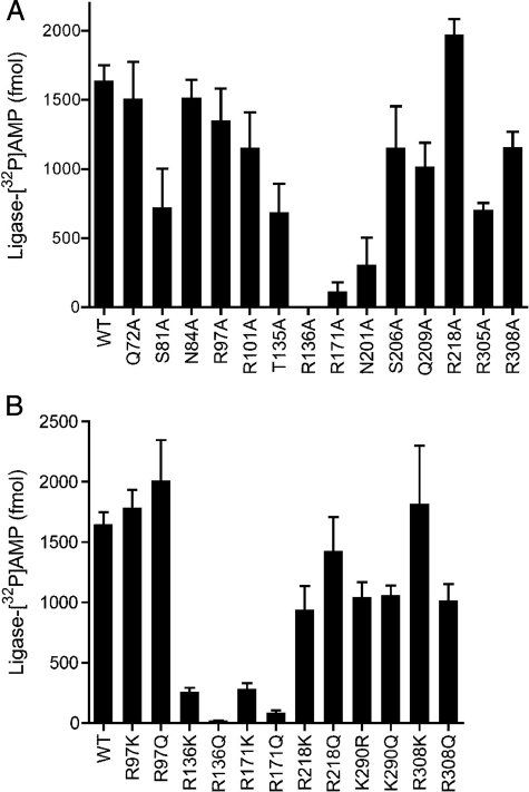
NAD(+)-dependent DNA ligases (LigA) are ubiquitous in bacteria, where they are essential for growth and present attractive targets for antimicrobial drug discovery. LigA has a distinctive modular structure in which a nucleotidyltransferase catalytic domain is flanked by an upstream NMN-binding module and by downstream OB-fold, zinc finger, helix-hairpin-helix, and BRCT domains. Here we conducted a structure-function analysis of the nucleotidyltransferase domain of Escherichia coli LigA, guided by the crystal structure of the LigA-DNA-adenylate intermediate. We tested the effects of 29 alanine and conservative mutations at 15 amino acids on ligase activity in vitro and in vivo. We thereby identified essential functional groups that coordinate the reactive phosphates (Arg(136)), contact the AMP adenine (Lys(290)), engage the phosphodiester backbone flanking the nick (Arg(218), Arg(308), Arg(97) plus Arg(101)), or stabilize the active domain fold (Arg(171)). Finer analysis of the mutational effects revealed step-specific functions for Arg(136), which is essential for the reaction of LigA with NAD(+) to form the covalent ligase-AMP intermediate (step 1) and for the transfer of AMP to the nick 5'-PO(4) to form the DNA-adenylate intermediate (step 2) but is dispensable for phosphodiester formation at a preadenylylated nick (step 3).


Figures

FIGURE 1.
FIGURE 1.
Schematic summary of E. coli LigA contacts to DNA. The nicked duplex DNA is depicted as a two-dimensional cartoon, with the continuous template strand on the left and the nicked strands on the right. The extrahelical 5′-adenylate is shown at right. The DNA contacts of LigA side chains (residue identity in plain text) and main chain amides (residue identity in italics) are indicated by arrows. Amino acids making both main chain and side chain contacts to DNA are in bold font. Water-mediated interactions are shown with waters as red spheres. LigA residues that penetrate the DNA helix and interact with the bases are indicated within the DNA base pair ladder. Residues Lys115, Glu173, Arg200, and Arg208 that were shown previously to be critical for LigA activity (19) are highlighted in red.
FIGURE 2.
FIGURE 2.
DNA interface of the NTase domain. A and B show stereo views of the NTase domain of E. coli LigA bound to the nicked DNA, which is rendered as a transparent surface over a stick model. Waters are depicted as red spheres. C shows a stereo view of the active site of Enterococcus faecalis LigA bound to NAD+ (7). The NTase domain is colored cyan, and the Ia domain is colored yellow. The amino acid numbers indicate the equivalent positions in E. coli LigA. Ionic and hydrogen bonding interactions are denoted by dashed lines in all panels.
FIGURE 3.
FIGURE 3.
LigA mutants. Aliquots (8 μg) of the nickel-agarose preparations of wild type (WT) LigA and the indicated alanine mutants (A) or conservative mutants (B) were analyzed by SDS-PAGE. The Coomassie Blue-stained gels are shown. The positions and sizes (kDa) of marker polypeptides are indicated on the left.
FIGURE 4.
FIGURE 4.
Mutational effects on the rate of sealing of a preadenylated nicked DNA. Reaction mixtures were constituted as described under “Experimental Procedures.” Each datum is the average of three (A) or two (B–D) separate experiments in which wild type LigA was assayed in parallel with the mutants specified in each graph. The error bars denote the standard deviation.
FIGURE 5.
FIGURE 5.
Effects of double-alanine mutations. A, aliquots (8 μg) of the nickel-agarose preparations of wild type (WT) LigA and the indicated double-alanine mutants were analyzed by SDS-PAGE. The Coomassie Blue-stained gel is shown. The positions and sizes (kDa) of marker polypeptides are indicated on the left. B, ligase adenylylation. Reaction mixtures contained 1 μm [32P-adenylate]-NAD and 8 pmol of LigA. The extents of label transfer to LigA are shown. Each datum is the average of triplicate assays from a single experiment in which wild type LigA was tested in parallel with the mutants specified. The error bars denote the standard deviation. C, sealing a preadenylylated nick. Reaction mixtures were constituted as described under “Experimental Procedures.” Each datum is the average of three separate experiments in which wild type LigA was assayed in parallel with the mutants specified. The error bars denote the standard deviation.
FIGURE 6.
FIGURE 6.
Mutational effects on ligase adenylylation. Reaction mixtures contained 1 μm [32P-adenylate]NAD and 8 pmol of LigA. The extents of label transfer to LigA are shown. Each datum is the average of three separate experiments in which wild type LigA was assayed in parallel with the mutants specified. The error bars denote the standard deviation.

Similar articles

Cited by

References

    1. Zhao, A., Gray, F., and MacNeill, S. A. (2006) Mol. Microbiol. 59 743–752 - PubMed
    1. Little, J. W., Zimmerman, S. B., Oshinsky, C. K., and Gellert, M. (1967) Proc. Natl. Acad. Sci. U. S. A. 58 2004–2011 - PMC - PubMed
    1. Gumport, R. I., and Lehman, I. R. (1971) Proc. Natl. Acad. Sci. U. S. A. 68 2559–2563 - PMC - PubMed
    1. Olivera, B. M., Hall, Z. W., and Lehman, I. R. (1968) Proc. Natl. Acad. Sci. U. S. A. 61 237–244 - PMC - PubMed
    1. Singleton, M. R., Håkansson, K., Timson, D. J., and Wigley, D. B. (1999) Structure 7 35–42 - PubMed

Publication types

MeSH terms

LinkOut - more resources