. 2009 Mar 20;284(12):7875-88.
doi: 10.1074/jbc.M807827200. Epub 2009 Jan 16.

Identification and functional analysis of a novel cyclin e/cdk2 substrate ankrd17

Affiliations

Identification and functional analysis of a novel cyclin e/cdk2 substrate ankrd17

Min Deng et al. J Biol Chem. .

Abstract

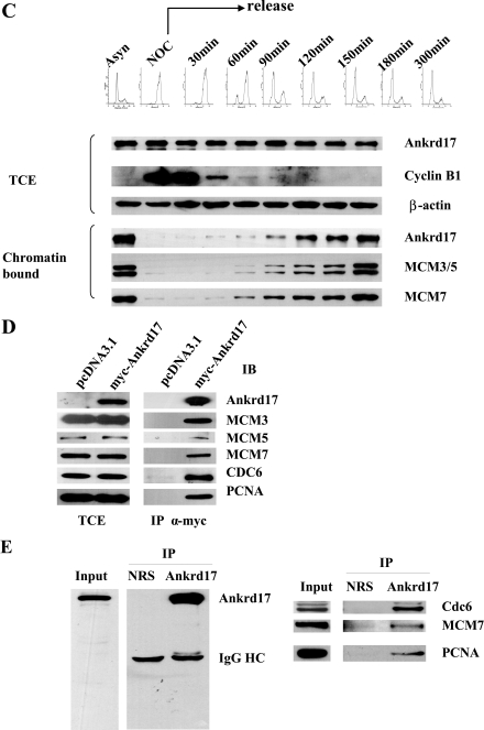
Cyclin E/Cdk2 is a key regulator in G(1)-S transition. We have identified a novel cyclin E/Cdk2 substrate called Ankrd17 (ankyrin repeat protein 17) using the TAP tag purification technique. Ankrd17 protein contains two clusters of a total 25 ankyrin repeats at its N terminus, one NES (nuclear exporting signal) and one NLS (nuclear localization signal) in the middle, and one RXL motif at its C terminus. Ankrd17 is expressed in various tissues and associates with cyclin E/Cdk2 in an RXL-dependent manner. It can be phosphorylated by cyclin E/Cdk2 at 3 phosphorylation sites (Ser(1791), Ser(1794), and Ser(2150)). Overexpression of Ankrd17 promotes S phase entry, whereas depletion of Ankrd17 expression by small interfering RNA inhibits DNA replication and blocks cell cycle progression as well as up-regulates the expression of p53 and p21. Ankrd17 is localized to the nucleus and interacts with DNA replication factors including MCM family members, Cdc6 and PCNA. Depletion of Ankrd17 results in decreased loading of Cdc6 and PCNA onto DNA suggesting that Ankrd17 may be directly involved in the DNA replication process. Taken together, these data indicate that Ankrd17 is an important downstream effector of cyclin E/Cdk2 and positively regulates G(1)/S transition.


Figures

FIGURE 1.
FIGURE 1.
Identification of Cdk2-associated proteins. A, 293T cells were transduced with Cdk2 expression retrovirus pMSCV-c-FLAG-HA-Cdk2. Empty retrovirus pMSCV-c-FLAG-HA was used as a control. The transduced cells were lysed and subjected to double immunoprecipitation, first with anti-FLAG beads and then with anti-HA beads. The elution was separated on a 4–12% Nupage SDS-PAGE and resolved proteins were visualized by staining with Sigma EZ blue and excised and subjected to mass spectrometry analysis. B, the list of Cdk2-associated proteins were identified by mass spectrometry. C, GST pull-down assay. 293T cells were transfected with Myc-tagged cyclin E and Myc-tagged Cdk2 plasmids. The cell lysates were incubated with GST, GST-Ankrd17, and GST-Ankrd17AQA, respectively. The associated proteins were pulled down by glutathione beads and analyzed by immunoblotting (IB) with Myc antibody. D, 293T cells were transfected with FLAG-tagged Ankrd17 or Ankrd17AQA, in which the RQL motif was mutated to AQA. The cell lysates were immunoprecipitated (IP) with FLAG antibody and immunoblotted with Cdk2 and cyclin E antibodies, respectively. E, the lysates from 293T cells were immunoprecipitated with normal rabbit serum and anti-Ankrd17 antibody, respectively, and immunoblotted with Cdk2 and Ankrd17 antibodies. F, the cell lysates from 293T cells were immunoprecipitated with normal rabbit serum or cyclin E antibody and immunoblotted with Ankrd17 antibody.
FIGURE 2.
FIGURE 2.
Characteristics of Ankrd17 protein. A, structure of ankyrin repeat protein Ankrd17. B, Ankrd17 expressed in different tissues. RNA from different human tissues were isolated and 1 μg of RNA was used for reverse transcriptase-PCR with Ankrd17 primers and glyceraldehyde-3-phosphate dehydrogenase (GAPDH) primers as internal control. C, Ankrd17 was localized in the nucleus in U2OS and HeLa cells. U2OS and HeLa cells were fractionated as cytosolic supernatant (C) and nuclear fraction (N). The lysates were subjected to immunoblot with Ankrd17, β-tubulin, and MCM antibodies. TCE refers to total cell lysate. D, the phosphorylation sites of Ankrd17 identified by mass spectral analysis. 293T cells were transfected with pCMV-FLAG-Ankrd17, pCS2mt-cyclin E, and pCMV-Myc-Cdk2. Cell lysates were harvested and Ankrd17 protein was immunoprecipitated with FLAG beads and subjected for mass spectrometry analysis.
FIGURE 3.
FIGURE 3.
In vitro kinase assay. A, 1 μg of GST, GST-Ankrd17, or GST-Ankrd17AQA were incubated with [γ-32P]ATP in the absence (lanes 1, 3, and 5) or presence (lanes 2, 4, and 6) of purified GST-cyclin E and GST-Cdk2. The reactions were subjected for SDS-PAGE and kinase activity was visualized by autoradiography of the gel. B, GST, GST-Ankrd17, or GST-Ankrd17AQA were incubated with [γ-32P]ATP in the presence of GST-cyclin E and GST-Cdk2 (Lanes 1–3) or GST-cyclin A and GST-Cdk2 (lanes 4–6). The reactions were subjected to SDS-PAGE and the phosphorylation of Ankrd17 was visualized by autoradiography of the gel. C, the GST-Ankrd17 (amino acids 1729–1795) and its Ser/Ala mutants were subjected to in vitro kinase assay as described in A. Lanes 1–7 were GST, GST-Ankrd17, GST-Ankrd17 S1740A/S1791A/S1794A, GST-Ankrd17 S1737A/S1791A/S1794A, GST-Ankrd17 S1737A/S1740A/S1794A, GST-Ankrd17 S1737A/S1740A/S1791A, and GST-Ankrd17 S1737A/S1740A/S1791A/S1794A, respectively.
FIGURE 4.
FIGURE 4.
Overexpression of Ankrd17 promotes cell cycle progression. A, the U2OS cells were transfected with pcDNA3.1 as control or the indicated amount of pCMV-FLAG-Ankrd17 together with pCMV-CD20 plasmid for the selection of transfected cells. Cells were harvested at 48 h after transfection and subjected for flow cytometry analysis. The results represent the means from triplicate samples. B, U2OS cells were transfected with 7.5 μg of pCMV-FLAG-Ankrd17 or pcDNA3.1 as control. 24 h after transfection, cells were then treated with DMSO (mock) or 100 ng/ml nocodazole for 22 h and harvested for flow cytometry. C, U2OS cells were transfected with pCMV-FLAG-Ankrd17 alone or with pCMV-Myc-Cdk2 and pCMV-Myc-cyclin E as indicated and harvested 48 h after transfection and subjected to flow cytometry analysis.
FIGURE 5.
FIGURE 5.
Effects of depletion of Ankrd17 on U2OS cell cycle distribution. A, analysis of the knockdown efficiency of three Ankrd17 siRNAs. U2OS cells were transfected with control siRNA and Ankrd17 siRNA1, -2, and -3. Cells were harvested at 12 or 48 h after transfection and the protein level of Ankrd17 were assessed by immunoblotting. B, U2OS cells were transfected with control siRNA and Ankrd17 siRNA1, -2, and -3 as in A, 48 h after transfection cells were harvested and analyzed for flow cytometry. C, U2OS cells were transfected with control siRNA and Ankrd17 siRNA1 for 24 h and arrested by serum starvation for 48 h and released for 16 h, and flow cytometry was performed before and after serum stimulation. The results represent the means from triplicate samples. D, depletion of Ankrd17 regulates the expression of p53 and p21. U2OS cells were transfected with control siRNA and Ankrd17 siRNA. The cell lysates were harvested and subjected to immunoblot with the antibodies as indicated.
FIGURE 6.
FIGURE 6.
Depletion of Ankrd17 inhibits DNA replication. U2OS cells were transfected with control siRNA and Ankrd17 siRNA1. The cells were pulse labeled with BrdUrd for 10 min at 48 or 72 h after transfection. The cells were fixed and stained with anti-BrdUrd antibody (red) and DNA was stained with 4′,6-diamidino-2-phenylindole (DAPI) (blue) (A for 48 h, B for 72 h). The percentage of BrdUrd positive cells were counted from 600 cells (C). The mean results from two independent experiments are depicted. D, the U2OS cells were transfected with siRNA and starved for 48 h, then released for 2–20 h as indicated and labeled with BrdUrd. The BrdUrd-positive cells were counted.
FIGURE 7.
FIGURE 7.
Ankrd17 bound to chromatin and interacted with proteins in DNA pre-replication complex. A, U2OS cells were lysed in CSK buffer; the CSK-insoluble (CSKP) fraction was then treated with micrococcal nuclease (MNase) or the indicated concentration of NaCl. The soluble (S) and insoluble (P) fractions were separated by centrifugation. The total cell extract (TCE) and all fractions were subjected to immunoblot with Ankrd17 and MCM3 antibodies. B, U2OS cells were synchronized at G1, G1/S, S, S/G2, and M phases assessed by flow cytometry (upper panel) as described under “Experimental Procedures.” Total cell extracts and chromatin-bound proteins were prepared for immunoblotting with the indicated antibodies. C, HeLa cells were synchronized at M phase with nocodazole and harvested by mitotic shake-off. Cells were collected at different time points after release from the block and the total cell extract and CSK-insoluble fraction (chromatin-bound) were prepared and subjected to immunoblot with the indicated antibodies and cyclin B as the G2/M phase marker. The DNA contents of cells were determined by flow cytometry (upper panel). D, 293T cells were transfected with pCMV-Myc-Ankrd17 or pcDNA3.1 as control. After 48 h the cells were harvested and cell lysates were immunoprecipitated with Myc antibody and immunoblotted with MCM3, MCM5, MCM7, Cdc6, and PCNA antibodies, respectively. E, 293T cell lysates were immunoprecipitated (IP) with Ankrd17 antibody and normal rabbit serum as control and immunoblotted with indicated antibodies.
FIGURE 7.
FIGURE 7.
Ankrd17 bound to chromatin and interacted with proteins in DNA pre-replication complex. A, U2OS cells were lysed in CSK buffer; the CSK-insoluble (CSKP) fraction was then treated with micrococcal nuclease (MNase) or the indicated concentration of NaCl. The soluble (S) and insoluble (P) fractions were separated by centrifugation. The total cell extract (TCE) and all fractions were subjected to immunoblot with Ankrd17 and MCM3 antibodies. B, U2OS cells were synchronized at G1, G1/S, S, S/G2, and M phases assessed by flow cytometry (upper panel) as described under “Experimental Procedures.” Total cell extracts and chromatin-bound proteins were prepared for immunoblotting with the indicated antibodies. C, HeLa cells were synchronized at M phase with nocodazole and harvested by mitotic shake-off. Cells were collected at different time points after release from the block and the total cell extract and CSK-insoluble fraction (chromatin-bound) were prepared and subjected to immunoblot with the indicated antibodies and cyclin B as the G2/M phase marker. The DNA contents of cells were determined by flow cytometry (upper panel). D, 293T cells were transfected with pCMV-Myc-Ankrd17 or pcDNA3.1 as control. After 48 h the cells were harvested and cell lysates were immunoprecipitated with Myc antibody and immunoblotted with MCM3, MCM5, MCM7, Cdc6, and PCNA antibodies, respectively. E, 293T cell lysates were immunoprecipitated (IP) with Ankrd17 antibody and normal rabbit serum as control and immunoblotted with indicated antibodies.
FIGURE 8.
FIGURE 8.
Depletion of Ankrd17 decreases Cdc6 and PCNA loading on chromatin. A, U2OS cells were transfected with control siRNA and Ankrd17 siRNA1 and extracted with CSK buffer. Total cell extract and CSK-insoluble proteins were subjected for immunoblotting with the indicated antibodies. B, the untransfected or transfected cells were extracted with or without CSK buffer (unextracted), fixed, and immunostained for PCNA (red) and MCM3 (green) antibodies, DNA was stained with 4′,6-diamidino-2-phenylindole (DAPI) (blue). C, untransfected and transfected cells were subjected to in situ extraction with CSK buffer or mock extraction (unextracted), fixed, and immunostained for PCNA (left) or MCM3 (right) antibodies as B. The percentages of nuclei staining positive cells for PCNA or MCM3 were counted.

Similar articles

Cited by

References

    1. Hwang, H. C., and Clurman, B. E. (2005) Oncogene 24 2776-2786 - PubMed
    1. Prior, M., Lehmann, S., Sy, M. S., Molloy, B., and McMahon, H. E. (2007) J. Virol. 81 11195-11207 - PMC - PubMed
    1. Reed, S. I. (1997) Cancer Surv. 29 7-23 - PubMed
    1. Sherr, C. J. (1994) Cell 79 551-555 - PubMed
    1. Brown, N. R., Nobel, M., Endicott, J. A., and Johnson, L. N. (1999) Nat. Cell Biol. 1 438-443 - PubMed

Publication types

MeSH terms