. 2009 Apr;37(5):1616-27.
doi: 10.1093/nar/gkp001. Epub 2009 Jan 16.

Unconventional decoding of the AUA codon as methionine by mitochondrial tRNAMet with the anticodon f5CAU as revealed with a mitochondrial in vitro translation system

Affiliations

Unconventional decoding of the AUA codon as methionine by mitochondrial tRNAMet with the anticodon f5CAU as revealed with a mitochondrial in vitro translation system

Chie Takemoto et al. Nucleic Acids Res. 2009 Apr.

Abstract

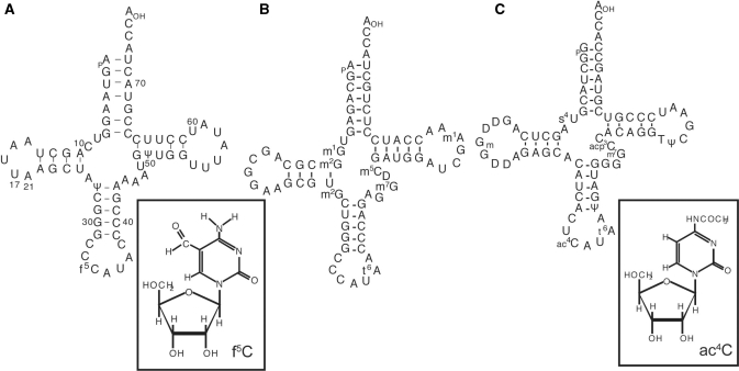
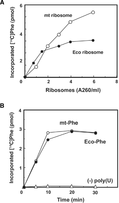
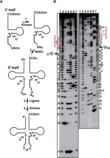
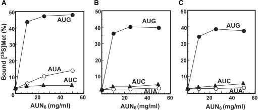
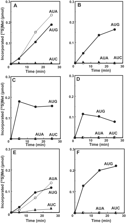
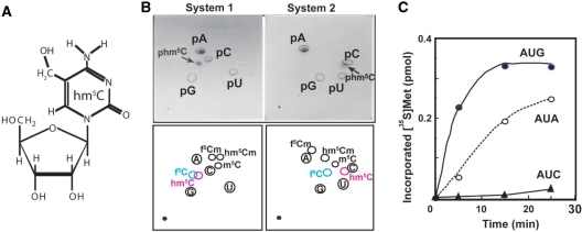
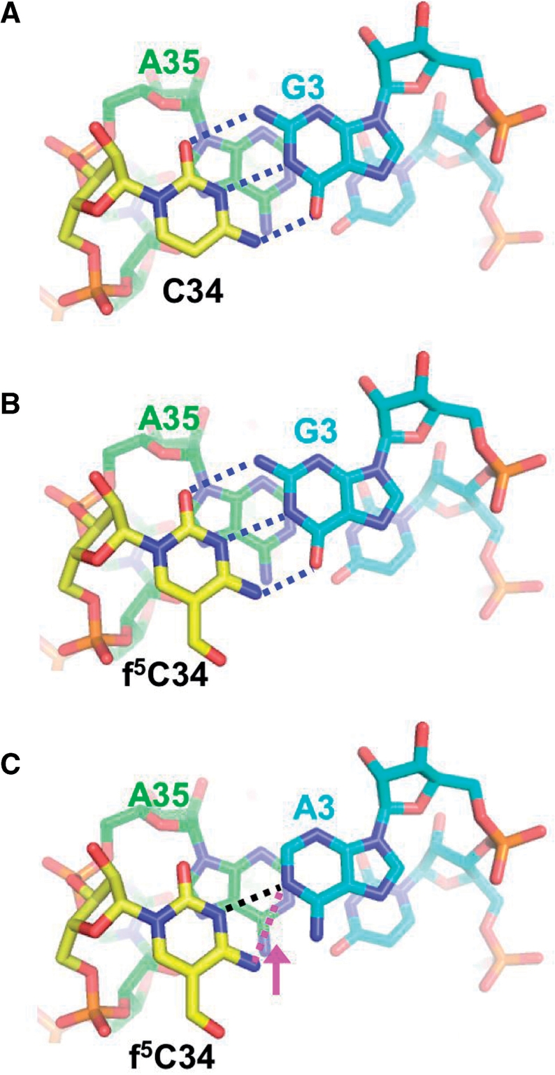
Mitochondrial (mt) tRNA(Met) has the unusual modified nucleotide 5-formylcytidine (f(5)C) in the first position of the anticodon. This tRNA must translate both AUG and AUA as methionine. By constructing an in vitro translation system from bovine liver mitochondria, we examined the decoding properties of the native mt tRNA(Met) carrying f(5)C in the anticodon compared to a transcript that lacks the modification. The native mt Met-tRNA could recognize both AUA and AUG codons as Met, but the corresponding synthetic tRNA(Met) lacking f(5)C (anticodon CAU), recognized only the AUG codon in both the codon-dependent ribosomal binding and in vitro translation assays. Furthermore, the Escherichia coli elongator tRNA(Met)(m) with the anticodon ac(4)CAU (ac(4)C = 4-acetylcytidine) and the bovine cytoplasmic initiator tRNA(Met) (anticodon CAU) translated only the AUG codon for Met on mt ribosome. The codon recognition patterns of these tRNAs were the same on E. coli ribosomes. These results demonstrate that the f(5)C modification in mt tRNA(Met) plays a crucial role in decoding the nonuniversal AUA codon as Met, and that the genetic code variation is compensated by a change in the tRNA anticodon, not by a change in the ribosome. Base pairing models of f(5)C-G and f(5)C-A based on the chemical properties of f(5)C are presented.


Figures

Figure 1.
Figure 1.
Nucleotide sequences of tRNAs used in this study and chemical structures of the modified nucleotide in the wobble position. (A) Bovine mt tRNAMet and 5-formylcytidine, (B) bovine cytoplasmic initiator tRNAMet and (C) E. coli elongator tRNAMet and 4-acetylcytidine. The numbering of the residues in the cloverleaf structure of the mt tRNA conforms to that in the previous report (71).
Figure 2.
Figure 2.
In vitro translation system prepared from bovine liver mitochondria. (A) Dose–response of poly(Phe) synthesis, as a function of the amount of mitochondrial (open circle) or E. coli (filled circle) ribosomes, using E. coli Phe-tRNA and mt EF-Tu/Ts. The reactions were carried out at 37°C for 8 min. (B) Poly(U)-dependent poly(Phe) synthesis activity of the complete mitochondrial system under the optimized conditions for Phe-tRNA from mitochondria (open circle) and E. coli (filled circle), respectively, as described in the Materials and methods section.
Figure 3.
Figure 3.
Preparation of synthetic tRNAMet variants. (A) Process of synthesizing mt tRNAMet by a combination of chemical synthesis and ligation (33). The synthetic tRNAs have sequences identical to the respective sequences of native tRNAMet, except for f5C and pseudouridine (Ψ). (B) Sequencing analysis using the Donis–Keller′s method of the synthetic mt tRNAMet labeled with [32P] at the 5′-end (left) and the 3′-end (right), respectively (30,32). Electrophoresis was performed on a 15% polyacrylamide–7 M urea–10% glycerol gel. Lanes: control without ribonuclease (RNase) (lane 1); limited alkaline hydrolysis (lanes 2 and 7); digestion by RNase T1 (specific for G: lane 3), RNase U2 (specific for A: lane 4), RNase PhyM (specific for A and U: lane 5) and RNase CL3 (specific for C: lane 6). As indicated by the arrows, Ψ was not digested by RNase PhyM (12). The numbering of the residues in the cloverleaf structure of the mt tRNA conforms to that in the previous report (71).
Figure 4.
Figure 4.
Codon-dependent ribosome binding assays of bovine mt tRNAMet variants. (A) Native mt tRNAMet purified from bovine liver mitochondria. (B) Synthetic mt tRNAMet with no modified bases. (C) Synthetic mt tRNAMet with two Ψ's. Oligonucleotides AUG6 (filled circle), AUA6 (open circle) and AUC6 (filled triangle) were used as mRNA. The presented values are the averages of three independent experiments and were reproducible within ± 1.2%.
Figure 5.
Figure 5.
The codon recognition abilities of tRNAsMet possessing different anticodons. Time courses of oligo(AUN)-dependent oligo(Met) synthesis in an in vitro translation system with mitochondrial (A–D) or E. coli (E and F) ribosomes. (A and E) Native bovine mt tRNAMet purified from bovine liver mitochondria. (B) Synthetic mt tRNAMet with two Ψ's. (C) Bovine cytoplasmic initiator tRNAMet. (D and F) E. coli elongator tRNAMet. Oligonucleotides AUA11 (open circle), AUC11 (filled triangle) and AUG11 (filled circle) were used as mRNA. The incorporation of [35S]Met was normalized by subtracting the value without mRNA, which was <2% of the input c.p.m. (about 0.03 pmol).
Figure 6.
Figure 6.
Reduction of 5-formylcytidine to 5-hydroxylcytidine (hm5C) of mt tRNAMet. (A) The chemical structure of hm5C. (B) 2D-TLC analysis of the nucleotide at the anticodon wobble position of mt tRNAMet after reduction (upper panels), and diagrams of chromatographic mobility of modified cytosine 5′-monophospates (lower panels) (49,50). The detailed procedure is described in Materials and Methods section. The solvent used for the first dimension in both systems was isobutyric acid/concentrated ammonia/water (66:1:33 v/v/v). For the second dimension, 2-propanol/HCl/water (70:15:15 v/v/v) and ammonium sulfate/0.1 M sodium phosphate (pH 6.8)/1-propanol (60 g:100 ml:2 ml) was used in the systems 1 and 2, respectively. (C) The codon recognition ability of the reduced mt tRNAMet (anticodon hm5CAU) on mt ribosomes. The incorporation of [35S]-Met was normalized by subtracting the value without mRNA.
Figure 7.
Figure 7.
Base pairing model of f5C-G and f5C-A. (A) C34-G1 base pair of the initiator tRNAMet and the AUG codon (PDB: 2J00). The first letter of the anticodon C34 (yellow) base pairs with the third letter of the codon G3 (cyan) and stacks on the 3′-adjacent nucleotide A35 (green). Hydrogen bonds between C34 and G3 are shown as blue dotted lines. (B) f5C-G base pair model. The coordinates of f5C were generated with the PRODRG server (http://davapc1.bioch.dundee.ac.uk/prodrg) (72) and superposed to the C34 in (A). Putative hydrogen bonds between f5C34 and G3 are shown as blue dotted lines. (C) The f5C-A base pair model. The coordinates for adenine were also generated with the PRODRG server and superposed on G3 in (B). Putative hydrogen bonds between f5C34 and A3 are shown as dotted lines. The distance between N3 of f5C34 and N1 of A3 or N4 of f5C34 and N1 of A3 is 2.95 Å (black dotted line) or 3.44 Å (magenta dotted line), respectively. An arrow (magenta) represents an expected shift to form a hydrogen bond between N4 of f5C34 and N1 of A3. These figures were prepared with PyMOL, from DeLano Scientific (http://www.pymol.sourceforge.net/).

References

    1. Anderson S, Bankier AT, Barrell BG, de Bruijn MHL, Coulson AR, Drouin J, Eperon IC, Nierlich DP, Roe BA, Sanger F, et al. Sequence and organization of the human mitochondrial genome. Nature. 1981;290:457–465. - PubMed
    1. Himeno H, Masaki H, Kawai T, Ohta T, Kumagai I, Miura K, Watanabe K. Unusual genetic codes and a novel gene structure for tRNA(AGYSer) in starfish mitochondrial DNA. Gene. 1987;56:219–230. - PubMed
    1. Wolstenholme DR. Animal mitochondrial DNA: structure and evolution. Int. Rev. Cytol. 1992;141:173–216. - PubMed
    1. Xu X, Arnason U. The complete mitochondrial DNA sequence of the horse, Equus caballus: extensive heteroplasmy of the control region. Gene. 1994;148:357–362. - PubMed
    1. Krettek A, Gullberg A, Arnason U. Sequence analysis of the complete mitochondrial DNA molecule of the hedgehog, Ernaceus europaeus, and the phylogenic position of the Lipotyphla. J. Mol. Evol. 1995;41:952–957. - PubMed

Publication types

MeSH terms