. 2009 Jan;21(1):90-105.
doi: 10.1105/tpc.108.061812. Epub 2009 Jan 16.

ENDOSPERM DEFECTIVE1 Is a Novel Microtubule-Associated Protein Essential for Seed Development in Arabidopsis

Affiliations

ENDOSPERM DEFECTIVE1 Is a Novel Microtubule-Associated Protein Essential for Seed Development in Arabidopsis

Cristina Pignocchi et al. Plant Cell. 2009 Jan.

Abstract

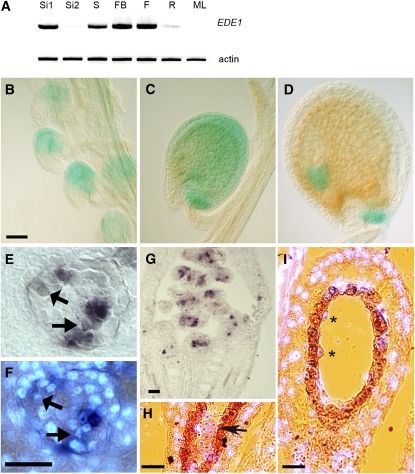
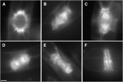
Early endosperm development involves a series of rapid nuclear divisions in the absence of cytokinesis; thus, many endosperm mutants reveal genes whose functions are essential for mitosis. This work finds that the endosperm of Arabidopsis thaliana endosperm-defective1 (ede1) mutants never cellularizes, contains a reduced number of enlarged polyploid nuclei, and features an aberrant microtubule cytoskeleton, where the specialized radial microtubule systems and cytokinetic phragmoplasts are absent. Early embryo development is substantially normal, although occasional cytokinesis defects are observed. The EDE1 gene was cloned using a map-based approach and represents the pioneer member of a conserved plant-specific family of genes of previously unknown function. EDE1 is expressed in the endosperm and embryo of developing seeds, and its expression is tightly regulated during cell cycle progression. EDE1 protein accumulates in nuclear caps in premitotic cells, colocalizes along microtubules of the spindle and phragmoplast, and binds microtubules in vitro. We conclude that EDE1 is a novel plant-specific microtubule-associated protein essential for microtubule function during the mitotic and cytokinetic stages that generate the Arabidopsis endosperm and embryo.


Figures

Figure 1.
Figure 1.
EDE1 Is Essential for Seed Development. (A) to (D) Confocal micrographs of Feulgen- stained seeds of the wild type (A) and ede1-1 mutant ([B] to [D]) at 6 DAP. Note the NCDs that invade the central vacuole (CV) of the endosperm. Only few enlarged NCDs are present in the ede1-1 mutant when compared with the wild type. The range of ede1-1 phenotypes is also shown: mild (B), moderate (C), and extreme (D). EM, embryo; CE, chalazal endosperm. Bars = 20 μm. (E) Developmental stages of the wild type (top row) and of ede1-1 mutant endosperm (bottom row). Whole mounts of cleared seeds were observed at different developmental stages (0 to 8 DAP) using differential interference contrast optics. The enlarged nuclei in the mutant are marked by asterisks. Bar = 20 μm (0 to 2 DAP) and 50 μm (4 to 8 DAP).
Figure 2.
Figure 2.
EDE1 Is Required for Microtubule Function in Endosperm. Wild-type ([A] to [C]) and mutant ([D] to [F]) endosperm stained with DAPI (red) and immunolabeled with antitubulin (green). Bar = 20 μm in (A), (B), (D), (E), and (F) and 40 μm in (C). (A) and (B) Syncytial phase of wild-type endosperm development at 4 DAP with microtubules radiating from evenly spaced nuclei around the periphery of the embryo sac. (C) Cytokinesis during endosperm cellularization at 6 DAP. The asterisks indicate phragmoplasts. (D) The mutant syncytial endosperm at 4 DAP is characterized by few enlarged nuclei displaying numerous nucleoli and lacking organized microtubule structures. (E) At 6 DAP, the mutant endosperm displays giant nuclei without radial microtubule systems and that never cellularize. (F) At 8 DAP, when the wild-type endosperm is fully cellularized, ede1-1 endosperm contains only giant nuclei with several nucleoli.
Figure 3.
Figure 3.
Structure of the EDE1 Gene. (A) Blocks denote exons, and lines denote introns. The ede1-1 allele contains a G-to-A base transition at +1066, ede1-2 contains an insertion of T-DNA into exon 1 (+414), and ede1-3 contains an insertion of T-DNA into intron 2 (+1231). The sequence of a cDNA (GenBank BX818775) was used to define the 5′ and 3′ untranslated regions, shown as gray boxes. (B) Detail of DNA and amino acid sequences surrounding the ede1-1 mutation in Columbia (Col) and ede1-1. Comparison of ede1-1 and Col cDNA sequences shows that the G-to-A polymorphism at +1066 removes the exon1/intron1 splice site and that the ede1-1 mutant plant uses a cryptic splice site at +1120 instead. (C) RT-PCR of At2g44190 transcript from total RNA isolated from young siliques of Col wild-type and homozygous ede1-1 plants. Product from genomic DNA is shown as control. mkr, marker (kb).
Figure 4.
Figure 4.
ede1 Null Mutant Embryos Have Cell Division Defects: Histological Sections Reveal Multinucleate Cells and Cell Wall Stubs. (A) and (B) ede1-2 embryos at heart stage. Arrowheads point to enlarged nuclei containing multiple nucleoli. (C) ede1-3 globular embryo revealing multinucleate cells (arrowheads) and cell wall stubs (white arrows). (D) Section across a wild-type embryo at heart stage. Bars = 50 μm in (A) and (D) and 20 μm in (B) and (C).
Figure 5.
Figure 5.
The EDE1 Gene Defines a Novel Family of Plant-Specific Proteins. (A) Multiple sequence alignment of the C-terminal region of all EDE1-like proteins found in Arabidopsis (At), O. sativa (NM_), and P. patens (PhyP_). Shading indicates amino acid conservation: black (100%), dark gray (80 to 99%), light gray (50 to 80%), and white (<50%). The sequences were aligned using ClustalW. (B) Predicted evolutionary relationship among members of the EDE1 family. The phylogenetic tree was generated using MEGA version 4. Bootstrap values from 1000 trials are indicated.
Figure 6.
Figure 6.
Expression of EDE1 in Tissues of Arabidopsis. (A) RT-PCR of EDE1 transcript from total RNA isolated from 2- to 4-DAP siliques (Si1), 8- to 10-DAP siliques (Si2), seedlings (S), flower buds (FB), flowers (F), roots (R), and mature leaves (ML). Actin was used as control. (B) to (D) GUS expression under the control of EDE1 promoter in ovules and developing seeds. Prefertilization ovules (B), ovule at 4 DAP (C), and ovule at 8 DAP (D). Bar = 40 μm. (E) to (G) Expression of EDE1 in unfertilized ovules by in situ hybridization. Bar = 40 micron. (E) Unfertilized ovule expressing EDE1 (purple brown signal) in a patchy pattern. (F) DAPI counterstaining of the same section as in (E) to reveal the nuclei. Early mitotic nuclei are indicated by arrows in (E) and (F). (G) Prefertilization silique showing patches of purple signal within unfertilized ovules. (H) Signal (purple) on an embryo (arrow) at 4 DAP with DAPI counterstaining (light blue). Bar = 40 micron. (I) Section through a 4-DAP ovule showing signal in the endosperm nuclei (asterisks). DAPI counterstaining is in light blue. The seed coat is shown in orange-brown. Bar = 40 μm.
Figure 7.
Figure 7.
GFP-EDE1 Localizes to Microtubules during Mitosis. Living tobacco BY-2 suspension cells transformed with EDE1 promoter:GFP-EDE1. Bar = 10 μm. (A) GFP-EDE1 accumulates in nuclear caps from which radial microtubules emanate in premitotic cells. (B) During mitosis, fluorescence accumulates in metaphase spindle and spindle poles. (C) and (D) Labeling of the kinetochore microtubules in anaphase, together with labeling around the spindle poles. (E) GFP-EDE1 strongly associated with midzone microtubules from which the early columnar phragmoplast develops. (F) Fluorescence remains associated with the phragmoplast throughout cell plate formation.
Figure 8.
Figure 8.
EDE1 Is a Microtubule Binding Protein. (A) GFP-EDE1 colocalizes with spindle microtubules in tobacco BY-2 cells. Left panel, GFP-EDE; middle panel, antitubulin indirect immunofluorescence; right panel, merged image. Bar = 10 μm. (B) EDE1 cosediments with microtubules in vitro. Equal amounts of radiolabeled EDE1 were used in pull-down experiments with (+) or without (−) microtubules that had been stabilized by taxol treatment. EDE1 protein preferentially copurified with microtubules (found in the pellet [P] rather than the supernatant [S] after centrifugation). A control protein (At5g16050) showed no preferential copurification with microtubules.

References

    1. Alonso, J.M., et al. (2003). Genome-wide insertional mutagenesis of Arabidopsis thaliana. Science 301 653–657. - PubMed
    1. Altschul, S.F., Gish, W., Miller, W., Myers, E.W., and Lipman, D.J. (1990). Basic local alignment search tool. J. Mol. Biol. 215 403–410. - PubMed
    1. Ambrose, J.C., Shoji, T., Kotzer, A.M., Pighin, J.A., and Wasteneys, G.O. (2007). The Arabidopsis CLASP gene encodes a microtubule-associated protein involved in cell expansion and division. Plant Cell 19 2763–2775. - PMC - PubMed
    1. An, G. (1985). High efficiency transformation of cultured tobacco cells. Plant Physiol. 79 568–570. - PMC - PubMed
    1. Assaad, F.F., Mayer, U., Wanner, G., and Jürgens, G. (1996). The KEULE gene is involved in cytokinesis in Arabidopsis. Mol. Gen. Genet. 253 267–277. - PubMed

Publication types

MeSH terms