. 2009 Sep;41(3):332-8.
doi: 10.1165/rcmb.2008-0288OC. Epub 2009 Jan 16.

Critical role of serum response factor in pulmonary myofibroblast differentiation induced by TGF-beta

Affiliations

Critical role of serum response factor in pulmonary myofibroblast differentiation induced by TGF-beta

Nathan Sandbo et al. Am J Respir Cell Mol Biol. 2009 Sep.

Abstract

Transforming growth factor-beta (TGF-beta) is a cytokine implicated in wound healing and in the pathogenesis of pulmonary fibrosis. TGF-beta stimulates myofibroblast differentiation characterized by expression of contractile smooth muscle (SM)-specific proteins such as SM-alpha-actin. In the present study, we examined the role of serum response factor (SRF) in the mechanism of TGF-beta-induced pulmonary myofibroblast differentiation of human lung fibroblasts (HLF). TGF-beta stimulated SM-alpha-actin expression in HLF, which paralleled with a profound induction of SRF expression and activity. Inhibition of SRF by the pharmacologic SRF inhibitor (CCG-1423), or via adenovirus-mediated transduction of SRF short hairpin RNA (shSRF), blocked the expression of both SRF and SM-alpha-actin in response to TGF-beta without affecting Smad-mediated signaling of TGF-beta. However, forced expression of SRF on its own did not promote SM-alpha-actin expression, whereas expression of the constitutively transactivated SRF fusion protein (SRF-VP16) was sufficient to induce SM-alpha-actin expression, suggesting that both expression and transactivation of SRF are important. Activation of protein kinase A (PKA) by forskolin or iloprost resulted in a significant inhibition of SM-alpha-actin expression induced by TGF-beta, and this was associated with inhibition of both SRF expression and activity, but not of Smad-mediated gene transcription. In summary, this is the first direct demonstration that TGF-beta-induced pulmonary myofibroblast differentiation is mediated by SRF, and that inhibition of myofibroblast differentiation by PKA occurs through down-regulation of SRF expression levels and SRF activity, independent of Smad signaling.


Figures

<b>Figure 1.</b>
Figure 1.
Transforming growth factor (TGF)-β–induced expression of smooth muscle (SM)–α-actin is accompanied by expression and activation of serum response factor (SRF). (A) Human lung fibroblasts were grown to subconfluence, serum-starved overnight, and stimulated with 2 ng/ml TGF-β for the indicated times. The cell extracts were analyzed by Western blotting with antibodies against SM–α-actin, SRF, or β-actin as indicated. (B) Subconfluent human lung fibroblasts cells were transfected with SRF-firefly luciferase reporter (SRF-luc) along with thymidine kinase-driven renilla luciferase (TK-RL) control reporter and serum-starved overnight, followed by stimulation with vehicle (C) or 2 ng/ml TGF-β for 24 hours. The activity of firefly luciferase (SRF-Luc) was then measured in cell lysates and normalized to the activity of renilla luciferase (TK-RL). Data represent the results of three experiments performed in triplicate.
<b>Figure 2.</b>
Figure 2.
Down-regulation of SM–α-actin and SRF expression by pharmacologic SRF inhibitor, CCG-1423. (A) Serum-starved human lung fibroblasts were pretreated with 10 μM CCG-1423 for 1 hour, followed by stimulation with 2 ng/ml TGF-β for 48 hours. The cell extracts were analyzed by Western blotting with antibodies against SM–α-actin, SRF, Smad2, Smad4, or β-actin as indicated. (B) Serum-starved human lung fibroblasts were pretreated with 10 μM CCG-1423 for 1 hour, followed by stimulation with 2 ng/ml TGF-β for indicated times. The cell extracts were analyzed by Western blotting with antibodies against phospho-Smad2 (Ser465/Ser467), total Smad2 or β-actin as indicated.
<b>Figure 3.</b>
Figure 3.
Down-regulation of SM–α-actin expression by SRF knockdown. Subconfluent human lung fibroblasts were transduced with recombination-deficient adenovirus expressing short hairpin RNA against SRF (Ad-shSRF), or with control adenovirus expressing shRNA against GFP (Ad-shGFP) in 0.1% bovine serum albumin for 48 hours, followed by stimulation of cells with 2 ng/ml TGF-β for an additional 48 hours. The cell extracts were analyzed by Western blotting with antibodies against SRF, SM–α-actin, Smad2, Smad4, or β-actin as indicated.
<b>Figure 4.</b>
Figure 4.
SRF overexpression is not sufficient on its own to drive SM–α-actin expression. HT-1080 cells were transfected with empty vector or with cDNAs for control vector (pcDNA3.1), GFP-tagged wild-type SRF, constitutively active SRF-VP16 fusion protein, or the GAL4-VP16 fusion protein for 48 hours. The cell extracts were analyzed by Western blotting with antibodies against SRF, SM–α-actin, or β-actin as indicated. Note that SRF antibodies against C-terminus of SRF do not recognize SRF-VP16, because the C-terminal trans-activation domain of SRF was replaced by the trans-activation domain of the viral co-activator VP16 (25).
<b>Figure 5.</b>
Figure 5.
Inhibition of TGF-β–induced SM–α-actin and SRF expression by cAMP-elevating agents. (A) Serum-starved human lung fibroblasts were pretreated with 10 μM forskolin (FSK) for 15 minutes, followed by stimulation with 2 ng/ml TGF-β for 48 hours. The cell extracts were analyzed by Western blotting with antibodies against SRF, SM–α-actin, or β-actin as indicated. (B) Serum-starved human lung fibroblasts were pretreated with 10 μM FSK for 15 minutes, followed by stimulation with 2 ng/ml TGF-β for 30 minutes. The cell extracts were analyzed by Western blotting with antibodies against phospho-Smad2 (Ser465/Ser467), total Smad2, vasodilator stimulated phosphoprotein (VASP), or β-actin as indicated. Electorphorectic mobility shift of VASP occurs with its phosphorylation by PKA (21). (C) Serum-starved human lung fibroblasts were pretreated with 10 μM Iloprost (Ilo) for 15 minutes, followed by stimulation with 2 ng/ml TGF-β for 48 hours. The cell extracts were analyzed by Western blotting with antibodies against SRF, SM–α-actin, or β-actin as indicated.
<b>Figure 6.</b>
Figure 6.
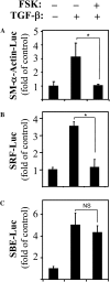
PKA regulates TGF-β–induced SM–α-actin promoter activation through inhibition of SRF, but not of Smad-binding elements (SBE). Human lung fibroblasts were transfected with luciferase reporters for −764 base pairs (A) SM–α-actin promoter, (B) SRF-luciferase reporter, or (C) SBE-luciferase reporter, along with thymidine kinase-driven renilla (TK-RL) control reporter. Serum-starved cells were pretreated with 10 μM FSK for 15 minutes, followed by stimulation with 2 ng/ml TGF-β for 24 hours. The activity of luciferase was then measured in cell lysates and normalized to the activity of renilla. Data represent the results of at least three experiments performed in triplicate (*P < 0.05).

Similar articles

Cited by

References

    1. Leask A, Abraham DJ. TGF-beta signaling and the fibrotic response. FASEB J 2004;18:816–827. - PubMed
    1. Phan SH. Fibroblast phenotypes in pulmonary fibrosis. Am J Respir Cell Mol Biol 2003;29:S87–S92. - PubMed
    1. Hinz B, Phan SH, Thannickal VJ, Galli A, Bochaton-Piallat ML, Gabbiani G. The myofibroblast: one function, multiple origins. Am J Pathol 2007;170:1807–1816. - PMC - PubMed
    1. Powell DW, Mifflin RC, Valentich JD, Crowe SE, Saada JI, West AB. Myofibroblasts: I. Paracrine cells important in health and disease. Am J Physiol 1999;277:C1–C9. - PubMed
    1. Broekelmann TJ, Limper AH, Colby TV, McDonald JA. Transforming growth factor beta 1 is present at sites of extracellular matrix gene expression in human pulmonary fibrosis. Proc Natl Acad Sci USA 1991;88:6642–6646. - PMC - PubMed

Publication types

MeSH terms