

doi: 10.1038/nbt.1520.
Epub 2009 Jan 18.
Transgenic mice with defined combinations of drug-inducible reprogramming factors
Affiliations
- PMID: 19151700
- PMCID: PMC2654270
- DOI: 10.1038/nbt.1520
Item in Clipboard
Transgenic mice with defined combinations of drug-inducible reprogramming factors
Nat Biotechnol.
2009 Feb.
Abstract
Proviruses carrying drug-inducible Oct4, Sox2, Klf4 and c-Myc used to derive 'primary' induced pluripotent stem (iPS) cells were segregated through germline transmission, generating mice and cells carrying subsets of the reprogramming factors. Drug treatment produced 'secondary' iPS cells only when the missing factor was introduced. This approach creates a defined system for studying reprogramming mechanisms and allows screening of genetically homogeneous cells for compounds that can replace any transcription factor required for iPS cell derivation.
Conflict of interest statement
Figures
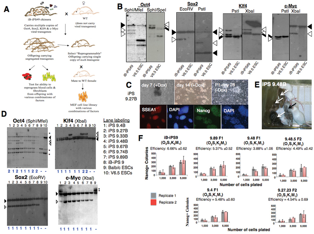
A) Experimental outline. iB-iPS#9 chimera is mated to generate offspring with different transgene copy number. Blood and tail fibroblasts were collected from adult offspring and MEF cultures were established from day E13.5 embryos. B) Southern analysis of iBiPS#9 line and V6.5 ESCs as controls. Filled arrowheads: endogenous bands; open arrowheads: proviral integrations. C) Top Panels: iPS colony formation from F1 offspring 9.27 (O1S1K1M1). Immuno-fluorescent analysis of the same iPS cell line that grew independently of Dox is shown in the lower panel. D) Southern analysis of F1 progeny blood derived iPS lines *: non-specific background bands. E) iPS cells contribute to chimeras (black arrow) that exhibit germline transmission (transgenic offspring: white arrows). F) Reprogramming efficiency of CD11b+ cells, 28 days after Dox induction. Efficiencies calculated as the fraction of Nanog positive colonies to cells seeded. Error bars: SD in duplicate wells. The generation (F1 or F2) and transgene copy number (subscript) are shown. “B” indicates iPS-line derived from peripheral blood.

A) PCR genotyping of select independent M2-rtTa+ MEF lines from mating offspring 9.27 (O1S1K1M1) to wild-type females. Genotype is indicated at the bottom. B) iPS cell derivation from MEF lines carrying three or more factor combinations. Missing factor was introduced by infection with TetO-FUW lentivirus (FUW) carrying the missing transcription factor. NA: not applicable, ND: not determined. The efficiencies reported are based on Nanog+ colonies fixed 30 days after plating 10,000 cells and addition of Dox. C) iPS cells from three factor MEF lines lacking c-Myc after transduction with Klf4. 200,000 O1S1K1 MEFs were infected with the indicated control virus and cultured in the present of Dox without passaging. Image of primary colony on Day 42 of Dox induction after infection with FUW-Klf4. Primary colonies were picked and passaged without Dox and expressed Nanog. Nine independent lines derived from two experiments. D) Kinetics of Nanog-GFP knock-in allele expression in two-factor lines, pre-treated or not with Dox, after transduction of the missing factors. 20,000 infected cells were seeded per well. Two wells were harvested every 48 hours for detection of Nanog-GFP by FACS. Nanog-GFP was defined by achieving >0.8% GFP positive cells. Blue dashed line: day of infection (d0). Pretreatment with Dox was done for 16 days. Two independent experimental sets are shown. Efficiency was determined after 28 days of Dox treatment as number of Nanog-GFP+ colonies per 10,000 cells initially seeded.
References
Publication types
MeSH terms
Substances
Grants and funding
LinkOut - more resources
Full Text Sources
Other Literature Sources
Molecular Biology Databases
