. 2009 Feb;11(2):219-25.
doi: 10.1038/ncb1830. Epub 2009 Jan 18.

Cytoplasmic penetration and persistent infection of mammalian cells by polyglutamine aggregates

Affiliations

Cytoplasmic penetration and persistent infection of mammalian cells by polyglutamine aggregates

Pei-Hsien Ren et al. Nat Cell Biol. 2009 Feb.

Abstract

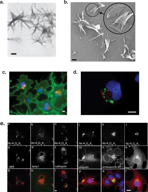
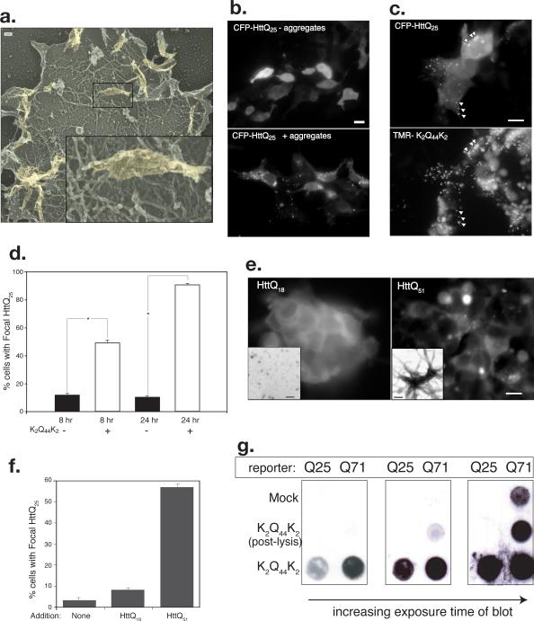
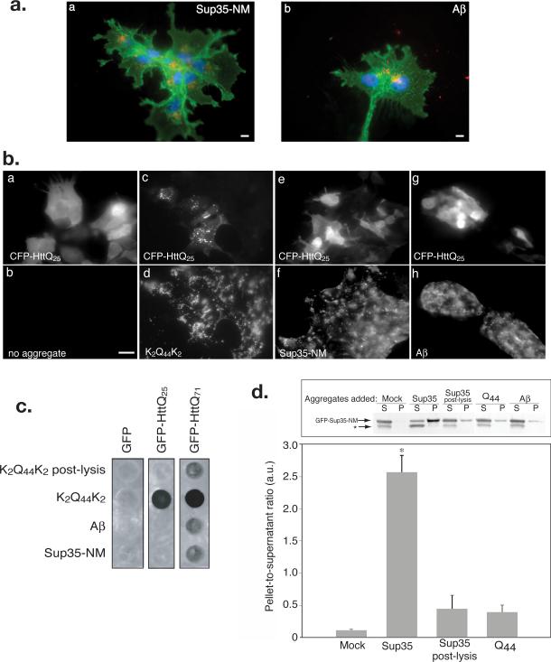
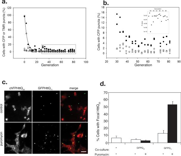
Sequence-specific nucleated protein aggregation is closely linked to the pathogenesis of most neurodegenerative diseases and constitutes the molecular basis of prion formation. Here we report that fibrillar polyglutamine peptide aggregates can be internalized by mammalian cells in culture where they gain access to the cytosolic compartment and become co-sequestered in aggresomes together with components of the ubiquitin-proteasome system and cytoplasmic chaperones. Remarkably, these internalized fibrillar aggregates are able to selectively recruit soluble cytoplasmic proteins with which they share homologous but not heterologous amyloidogenic sequences, and to confer a heritable phenotype on cells expressing the homologous amyloidogenic protein from a chromosomal locus.


Figures

Figure 1
Figure 1. Synthetic polyglutamine peptides form filamentous aggregates that are internalized by mammalian cells in culture
A. Transmission electron micrograph of K2Q44K2 aggregates stained with uranyl acetate. Bar = 200 nm. B. “Deep-etch” transmission electron micrograph of K2Q44K2 aggregates. Bar = 100 nm. C. Juxtanuclear localization of K2Q44K2 aggregates (red). Plasma membrane was labeled with wheat germ agglutinin (green) and nuclei with bisbenzimide (blue). Bar = 2 μm. D. Pericentriolar distribution of K2Q44K2 aggregates. COS7 cells were incubated with FITC-K2Q44K2 aggregates (green). Fresh medium was replenished after 1 hour and cells chased overnight. Following fixation cells were stained with antibody to γ-tubulin (red) and bisbenzimide (blue). Scale bars = 2 μm. E. Comparison of internalized K2Q44K2 aggregates with intracellular markers. Confocal micrographs of COS7 cells exposed to FITC-K2Q44K2 for 10 hours. Cells were fixed and stained with antibodies against the indicated endolysosomal markers (d-f) or cytoplasmic quality control components (m-p). Cell nuclei were stained with bisbenzimide. Scale bar for panels a-i = 10 μm; for panels j-s = 2 μm.
Figure 2
Figure 2. Internalized polyglutamine peptide aggregates access the cytosolic compartment
A. Internalized K2Q44K2 aggregates are visible attached to the actin cortex of unroofed HEK293 cells visualized by deep-etch TEM. Inset higher magnification view of region indicated by box. B. K2Q44K2 aggregates induce focal redistribution of intracellular HttQ25. Diffuse distribution of CFP-HttQ25 fluorescence in cells incubated without aggregates (top) is converted to a punctate pattern following overnight exposure to aggregated TMR-K2Q44K2 (bottom). Scale bar = 2 μm. C. Colocalization of CFP-HttQ25 puncta and TMR-K2Q44K2. Scale bars = 2 μm. D. Coalescence of CFP-HttQ25 fluorescence into puncta following infection with K2Q44K2 aggregates. Cells (n ≥ 250) exposed to TMR-K2Q44K2 aggregates (white bars) or control (black bars) were scored for punctate CFP-HttQ25 appearance. Asterisk, P- ≤ 0.001. Data representative of three independent experiments. E. Fibrillar aggregates composed of bacterially expressed, purified HttQ51 are able to induce conversion of cytosolic chFP-Q25 reporter. Scale bar = 2 μm. Insets, appearance of protein assemblies of HttQ18 (left) or HttQ51 (right) by negative stain EM. Scale bars = 20 nm. F. Quantification of data from experiment in Fig. 2D. Number of cells counted in each experiment ≥300 cells. G. K2Q44K2 aggregates induce aggregation of intracellular Htt. HEK293 cells stably expressing GFP-HttQ25 or Q71 were infected with K2Q44K2 aggregates for 24 hours prior to filter retardation assay. Post-lysis control is explained in Methods. Data representative of 3 independent experiments.
Figure 3
Figure 3. Internalized fibrillar aggregates induce homotypic but not heterotypic aggregation of cytoplasmic reporters
A. Sup35-NM and Aβ aggregates are internalized by mammalian cells. COS7 cells were infected with aggregated Sup35-NM (red; a) or Aβ (1-40) (red; b). Cells are labeled as in Fig. 1D. Scale bars = 2 μm. B. K2Q44K2 aggregates induce selective redistribution of CFP-HttQ25. HEK293 cells transfected with CFP-HttQ25, were incubated with aggregated K2Q44K2 (c and d), Sup35-NM (e and f), or Aβ(1-40) (g and h). Scale bar = 2 μm. C. Internalized K2Q44K2 aggregates induce selective aggregation of cytoplasmic polyQ reporters. Filter retardation assay of HEK293 cells stably expressing the indicated reporters following incubation with the indicated aggregates (1 μM) for ~ 24 hr or K2Q44K2 (0.2 μM) as post-lysis control. Data representative of three independent experiments. D. Infection with Sup35-NM induces selective aggregation of cytoplasmic GFPSup35-NM. Upper panel: immunoblot of Sup35-NM sedimentation analysis from HEK293 cells expressing GFP-Sup35-NM exposed to protein aggregates as noted (1μM) prior to lysis and sedimentation into pellet (P) and supernatant (S) fractions. Asterisk, probable proteolysis product. Bottom panel: quantification (mean ± SEM) of data from 4 immunoblot experiments. Asterisk, p≤ 0.0022.
Figure 4
Figure 4. Propagation of homotypic polyQ aggregation in cell culture
A. HEK293 cells stably expressing CFP-HttQ25 were exposed to TMR-K2Q44K2 aggregates (1 μM, solid circles; 0.1 μM, solid squares) or controls: mock infection (open circles), dextran (open triangles), or Aβ aggregates (open squares). Following the indicated number of generations in culture, cells were scored for CFP fluorescence distribution and for the presence of TMR (open diamonds) fluorescence. Each point represents counts of ≥400 cells. The solid line represents the theoretical decay of TMR fluorescence. B. Blow-up of boxed region from Fig. 4a. Symbols as in Fig. 4a. Data representative of three independent experiments. Inset, P-values determined from binomial test of proportions for generations 24-87. C. Intercellular transfer of polyQ aggregates in mammalian cell co-culture. Puror HEK293 cells stably expressing chFP-HttQ25 were cultured together with puromycin-sensitive cells stably expressing GFP-HttQ71. Cells were imaged after 72 hr of co-culture in DMEM (top row) or DMEM containing 4μg/ml puromycin (bottom row). Scale bars = 2 μm. D. Quantification of chFP-HttQ25 puncta formation in puror reporter cells cultured alone or co-cultured with cells stably expressing GFP-HttQ25 or GFP-HttQ71 in the presence or absence of puromycin (4μg/ml). Number of cells counted, n = 699-1276.

References

    1. Chiti F, Dobson CM. Protein misfolding, functional amyloid, and human disease. Annu Rev Biochem. 2006;75:333–366. - PubMed
    1. Taylor JP, Hardy J, Fischbeck KH. Toxic proteins in neurodegenerative disease. Science. 2002;296:1991–1995. - PubMed
    1. Carrell RW, Lomas DA. Conformational disease. Lancet. 1997;350:134–138. - PubMed
    1. Pepys MB. Amyloidosis. Annu Rev Med. 2006;57:223–241. - PubMed
    1. Sousa MM, Cardoso I, Fernandes R, Guimaraes A, Saraiva MJ. Deposition of transthyretin in early stages of familial amyloidotic polyneuropathy: evidence for toxicity of nonfibrillar aggregates. Am J Pathol. 2001;159:1993–2000. - PMC - PubMed

Publication types

MeSH terms