. 2009 Feb;16(2):183-9.
doi: 10.1038/nsmb.1536. Epub 2009 Jan 18.

A distinct class of small RNAs arises from pre-miRNA-proximal regions in a simple chordate

Affiliations

A distinct class of small RNAs arises from pre-miRNA-proximal regions in a simple chordate

Weiyang Shi et al. Nat Struct Mol Biol. 2009 Feb.

Abstract

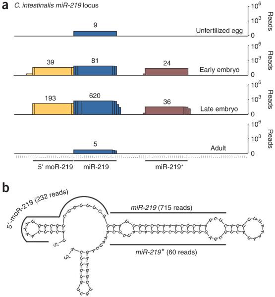
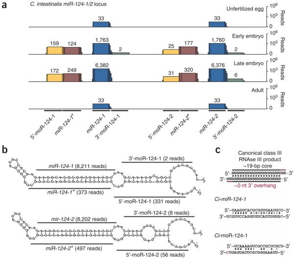
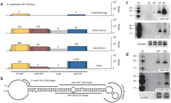
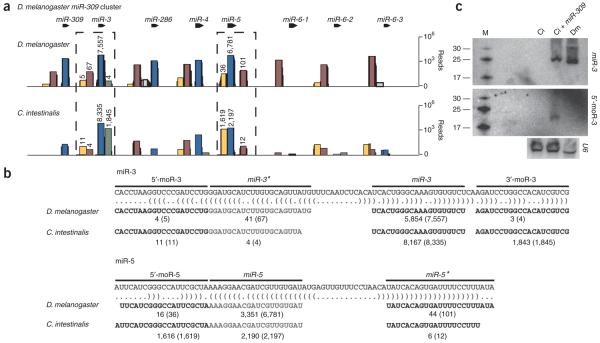
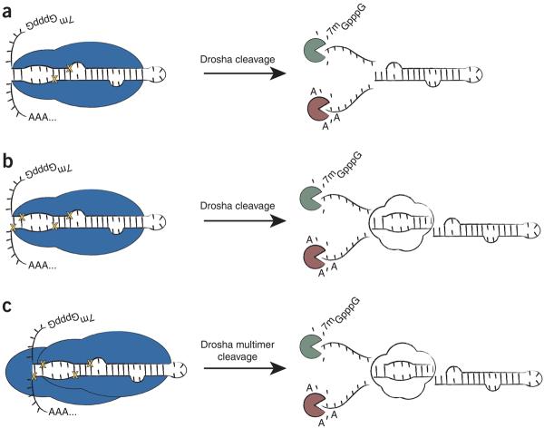
MicroRNAs (miRNAs) have been implicated in various cellular processes. They are thought to function primarily as inhibitors of gene activity by attenuating translation or promoting mRNA degradation. A typical miRNA gene produces a predominant approximately 21-nucleotide (nt) RNA (the miRNA) along with a less abundant miRNA(*) product. We sought to identify miRNAs from the simple chordate Ciona intestinalis through comprehensive sequencing of small RNA libraries created from different developmental stages. Unexpectedly, half of the identified miRNA loci encode up to four distinct, stable small RNAs. The additional RNAs, miRNA-offset RNAs (moRs), are generated from sequences immediately adjacent to the predicted approximately 60-nt pre-miRNA. moRs seem to be produced by RNAse III-like processing, are approximately 20 nt long and, like miRNAs, are observed at specific developmental stages. We present evidence suggesting that the biogenesis of moRs results from an intrinsic property of the miRNA processing machinery in C. intestinalis.


Figures

Figure 1
Figure 1
Developmental expression of small RNAs encoded by the C. intestinalis miR-219 locus. (a) Graphical depiction of small RNAs that map to the miR-219 locus at four developmental time points, indicated to the right. The histograms represent overlapping Illumina sequencing reads (numbered above stack) centered at each position (miRNA, blue; miRNA*, burgundy; 5′-moR, yellow). The y axis is plotted on a log scale. The secondary structure of the locus is presented in parenthetical format. (b) Locations of miRNA, miRNA* and moR sequences on the predicted secondary structure surrounding the pre–miR-219 hairpin. mFold was used to predict pre-miRNA secondary structure here and in the following figures,.
Figure 2
Figure 2
Coincident expression of 5′ and 3′ moR sequences from the C. intestinalis miR-124 locus. (a) Sequencing reads at each position of the miR-124 cluster are shown (miRNA, blue; miRNA*, burgundy; 5′-moR, yellow; 3′-miRNA, green). (b) miRNA and moR sequences aligned with sequence surrounding the predicted pre–miR-124-1 and pre–miR-124-2 stem-loop structures. A red ‘C’ in the pre–miR-124-1 structure indicates a shared base between multiple 5′-moR and miR-124-1* clones. (c) Standard class III RNAse III product is shown (above), depicting an ∼19-nt core of matched RNA bases, along with an ∼2-nt 3′ overhang. Aligned sequences are shown in the context of the predicted secondary structure of the pri-miRNA for miR-124-1 (top) and miR-124-1* (bottom), as well as 5′-moR-124-1 (bottom) and 3′-moR-124-1 (top). A shared base between loci is marked as a red “C”.
Figure 3
Figure 3
Direct detection of the 5′-moR-133 species. (a) Overlapping sequencing reads at each position along the miR-133 locus (miRNA, blue; miRNA*, burgundy; loop, gray; 5′-moR, yellow). (b) Alignment of sequenced reads on the predicted structure surrounding pre–miR-133. (c) Total RNA (∼30 μg per lane) was used for northern blots showing the ∼21-nt miR-133 (above) and 5′-moR-133 (middle) species throughout C. intestinalis development (M, size markers; Un, unfertilized eggs; EE, early embryos; LE, late embryos; Ad, adult animals). A northern blot for U6 RNA was used as a loading control (below). (d) As in c, comparing tailbud-stage C. intestinalis embryos that are unelectroporated (wild type, WT) or electroporated with a Ci-Brachyury enhancer:minimal Ci-miR-133 transgene (Bra). The Ci-Brachyury enhancer drives expression in the developing notochord.
Figure 4
Figure 4
Ectopic expression of Drosophila pri-miRNAs can induce moR production in C. intestinalis embryos. (a) Small RNAs were cloned from 2–4-hour-old D. melanogaster Toll10b mutant embryos (above), which contain only mesodermal cell types, or tailbud-stage C. intestinalis embryos expressing the entire D. melanogaster pri–miR-309 cluster (below), and were subjected to Illumina sequencing. The resulting sequencing reads are shown at each position along the D. melanogastermiR-309 locus (miRNA, blue; miRNA*, burgundy; 5′-moR, yellow; intervening loop, gray). (b) The most abundant reads overlapping the respective regions of the miR-3 (above) or miR-5 (below) loci are shown. The number of clones matching the exact sequence depicted is shown in comparison to the overall number of clones overlapping that segment (in parentheses). (c) Northern blots showing miR-3 (above) and 5′-moR-3 (middle) in C. intestinalis and D. melanogaster embryos. For each well, ∼50 μg total RNA was analyzed from tailbud-stage C. intestinalis embryo that were unelectroporated (Ci), similarly staged C. intestinalis embryos electroporated with D. melanogastermiR-309 expression plasmids (Ci + miR-309), or 2–4-hour-old Toll10b embryos. Below is shown a northern blot in which a cross-reactive probe for U6 RNA was used as a loading control.
Figure 5
Figure 5
A speculative model for the biogenesis of moRs. (a) Previous analysis of D. melanogaster and mouse small RNAs suggested that pre-miRNA-proximal sequences (analogous to moRs) were by-products of exonucleolytic degradation following excision of the pre-miRNA hairpin by Drosha (Drosha is represented in blue and yellow crosses indicate where Drosha cuts). (b) moR production may result via excision of an ∼20-nt, imperfectly paired duplex RNA at the immediate base of the pre-miRNA stem-loop, following two concurrent or sequential cuts by a single Drosha molecule. (c) Alternatively, a multimeric complex containing at least two Drosha molecules could associate with a substrate pri-miRNA. Here each Drosha molecule would cleave the pri-miRNA at a distinct position, liberating the pre-miRNA, as well as the ∼20-nt moR duplex.

References

    1. Ambros V. The functions of animal microRNAs. Nature. 2004;431:350–355. - PubMed
    1. Zamore PD, Haley B. Ribo-gnome: the big world of small RNAs. Science. 2005;309:1519–1524. - PubMed
    1. Bartel DP. MicroRNAs: genomics, biogenesis, mechanism, and function. Cell. 2004;116:281–297. - PubMed
    1. Lau NC, Lim LP, Weinstein EG, Bartel DP. An abundant class of tiny RNAs with probable regulatory roles in Caenorhabditis elegans. Science. 2001;294:858–862. - PubMed
    1. Pasquinelli AE, et al. Conservation of the sequence and temporal expression of let-7 heterochronic regulatory RNA. Nature. 2000;408:86–89. - PubMed

Publication types

Associated data