. 2009 Mar 12;28(10):1285-97.
doi: 10.1038/onc.2008.479. Epub 2009 Jan 19.

A non-Smad mechanism of fibroblast activation by transforming growth factor-beta via c-Abl and Egr-1: selective modulation by imatinib mesylate

Affiliations

A non-Smad mechanism of fibroblast activation by transforming growth factor-beta via c-Abl and Egr-1: selective modulation by imatinib mesylate

S Bhattacharyya et al. Oncogene. .

Abstract

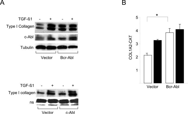
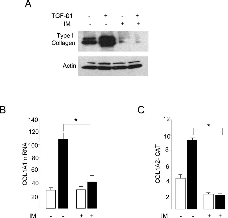
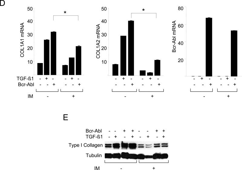
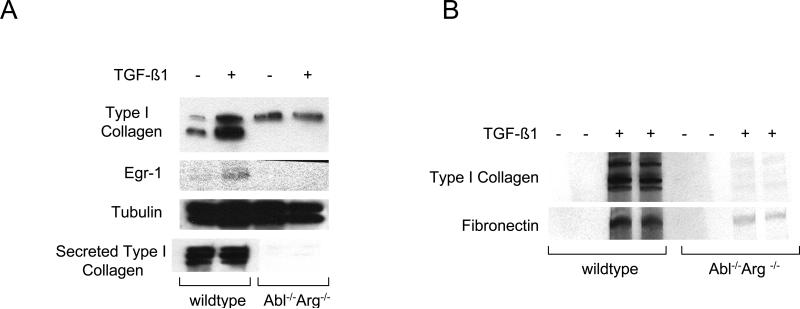
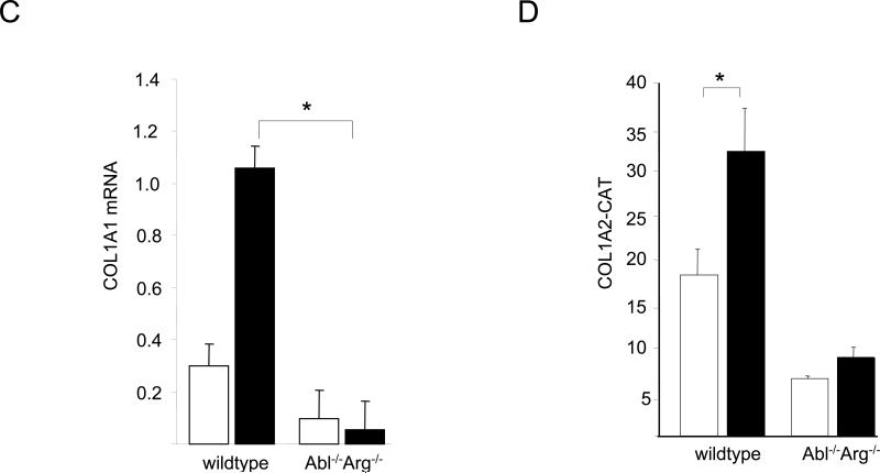
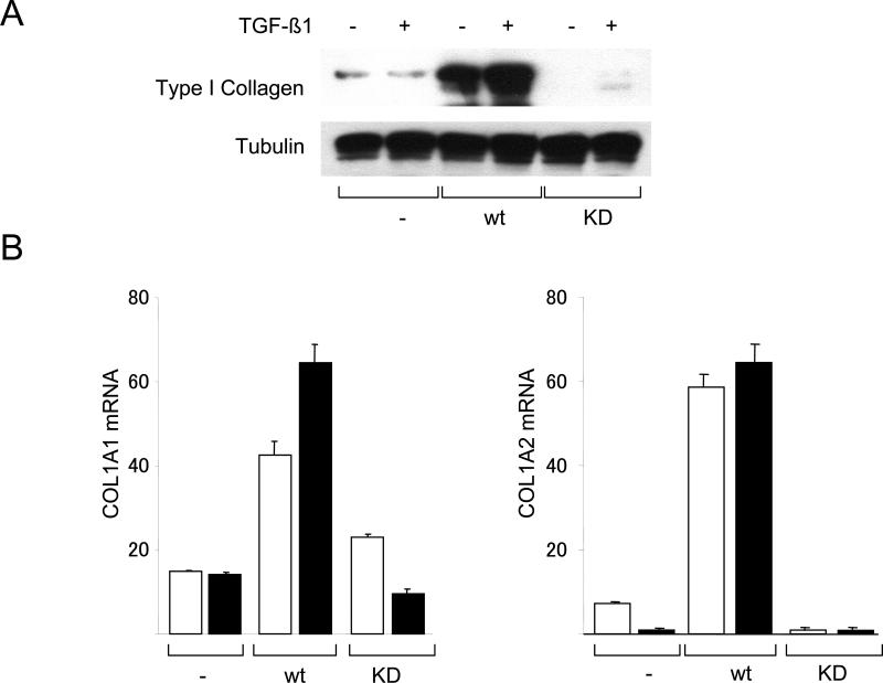
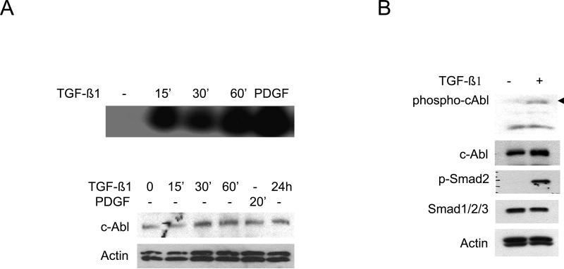
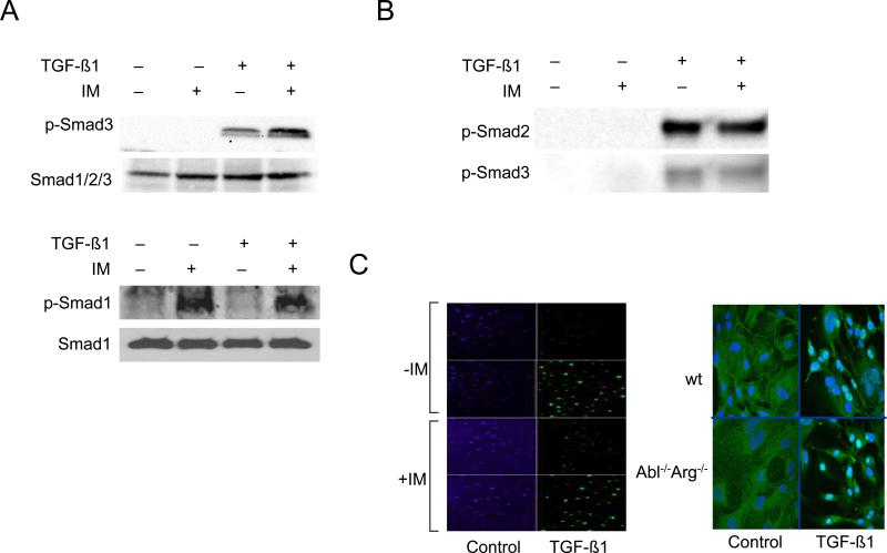
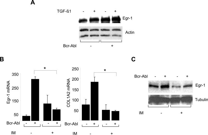
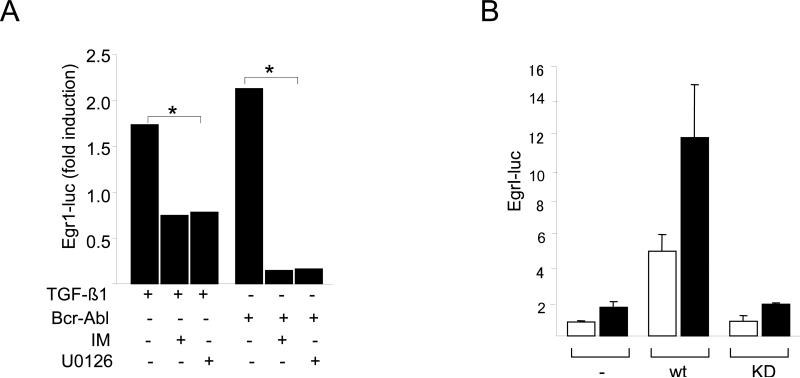
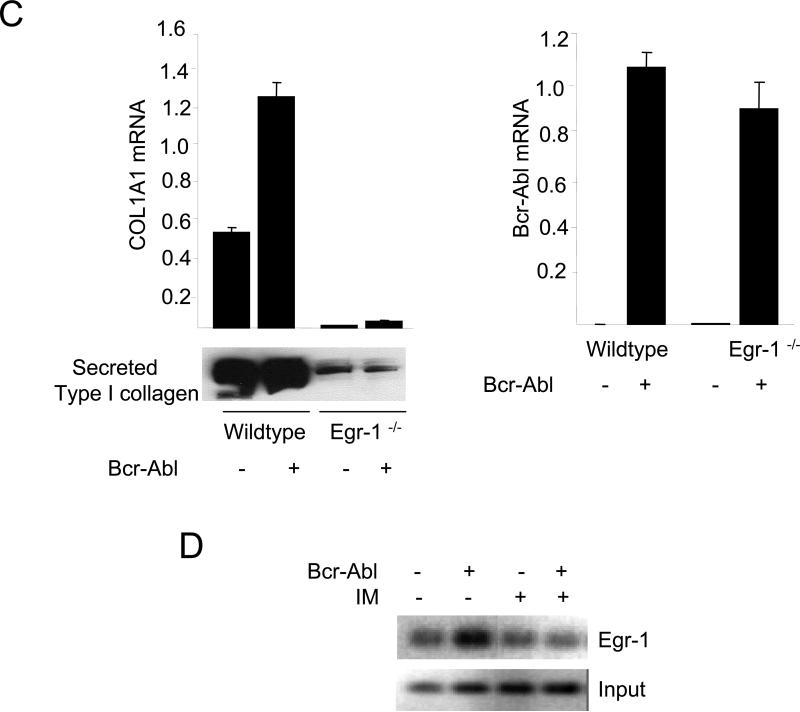
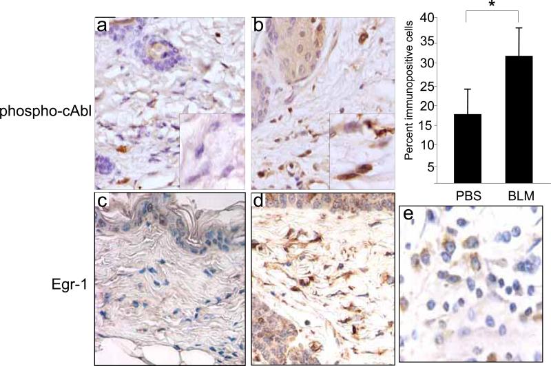
The nonreceptor protein tyrosine kinase c-Abl regulates cell proliferation and survival. Recent studies provide evidence that implicate c-Abl as a mediator for fibrotic responses induced by transforming growth factor-beta (TGF-beta), but the precise mechanisms underlying this novel oncogene function are unknown. Here, we report that when expressed in normal fibroblasts, a constitutively active mutant of Abl that causes chronic myelogenous leukemia (CML) stimulated the expression and transcriptional activity of the early growth response factor 1 (Egr-1). Mouse embryonic fibroblasts (MEFs), lacking c-Abl, were resistant to TGF-beta stimulation. Responsiveness of these MEFs to TGF-beta could be rescued by wild-type c-Abl, but not by a kinase-deficient mutant form of c-Abl. Furthermore, Abl kinase activity was necessary for the induction of Egr-1 by TGF-beta in normal fibroblasts, and Egr-1 was required for stimulation of collagen by Bcr-Abl. Lesional skin fibroblasts in mice with bleomycin-induced fibrosis of skin displayed evidence of c-Abl activation in situ, and elevated phospho-c-Abl correlated with increased local expression of Egr-1. Collectively, these results position Egr-1 downstream of c-Abl in the fibrotic response, delineate a novel Egr-1-dependent intracellular signaling mechanism that underlies the involvement of c-Abl in certain TGF-beta responses, and identify Egr-1 as a target of inhibition by imatinib. Furthermore, the findings show in situ activation of c-Abl paralleling the upregulated tissue expression of Egr-1 that accompanies fibrosis. Pharmacological targeting of c-Abl and its downstream effector pathways may, therefore, represent a novel therapeutic approach to blocking TGF-beta-dependent fibrotic processes.


Figures

Figure 1
Figure 1. Constitutively active Bcr-Abl stimulates collagen gene expression
Confluent foreskin fibroblasts were transiently transfected with Bcr-Abl (A, top panel; B) or wildtype c-Abl (A, lower panel) and incubated with TGF-β for 24 h. A. Whole cell lysates were subjected to Western analysis. Representative immunoblots are shown. B. Fibroblasts were cotransfected with 772COL1A2-CAT. Cell lysates were assayed for their CAT activities. Results, normalized with Renilla luciferase, are expressed as means ± S.D. of triplicate determinations. Open boxes, untreated fibroblasts; closed boxes, TGF-ß-treated fibroblasts. *p<0.005.
Figure 2
Figure 2. Imatinib blocks stimulation of collagen gene expression induced by Bcr-Abl or TGF-ß
Foreskin fibroblasts were pretreated with 10 μM imatinib (IM) for 30 min, followed by TGF-ß1 for a further 24 h. A. Whole cell lysates were subjected to Western analysis. Representative immunoblots. B. Total RNA was examined by real-time qPCR. Results normalized with actin are shown as means ± SD from triplicate determinations. Open boxes, untreated fibroblasts; closed boxes, TGF-ß-treated fibroblasts. *p<0.005. C. Fibroblasts were transiently transfected with 772COL1A2-CAT. Cell lysates were assayed for their CAT activities. The results are the means ± S.D. of triplicate determinations. Open boxes, untreated fibroblasts; closed boxes, TGF-ß-treated fibroblasts. D. Fibroblasts were transfected with Bcr-Abl and total RNA was subjected to realtime qPCR analysis. The results are representative of two independent experiments. * p<0.005. E. Whole cell lysates were subjected to Western analysis. Representative immunoblots are shown. IM, imatinib mesylate.
Figure 3
Figure 3. Impaired TGF-ß response in Abl−/− Arg−/− mouse embryonic fibroblasts
Fibroblasts from Abl −/−Arg −/− and wildtype mouse embryos in parallel were incubated with TGF-ß for 24 h. A. Whole cell lysates were subjected to Western analysis. Representative immunoblots. B. Following radiolabeling of cultures with [14C]-proline for 24 h, conditioned media were harvested and subjected to SDS-PAGE. Representative autoradiographs. C. Total RNA was subjected to real-time qPCR analysis. Results, expressed relative to actin, are shown as the means ± S.D.of triplicate determinations from a representative experiment. Open boxes, untreated fibroblasts; closed boxes, TGF-ß-treated fibroblasts. *p<0.005. D. Fibroblasts were transiently transfected with 772COL1A2-CAT. Following incubation with TGF-ß for 24 h, cell lysates were assayed for their CAT activities. The results are shown as the means ± S.D. of triplicate determinations. Open boxes, untreated fibroblasts; closed boxes, TGF-ß-treated fibroblasts. *p<0.005.
Figure 4
Figure 4. c-Abl rescues TGF-ß responses in Abl−/− Arg−/− MEFs
Abl −/−Arg −/− MEFs stably expressing c-Abl or a kinase-inactive mutant form of cAbl (KD) in parallel were incubated with TGF-ß for 24 h. A. Whole cell lysates were subjected to Western analysis. Representative immunoblots. B. Total RNA was subjected to real-time qPCR analysis. Results, expressed relative to actin are means ± S.D. from triplicate determinations from a representative experiment. Open boxes, untreated fibroblasts; closed boxes, TGF-ß-treated fibroblasts. *p<0.005.
Figure 5
Figure 5. TGF-ß induces c-Abl kinase activity
A. Confluent fibroblasts were incubated with TGF-ß1 (10 ng/ml) or PDGF (25 ng/ml) for indicated periods. Whole cell lysates were immunoprecipitated with antibodies to c-Abl and kinase activity was assayed using GST-Crk as substrate (upper panel). Western analysis of whole cell lysates (lower panel). B. Fibroblasts were incubated with TGF-β1 for 30 min and whole cell lysates were subjected to Western analysis.
Figure 6
Figure 6. Smad2/3-independent inhibition of TGF-ß responses by imatinib
A-C. Confluent foreskin fibroblasts were pretreated for 30 min with imatinib (10 μM) followed by incubation with TGF-ß1 for a further 30 min. A. Whole cell lysates were subjected to Western analysis. Representative immunoblots. B. Nuclear extracts were subjected to DNA affinity precipitation assays using biotin end-labeled oligonucleotides harboring the SBE sequence as described under “Materials and Methods.” A representative Western blot. C . Fibroblasts were immunostained with antibodies to Smad4, and examined by immunofluorescence confocal microscopy. Representative images. IM, Imatinib mesylate. D. Abl −/−/Arg −/− MEFs and wildtype MEFs in parallel were incubated with TGF-ß1 (30 min) and Smad2/3 localization was examined by confocal microscopy. Representative images (left panel). The nuclear localization of Smad2/3 was quantitated as described under methods (right panel). Results represents the means ± S.D. from separate fields. Open boxes, untreated fibroblasts; closed boxes, TGF-ß-treated fibroblasts. *p<0.005.
Figure 7
Figure 7. Bcr-Abl stimulates Egr-1
A. Confluent foreskin fibroblasts were transiently transfected with Bcr-Abl and incubated with TGF-β for 24 h. A. Whole cell lysates were subjected to Western analysis. Representative immunoblots are shown. B. Confluent fibroblasts were transiently transfected with Bcr-Abl in presence of imatinib. Total RNA normalized with actin was subjected to realtime qPCR analysis. The results are the means ± S.D. of triplicate determinations. *p<0.005. C. Whole cell lysates from confluent cultures treated with imatinib were subjected to Western analysis. Representative immunoblots are shown.
Figure 8
Figure 8. Egr-1 acts downstream of c-Abl in TGF-ß signaling
A. NIH3T3 fibroblasts were transiently cotransfected with indicated concentrations of Bcr-Abl along with Egr-1-luc. Following pretreatment with imatinib or U0126, cultures were incubated with TGF-ß1 for a further 24 h and cell lysates were assayed for their luciferase activities. The results, normalized with Renilla luciferase, are expressed as fold-induction relative to TGF-ß or Bcr-Abl. *p<0.005. B. Abl −/−Arg −/− MEFs stably expressing c-Abl or a kinase-deficient mutant form of c-Abl (KD) were transfected with Egr-1- luc, and following incubation with TGF-ß1 for 24 h, cell lysates were assayed for their luciferase activities. The results, normalized with Renilla luciferase, are expressed as means ± S.D. of triplicate determinations from two independent experiments. Open boxes, untreated MEFs; closed boxes, TGF-ß-treated MEFs. C. Wildtype and Egr-1−/− skin fibroblasts were transfected with Bcr-Abl and total RNA were subjected to realtime qPCR analysis (upper panel), and secreted Type I collagen was examined by Western blot analysis (lower panel). The results are the means ± S.D. of triplicate determinations. D. ChIP assays. Foreskin fibroblasts transfected with Bcr-Abl were cross-linked with formaledehyde, chromatin was immunoprecipitated with antibodies to Egr-1, followed by PCR amplification with COL1A2-specific primers. PCR products were analyzed by agarose gel electrophoresis. Input genomic DNA was used as positive control. IM, Imatinib mesylate.
Figure 9
Figure 9. Increased expression of phospho-cAbl and Egr-1 in lesional tissue in bleomycin-induced scleroderma in the mouse
6-weeks old BALB/C mice received daily s.c. injections of bleomycin (BLM) or PBS. At day 28 after injections were started, lesional skin was examined by immunohistochemistry using antibodies against phospho-cAbl or Egr-1. Representative photomicrographs are shown. Original magnification × 400 (upper panels) or × 1000 (lower panels). The proportion of fibroblasts in the lesional dermis immunopositive for phospho-cAbl was determined. Results represents the means ± S.D. from six separate fields from at least three mice/group. Human breast carcinoma tissue was used as positive control.

References

    1. Jimenez SA, Derk CT. Following the molecular pathways toward an understanding of the pathogenesis of systemic sclerosis. Ann Intern Med. 2004;140:37–45. - PubMed
    1. Varga J, Abraham D. Systemic sclerosis: a prototypic multisystem fibrotic disorder. J Clin Invest. 2007;117:557–567. - PMC - PubMed
    1. Varga JA, Trojanowska M. Fibrosis in systemic sclerosis. Rheum Dis Clin North Am. 2008;34(1):1, 115–43. - PMC - PubMed
    1. Massagué J, Seoane J, Wotton D. Smad transcription factors. Genes Dev. 2005;19:2783–2810. - PubMed
    1. Moustakas A, Heldin CH. Non-Javelaud D, Mauviel. A Crosstalk mechanisms between the mitogen-activated protein kinase pathways and Smad signaling downstream of TGF-beta: implications for carcinogenesis. Oncogene. 2005 Aug 29;24(37):5742–50. - PubMed

Publication types

MeSH terms