. 2009 Mar 5;28(9):1248-60.
doi: 10.1038/onc.2008.476. Epub 2009 Jan 19.

Hypoxia-associated p38 mitogen-activated protein kinase-mediated androgen receptor activation and increased HIF-1alpha levels contribute to emergence of an aggressive phenotype in prostate cancer

Affiliations

Hypoxia-associated p38 mitogen-activated protein kinase-mediated androgen receptor activation and increased HIF-1alpha levels contribute to emergence of an aggressive phenotype in prostate cancer

L Khandrika et al. Oncogene. .

Retraction in

Abstract

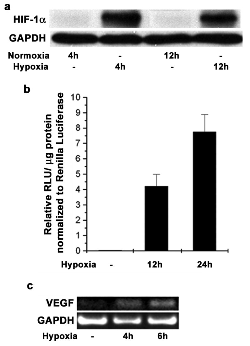
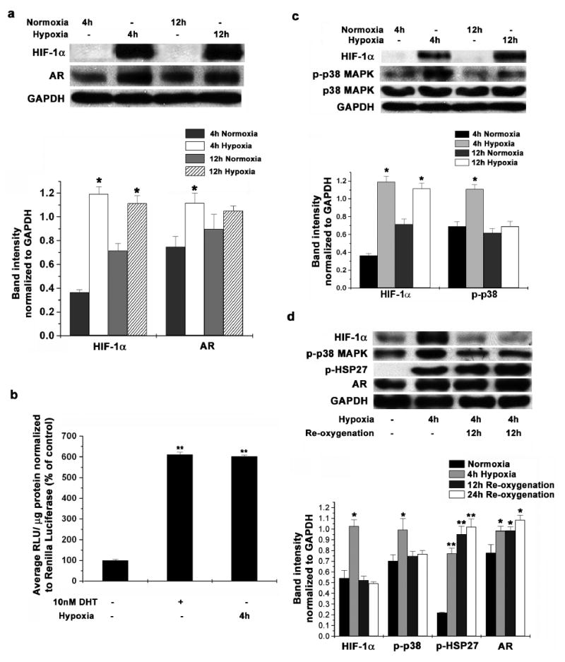
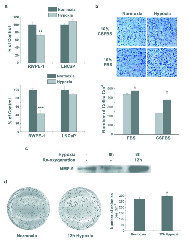
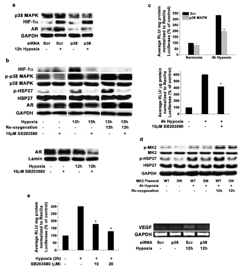
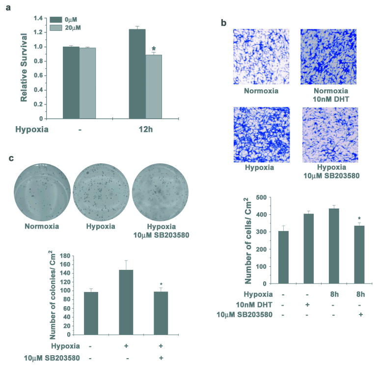
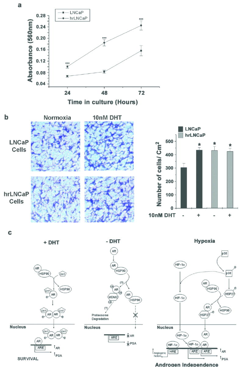
Androgen receptor (AR) signaling is involved in the development and progression of prostate cancer. Tumor microvasculature contributes to continual exposure of prostate cancer cells to hypoxia-reoxygenation, however, the role of hypoxia-reoxygenation in prostate cancer progression and modulation of AR signaling is not understood. In this study, we evaluated the effects of hypoxia-reoxygenation in LNCaP cells, a line of hormone responsive human prostate cancer cells. Our results demonstrate that hypoxia-reoxygenation resulted in increased survival, higher clonogenicity and enhanced invasiveness of these cells. Moreover, hypoxia-reoxygenation was associated with an increased AR activity independent of androgens as well as increased hypoxia inducible factor (HIF-1alpha) levels and activity. We also observed that the activation of p38 mitogen-activated protein (MAP) kinase pathway was an early response to hypoxia, and inhibition of p38 MAP kinase pathway by variety of approaches abolished hypoxia-reoxygenation induced increased AR activity as well as increased survival, clonogenicity and invasiveness. These results demonstrate a critical role for hypoxia-induced p38 MAP kinase pathway in androgen-independent AR activation in prostate cancer cells, and suggest that hypoxia-reoxygenation may select for aggressive androgen-independent prostate cancer phenotype.


Figures

Figure 1
Figure 1. Hypoxia stabilizes HIF-1α and VEGF in LNCaP cells
(a) LNCaP cells were subjected to hypoxia in a modular incubator for 4h and 12h. Western blot analysis for HIF-1α was done using GAPDH protein as a loading control. (b) LNCaP cells were transfected with pGL3-TK-3X HRE plasmid and pRL-CMV as a transfection control. 48h post transfection cells were incubated in hypoxia for 12h and 24h. Luciferase activity was measured with Dual luciferase assay kit and the firefly luciferase activity was normalized to Renilla luciferase activity. Each data point represents mean ± S.D of duplicate experiments done in duplicate. (c) LNCaP cells were subjected to hypoxia for 4h and 6h and total RNA was isolated. Reverse Transcriptase PCR was performed to determine the mRNA levels of VEGF; GAPDH was used as a loading control.
Figure 2
Figure 2. Hypoxia- reoxygenation increases Androgen Receptor protein levels and activity in LNCaP cells
(a) LNCaP cells were subjected to hypoxia in a modular incubator for 4h and 12h. Western blot analysis for protein levels of HIF-1α and Androgen Receptor was performed using GAPDH protein as a loading control (Upper Panel). Representative blots from 3 individual experiments are shown. Protein amounts were calculated by determining band intensities using densitometry and normalizing them to GAPDH levels used as a loading control (Lower Panel). Each data point is represented as mean ± S.D of triplicate experiments (* indicates p<0.05 compared to respective normoxia controls, n=3). (b) LNCaP cells maintained in androgen free medium (10% CSFBS) were transfected with pGL3- TARPp/ PSAe plasmid and pRL-CMV as a transfection control. 36 after transfection, cells were incubated either in 10nM DHT or in hypoxia for 4h. Luciferase activity was measured with Dual luciferase assay kit as before. Each data point is represented as mean ± S.D of duplicate experiments done in duplicate (** indicates p<0.01 compared to normoxic and un-stimulated control, n=4). (c) LNCaP cells were subjected to hypoxia in a modular incubator for 4h and 12h. Western blot analysis for protein levels of HIF-1α, total p38 MAPK and phospho-p38 MAPK was performed using GAPDH protein as a loading control (Upper Panel). Representative gel images from 3 individual experiments are shown. Protein amountss were calculated as before (Lower Panel). Each data point is represented as mean ± S.D of triplicate experiments (* indicates p<0.05 compared to respective normoxia controls, n=3). (d) LNCaP cells were incubated in hypoxia for 4h and also re-oxygenated for 12h and 24h after hypoxia. Western blot analysis was used to determine the protein levels of HIF-1α, phospho-p38 MAPK, phospho-HSP27 and Androgen Receptor in cell lysates with GAPDH protein as a loading control (Upper Panel). Representative images from 3 individual experiments are shown. Protein amounts were quantitated by densitometry as above (Lower Panel). Each data point is represented as mean ± S.D of triplicate experiments (* indicates p<0.05, ** indicates p<0.01 compared to respective normoxia controls, n=3).
Figure 3
Figure 3. Hypoxia-reoxygenation Promotes Aggressive phenotype
(a) RWPE-1 and LNCaP cells were incubated for 12h (Upper Panel) and 24h (Lower Panel) in hypoxia. Cells were stained with crystal violet to determine survival. Data points represent mean ± S.D of duplicate experiments done in triplicate (** indicates p<0.01 and *** indicates p<0.001 compared to normoxia control, n=6). (b) LNCaP cells were incubated in hypoxia for 8h in complete medium in the presence or absence of androgens. Equal numbers of cells were seeded in Boyden chambers and 48h later, invaded cells were stained with Crystal Violet. Cells that invaded through matrigel were quantified using ImageJ software (Lower Panel) and each data point represents mean ± S.D of duplicate experiments done in duplicate (* indicates p<0.05 compared to normoxia control, n=4). Representative images are shown. (c) LNCaP cells were subjected to hypoxia for 8h in serum free medium and cultured for further 12 hours in normoxia. Equal volume of conditioned medium was collected after hypoxia or after re-oxygenation, concentrated and equal volume of concentrated medium was used for western blot analysis to determine the secreted MMP-9 protein. (d) LNCaP cells were incubated in hypoxia for 12 hours and colony formation in soft agar was assayed (Left Panel). Representative images from duplicate experiments done in triplicate are shown. Number of colonies was counted using ImageJ software and quantified (Right Panel) Each data point represents mean ± S.D (* indicates p<0.05 compared to control, n=6).
Figure 4
Figure 4. Inhibition of p38 MAPK reduces AR levels and activity and also reduces HIF-1α activity in LNCaP cells
(a) LNCaP cells transfected with siRNA targeting p38 MAPK or scrambled sequence were incubated in hypoxia for 12 hours and total protein was extracted. Western blot analysis was performed to determine the levels of AR, p38 MAPK, HIF-1α and GAPDH. Representative blot from three individual experiments is shown. (b) LNCaP cells were pre-treated with 10μM SB203580 and then incubated in hypoxia for 12h and one set was reoxygenated for further 12h after hypoxia. Western blot analysis was used to determine the levels of HIF-1α, total p38, phospho-p38, HSP27, phospho-HSP27 and Androgen Receptor protein in cell lysates with GAPDH protein as a loading control (upper Panel). Representative blot from triplicate experiments is shown. LNCaP cells were pre-treated with 10μM SB203580 and then incubated in hypoxia for 12h and nuclear extracts were prepared. Western blot analysis was used to determine the protein levels of Androgen Receptor in nucleus with Lamin A protein as a loading control (Lower Panel). (c) LNCaP cells were transfected with pGL3- TARPp/ PSAe plasmid and pRL-CMV as a transfection control. 36h after transfection, cells were pre-treated for 1 hour with 10μM SB203580 (Upper Panel) and incubated in hypoxia for 4h or cells were transfected with siRNA targeting p38 MAPK or scrambled sequence (Scr) along with pGL3-TARPp/ PSAe plasmid and pRL-CMV as a transfection control (Lower Panel). 48h after transfection, cells were incubated in hypoxia for 4h in hypoxia and Luciferase activity was measured with Dual luciferase assay kit as above. Each data point is represented as mean ± S.D of duplicate experiments done in duplicate (* indicates p<0.05 compared to normoxic and un-stimulated control, n=4). (d) LNCaP cells were transfected with plasmids expressing MAPKAP K2 (MK2) wild type (WT) or dominant negative (DN) forms. 48 hours post transfection, cells were incubated for 4 hours in hypoxia and one set was re-oxygenated for 12 hours. Western blot analysis was performed to analyze protein levels of MK2, phospho-MK2, HSP27 and phospho-HSP27, with GAPDH as a loading control. Representative blot from duplicate experiments is shown. (e; Left Panel) LNCaP cells were transfected with pGL3-TK-3X HRE plasmid and pRL-CMV as a transfection control. 48h post transfection, cells were pre-incubated with SB203580 at the indicated concentrations for 1h and incubated in hypoxia for 2h. Luciferase activity was measured with Dual luciferase assay kit as before. Each data point represents mean ± S.D of duplicate experiments done in duplicate (* indicates p<0.05 compared to cells untreated with SB203580 but incubated in hypoxia, n=4). (e; Right Panel) LNCaP cells were transfected with siRNA designed against p38 MAPK or scrambled control (Scr). 48h post transfection, cells were incubated in hypoxia for further 24h. Total RNA was isolated from the cells and Reverse Transcriptase PCR was used to amplify VEGF mRNA. Representative gel image from duplicate experiments is shown.
Figure 5
Figure 5. Inhibition of p38 MAPK reduces survival and invasion of LNCaP cells
(a) LNCaP cells were pre-treated with 10μM SB203580 for 1 hour and then incubated in hypoxia for 12h. Cells were stained with crystal violet to determine survival. Data points represent mean ± S.D of duplicate experiments done in triplicate (* indicates p<0.05 compared to normoxia control, n=6). (b) LNCaP cells were incubated in hypoxia for 8h either in absence of androgens either with or without 10μM SB203580. Equal numbers of cells were seeded in Boyden chambers and one set of cells not subjected to hypoxia, were allowed to invade in presence of 10nM DHT. 48h later invaded cells were stained with Crystal Violet (Upper Panel). Representative images from two individual experiments done in duplicate are shown. Cells that invaded through matrigel were quantified (Lower Panel) as before and each data point represents mean ± S.D (* indicates p<0.05 compared to cells incubated in hypoxia in androgen free medium and uninhibited p38 MAPK, n=4). (c) LNCaP cells were incubated in hypoxia either with or without 10μM SB203580 for 12 hours and colony formation in soft agar was assayed (Upper Panel). Representative images from duplicate experiments done in triplicate are shown. Number of colonies was quantified as before (Lower Panel). Each data point represents mean ± S.D (* indicates p<0.05 compared to control, n=6).
Figure 6
Figure 6. Chronic hypoxia- reoxygenation selects for LNCaP cells with aggressive phenotype
(a) LNCaP cells were cultured in complete medium and incubated in hypoxia for 12h and re-oxygenated for 12h, for 3 weeks. Equal numbers of surviving cells (hrLNCaP) were seeded in androgen free medium and proliferation was measured by MTT assay over 3 days. Each data point represents mean ± S.D of two individual experiments done in quadruplicate (*** indicates p<0.001, n=8). (b) Equal number of hrLNCaP cells and LNCaP cells were seeded in Boyden chambers in androgen free medium and cells were allowed to invade either in the presence or absence of 10nM DHT. 48h later, invaded cells were stained with Crystal Violet (Left Panel). Representative images from two individual experiments done in duplicate are shown. Invaded cells were quantified as before and each data point represents mean ± S.D (* indicates p<0.05 compared to LNCaP cells in androgen free medium, n=4) (Right Panel). (c) Plausible mechanism of dual control of p38 MAPK to promote aggressive growth of prostate cancer cells in hypoxia. When subjected to hypoxia, AR can be activated in an androgen independent manner via p38 MAPK and HSP27 and can translocate into the nucleus. On the other hand, activated p38 MAPK can help stabilize HIF-1α and promote HIF-1 mediated gene transcription. Together the active AR and HIF-1 promote aggressive growth and may in due course lead to androgen independent prostate cancer.

Similar articles

Cited by

References

    1. Akakura N, Kobayashi M, Horiuchi I, Suzuki A, Wang J, Chen J, et al. Constitutive expression of hypoxia-inducible factor-1alpha renders pancreatic cancer cells resistant to apoptosis induced by hypoxia and nutrient deprivation. Cancer Res. 2001;61:6548–6554. - PubMed
    1. Asirvatham AJ, Schmidt M, Gao B, Chaudhary J. Androgens regulate the immune/inflammatory response and cell survival pathways in rat ventral prostate epithelial cells. Endocrinology. 2006;147:257–271. - PubMed
    1. Baek SH, Lee UY, Park EM, Han MY, Lee YS, Park YM. Role of protein kinase C delta in transmitting hypoxia signal to HSF and HIF-1. J Cell Physiol. 2001;188:223–235. - PubMed
    1. Berra E, Pagès G, Pouysségur J. MAP kinases and hypoxia in the control of VEGF expression. Cancer Metastasis Rev. 2000;19:139–145. - PubMed
    1. Bhattacharya S, Michels CL, Leung MK, Arany ZP, Kung AL, Livingston DM. Functional role of p35srj, a novel p300/CBP binding protein, during transactivation by HIF-1. Genes Dev. 1999;13:64–75. - PMC - PubMed

Publication types

Substances