. 2010 Sep;36(3):323-336.
doi: 10.1007/s10695-008-9297-z. Epub 2009 Jan 20.

Haemopoiesis in the head kidney of tilapia, Oreochromis niloticus (Teleostei: Cichlidae): a morphological (optical and ultrastructural) study

Affiliations

Haemopoiesis in the head kidney of tilapia, Oreochromis niloticus (Teleostei: Cichlidae): a morphological (optical and ultrastructural) study

El-Saydah H Abdel-Aziz et al. Fish Physiol Biochem. 2010 Sep.

Erratum in

  • Fish Physiol Biochem. 2012 Dec;38(6):1857. Ali, Tamer El-Sayed [corrected to El-Sayed Ali, Tamer]

Abstract

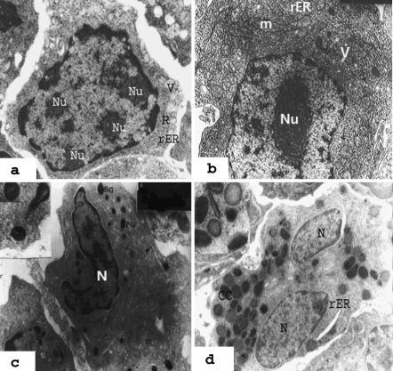
The present work focused on the histological and ultrastructural studies on haemopoiesis in the kidney of tilapia, Oreochromis niloticus. Haemopoietic tissue was found mainly in the head kidney and a small amount occurred in the mesonephros. The haemopoiesis of tilapia had the following series: erythropoiesis, granulopoiesis, thrombopoiesis, monopoiesis and lymphoplasmopoiesis. Erythropoiesis includes proerythroblasts, basophilic erythroblasts, polychromatic erythroblasts, acidophilic erythroblasts and young and mature erythrocytes. The proerythroblasts were the largest cells in the erythropoietic series. During the maturation process both the nuclear and cellular size decreased gradually due to the chromatin condensation and the progressive substitution of cytoplasmic matrix with a large amount of haemoglobin. Granulopoietic series consisted of cells with variable shape and size at different stages of maturity from myeloblasts to mature granulocytes. The promyelocytes were the largest cells in the series and were characterised by the appearance of primary (azoruphilic) granules. The maturation process involved the appearance of specific granules in the heterophilic, eosinophilic and basophilic series. It is important to mention that eosinophilic granulocytes were the dominant granulopoietic series in the haemopoietic tissue (Ht) of tilapia. Lymphopoietic series consisted of lymphoblasts, large lymphocytes, small lymphocytes and active and inactive plasma cells. Thrombopoietic series consisted of thromboblasts, prothromboblasts and thrombocytes. Thrombocytes of tilapia were nucleated and possessed a spindle shape. Melanomacrophage centres were dominant among the Ht of the head kidney. Also, monocytes were detected and shown to be large cells with an indented nucleus and cytoplasm containing numerous vesicles of different sizes and a few lysosomes.


Figures

Fig. 1
Fig. 1
Light micrographs of the cross sections of the head kidney in Oreochromis niloticus. a Head kidney: note the haemopoietic tissue (Ht), blood vessel (BV), kidney tubules (T) and sinusoids (s) (H&E, ×160). b Haemopoietic tissue in the head kidney: showing developing blood cells around sinusoids (s) and melanomacrophage centres (MMCs) (H&E, ×250). c Inter-renal tissue around blood vessels (BV) and haemopoietic tissue with scattered numerous mature acidophils (H&E, ×250. d Haemopoiesis in the HK: s sinusoids loaded with RBCs, E erythropoiesis, G granulopoiesis, B blast cell (toludine blue, ×1,000
Fig. 2
Fig. 2
Electron micrographs of developing erythrocytes (erythropoiesis) illustrating progressive reduction in cell size and cytoplasmic organelles, condensation of nuclear chromatin and cytoplasmic homogeneity with haemoglobin formation. a Proerythroblast has large spherical euchromatic nucleus with finely granulated chromatin and one large nucleolus (Nu) with reticular nucleolema and cytoplasm with numerous free ribosomes (R) with dense matrix and disorganised cristae, Golgi complexes (G) and small vacuoles (V) (×8,000). b Basophilic erythroblast: ovoid cell with cytoplasm loaded with free clustered ribosomes (R), large mitochondria (m), rER cisternae and oval nucleus (N) with marginal chromatin condensation (×8,000). c Polychromatic erythroblast (P): elongated cell with a dense nucleus (N), containing heterochromatin in coarse blocks and a dense ring around the edge, nearly homogeneous cytoplasm with plenty of polyribosomes (arrow) and acidophilic erythroblast (A), scattered ribosomes (R) in rosettes, deformed mitochondria (m) and atrophoid nucleus. G granulocyte (×8,000). d Groups of young and mature erythrocytes. Note acidophilic erythroblast (A), young erythrocytes (YE) with homogeneous cytoplasm and remnant of ribosomal rosettes. Mature erythrocytes (ME) have an oval shape and homogeneous cytoplasm nearly devoid of organelles. Heterochromatic nucleus (N) (×8,000)
Fig. 3
Fig. 3
Electron micrographs of granulopoiesis in the haemopoietic tissue of the head kidney of Oreochromis niloticus. a Myeloblast: note the voluminous euchromatic nucleus, which contained 2–4 nucleoli (Nu). The cytoplasm characterised by scattered free ribosomes (R), rER strands and pinocytotic vesicles (V) (×8,000). b Promyelocytes: the largest cells in the granulopoietic series. Large euchromatic nucleus with prominent nucleolus (Nu), cytoplasm with numerous polyribosomes (arrow), mitochondria (m), primary granules (PG), rER and vesicles (V) (×8,000). c Eosinophilic myelocyte: with an oval band nucleus (N), mitochondria (m), polyribosomes, rER, small vesicles (v), primary granules (PG) and specific granules (SG) with light axial crystalloid core (×8,000). d Eosinophilic meta-myelocyte: note the numerous heterogenic granules, some with crystalline core (CC), predominant rER and bilobed nucleus (N) (×8,000). Inset: clarifies heterogeneous granules (×14,000)
Fig. 4
Fig. 4
Electron micrographs of granulopoiesis in the haemopoietic tissue of the head kidney of Oreochromis niloticus. a Heterophil myelocyte: nucleus with prominent nucleolus (Nu), cytoplasm with small primary granules and large dark homogeneous specific granules (G), mitochondria (m), rough endoplasmic granules (rER). M Macrophage (×8,000). b Neutrophil (heterophil): round cell with bilobed (constricted) nucleus (N) and homogeneously dense specific granules (g).m mitochondria, G Golgi (×8,000). c Basophilic myelocyte: with numerous large granules (g), developed cisternae of rER (arrow), numerous free ribosomes (R), large oval nucleus (N) with heterochromatin blocks. m mitochondria (×14,000). d Basophilic metamyelocyte: an elongated cell with an irregular surface with elongated bend nucleus (N) and condensed marginal heterochromatin. The cytoplasm contained few large mitochondria, lamellated cisternae of rER and numerous granules (g) variable in electron density. (×8,000). Inset: showing the fibrillary content of specific granules. (×14,000)
Fig. 5
Fig. 5
Electron micrographs showing thrombopoiesis in the head kidney of Oreochromis niloticus. a Thromboplast: note the euchromatic nucleus with a prominent nucleolus (Nu), marginal band of microtubules (mt), polyribosomes (R), pseudopodia (P) and vesicles (v) (×10,000). b Prothrombocyte: irregular-shaped cell with surface-connected canalicular system (SCCS), large granules (G) with homogeneous or fibrillary content, rER cisternae (arrow), large and irregularly shaped nucleus (N) and marginal microtubules (mt) (×10,000). c Thrombocyte (B): spindle-shaped cell with large oval heterochromatic nucleus, homogeneous cytoplasm with medium electron density containing scattered mitochondria. Prothrombocyte (A): irregularly shaped cell contains large euchromatic elongated nucleus (N), numerous vesicles through the cytoplasm (arrow), Golgi complex (G) and numerous small mitochondria (m) (×10,000)
Fig. 6
Fig. 6
Electron micrographs showing lymphoplasmopoiesis in the head kidney of Oreochromis niloticus. a Lymphoblast: round cells with voluminous nucleus (N) with finely granular chromatin, prominent nucleoli, cytoplasm with a few large mitochondria (m) and free ribosomes (R). L lysosome (×10,000). b Large lymphocyte: the cell had a irregular outline with a large central nucleus (N) containing large blocks of heterochromatin and a moderate amount of cytoplasm filled with numerous ribosomes (R), a few mitochondria (m) and scattered vesicles and vacuoles (×8,000). c Small lymphocyte: irregularly shaped cells with radially arranged chromatin blocks in large nucleus (N) and cytoplasm with scattered free ribosomes (R), vacuoles (V) and numerous microvilli. Plasma membrane exhibits numerous short microvilli (mv) (×10,000). d Plasma cell: note the eccentric nucleus (N) with chromatin blocks and cytoplasm with abundant dilated rER, mitochondria (m) and paranuclear expanded Golgi complex (G) (×10,000)
Fig. 7
Fig. 7
Electron micrographs of melanomacrophage centres and macrophages in the haemopoietic tissue (Ht) of the head kidney of Oreochromis niloticus. a Melanomacrophage centre: surrounded by a thin layer of connective tissue capsule (arrow). It contains numerous melanomacrophage cells with melanin granules and heterogeneous cytoplasmic contents (×1,400). b Melanomacrophage cell: note the irregular surface with pseudopodial processes (Pp), nucleus (N), melanin granules (MG), rER (arrow) and vesicles (v) (×10,000). c Erythrophagoytosis: note the engulfed senile or atretic (RBCs) by macrophage. Large eccentric nucleus (N), phagocytic vacuoles (v), residual bodies (r) in the macrophage (×5,000). d Macrophage cell: note the macrophage with euchromatic eccentric nucleus (N) and cytoplasm filled with phagolysosomes (PL), Golgi complex (G), pseudopodial processes (Pp) and ribosomes (R)

References

    1. Abdel-Rahman MA (1997) Toxicological studies on heavy metals on Siganus rivulatus. M. Sc. Thesis. Department of Oceanography, Faculty of Science, Alexandria University, Egypt, p 188
    1. Agius C. Phylogenetic development of melano-macrophages centers in fish. J Zool. 1980;191:11–31. doi: 10.1111/j.1469-7998.1980.tb01446.x. - DOI
    1. Agius C. Preliminary studies on the ontogeny of the melano-macrophages of teleost haemopoietic tissues and age-related changes. Dev Comp Immunol. 1981;5:597–606. - PubMed
    1. Agius C. The melano-macrophages centers of fish: a review. In: Maning MJ, Taner MF, editors. Fish immunology. London: Academic Press; 1985. pp. 85–105.
    1. Agius C, Agbede SA. An electron microscopical study of the genesis of lipofucsin, melanin and hemosiderin in haemopoietic tissues of fish. J Fish Biol. 1984;24:471–488. doi: 10.1111/j.1095-8649.1984.tb04818.x. - DOI

MeSH terms

LinkOut - more resources