. 2009 Apr;37(5):1650-62.
doi: 10.1093/nar/gkp004. Epub 2009 Jan 19.

Generation of single-chain LAGLIDADG homing endonucleases from native homodimeric precursor proteins

Affiliations

Generation of single-chain LAGLIDADG homing endonucleases from native homodimeric precursor proteins

Hui Li et al. Nucleic Acids Res. 2009 Apr.

Abstract

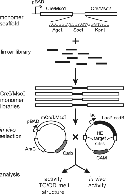
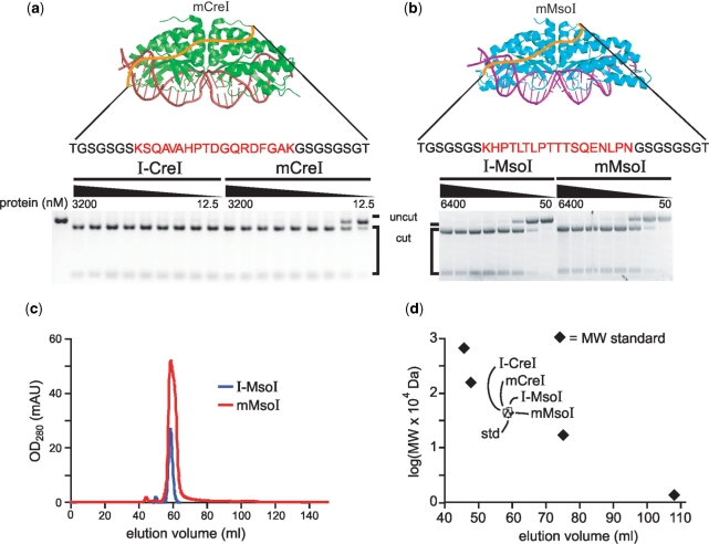
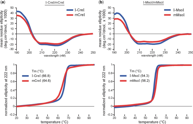
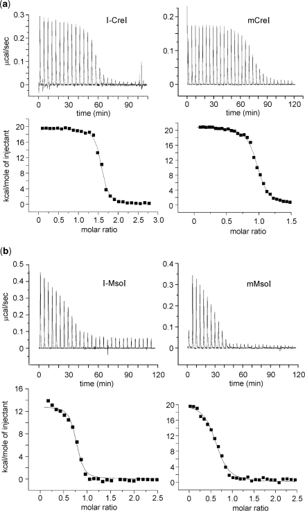
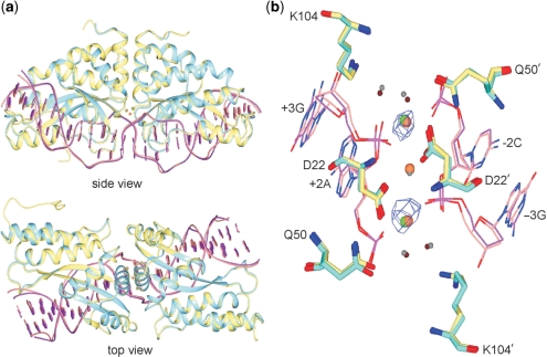
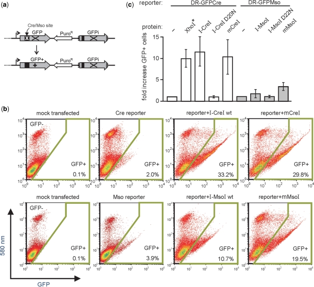
Homing endonucleases (HEs) cut long DNA target sites with high specificity to initiate and target the lateral transfer of mobile introns or inteins. This high site specificity of HEs makes them attractive reagents for gene targeting to promote DNA modification or repair. We have generated several hundred catalytically active, monomerized versions of the well-characterized homodimeric I-CreI and I-MsoI LAGLIDADG family homing endonuclease (LHE) proteins. Representative monomerized I-CreI and I-MsoI proteins (collectively termed mCreIs or mMsoIs) were characterized in detail by using a combination of biochemical, biophysical and structural approaches. We also demonstrated that both mCreI and mMsoI proteins can promote cleavage-dependent recombination in human cells. The use of single chain LHEs should simplify gene modification and targeting by requiring the expression of a single small protein in cells, rather than the coordinate expression of two separate protein coding genes as is required when using engineered heterodimeric zinc finger or homing endonuclease proteins.


Figures

Figure 1.
Figure 1.
Generation and characterization of catalytically active, monomeric versions of I-CreI and I-MsoI. Two divergent copies of the I-CreI or I-MsoI ORF were synthesized, and cloned into expression vectors together with an intervening multiple cloning site linker. A library of previously generated, randomized linkers was then PCR-amplified, cleaved and inserted at the multiple cloning site to generate libraries of monomeric mCre and mMso proteins with linker insertions. Active monomeric proteins were identified on the basis of an activity-dependent selection in E. coli, then characterized by a combination of biochemical, biophysical and functional assays. A high resolution co-crystal structure was also generated for one of the mMsoI proteins with a 33 residue linker (see Figure 5).
Figure 2.
Figure 2.
In vitro cleavage and purification of monomeric and native, homodimeric versions of I-CreI and I-MsoI. (a, b) In vitro cleavage analyses using a linearized plasmid DNA substrate revealed comparable cleavage activities of monomeric and homodimeric versions of I-CreI and I-MsoI. Nineteen additional monomeric variants were characterized in similar fashion (Supplementary Table 1). Protein concentrations were 12.5–3200 mM for the I-CreI/mCreI proteins and 50–6400 nM for I-MsoI/mMsoI proteins. (c) Elution profiles of I-MsoI (blue) and mMsoI (red) were superimposable in size exclusion chromatography analyses on a HiLoad 16/60 Superdex 75 column (GE healthcare, Piscataway, NJ). Gel filtration analysis of all four proteins revealed predicted molecular weights when compared with an equivalent size gel filtration standard (MW std = 44 kDa chicken ovalbumin; Biorad, Hercules, CA).
Figure 3.
Figure 3.
Far-ultraviolet CD and thermal denaturation profiles of homodimeric and monomeric I-CreI (a) and I-MsoI (b). All four curves in both CD and thermal denaturation analyses were generated by best fit analyses for a two-state transition, and had 50% transition temperatures of 66.8 (I-CreI), 64.6 (mCreI), 54.3 (I-MsoI) and 56.2°C (mMsoI), respectively.
Figure 4.
Figure 4.
DNA binding and thermodynamic profiles for homodimeric and monomeric I-CreI and I-MsoI as determined by ITC. Heat absorption upon the injection of native target site DNAs can be seen in upper panels in (a) for I-CreI/mCreI and in (b) for I-MsoI and mMsoI. The molar ratio of injected target site DNA to protein is shown in bottom panels. Results for all proteins could be fitted to standard saturation curves, and all four proteins displayed endothermic binding profiles. A summary of thermodynamic parameters for all four proteins is given in Table 3. Panel (b) I-MsoI data were adapted from ref. 32.
Figure 5.
Figure 5.
Superposition of the I-MsoI:DNA and newly determined mMsoI:DNA co-crystal structures. (a) side and top views of the native homodimeric I-MsoI and newly determined monomeric mCreI co-crystal structures reveals backbone superposition with a 0.47 Å RMSD for protein and DNA. I-MsoI and mMsoI structures, and their DNA molecules, are shown respectively in cyan, yellow, purple and pink. Three calcium ions in I-MsoI:DNA structure and two calcium ions in mMsoI:DNA structure are shown, respectively, in green and coral. (b) Superposition of the I-MsoI and mMsoI active sites. Three active site residues—D22, Q50 and K104—with two calcium ions and two nucleotides flanking the scissile phosphodiester bond are shown. Four water molecules from I-MsoI:DNA and mMsoI:DNA structures are shown as small spheres in grey or tan, respectively. Anomalous difference mapping analysis revealed two calcium ions in the mMsoI:DNA active site. Figures were prepared using the CCP4 Molecular Graphics software package (33).
Figure 6.
Figure 6.
mCreI and mMsoI induce catalytic activity-dependent recombination in human cells. (a) In vivo recombination activity was assayed by co-transfecting coding plasmids for I-CreI or I-MsoI proteins together with a recombination reporter plasmid that contains a direct repeat of two genetically inactive copies of GFP. In vivo cleavage of the I-CreI or I-MsoI target site initiates gene conversion and repair of the cleaved copy to generate GFP+ cells that can be detected and quantified by flow cytometry (Materials and methods section). (b) Flow histograms of cells mock-transfected, transfected with reporter plasmid alone, or co-transfected with reporter: endonuclease plasmid pairs. The gate for GFP+ cells is shown by the boxed area, and the GFP+ frequency is given in the lower right of each histogram. (c) Frequency and fold increase in GFP+ cells for different reporter/coding plasmid combination. The frequency of GFP+ cells generated by in vitro XhoI-linearized DR-GFPCre reporter DNA (*) indicates that a substantial fraction of reporter molecules are likely cleaved in vivo by I-CreI and mCreI. D20N I-CreI and D22N I-MsoI are catalytically inactive mutants of I-CreI and I-MsoI which fail to induce GFP+ cells when cotransfected with a Cre or Mso-specific reporter plasmid. Error bars are means ± SDs.

References

    1. Stoddard BL. Homing endonuclease structure and function. Quart. Rev. Biophys. 2005;38:49–95. - PubMed
    1. Belfort M, Perlman PS. Mechanisms of intron mobility. J. Biol. Chem. 1995;270:30237–30240. - PubMed
    1. Argast GM, Stephens KM, Emond MJ, Monnat RJ., Jr I-PpoI and I-CreI homing site sequence degeneracy determined by random mutagenesis and sequential in vitro enrichment. J. Mol. Biol. 1998;280:345–353. - PubMed
    1. Chevalier B, Turmel M, Lemieux C, Monnat RJ, Jr, Stoddard BL. Flexible DNA target site recognition by divergent homing endonuclease isoschizomers I-CreI and I-MsoI. J. Mol. Biol. 2003;329:253–269. - PubMed
    1. Silva GH, Dalgaard JZ, Belfort M, Van Roey P. Crystal structure of the thermostable archaeal intron-encoded endonuclease I-DmoI. J. Mol. Biol. 1999;286:1123–1136. - PubMed

Publication types

Associated data