. 2009 Jan 26;184(2):205-13.
doi: 10.1083/jcb.200806072. Epub 2009 Jan 19.

Histone hypercitrullination mediates chromatin decondensation and neutrophil extracellular trap formation

Affiliations

Histone hypercitrullination mediates chromatin decondensation and neutrophil extracellular trap formation

Yanming Wang et al. J Cell Biol. .

Abstract

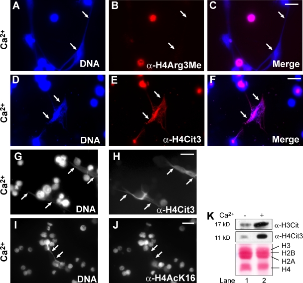
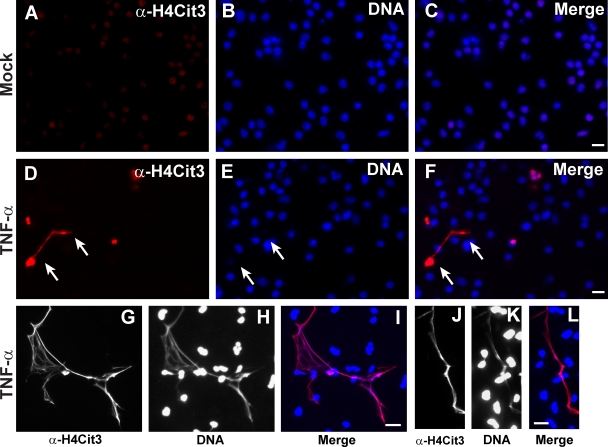
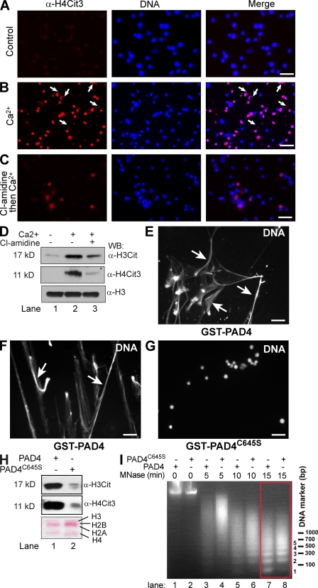
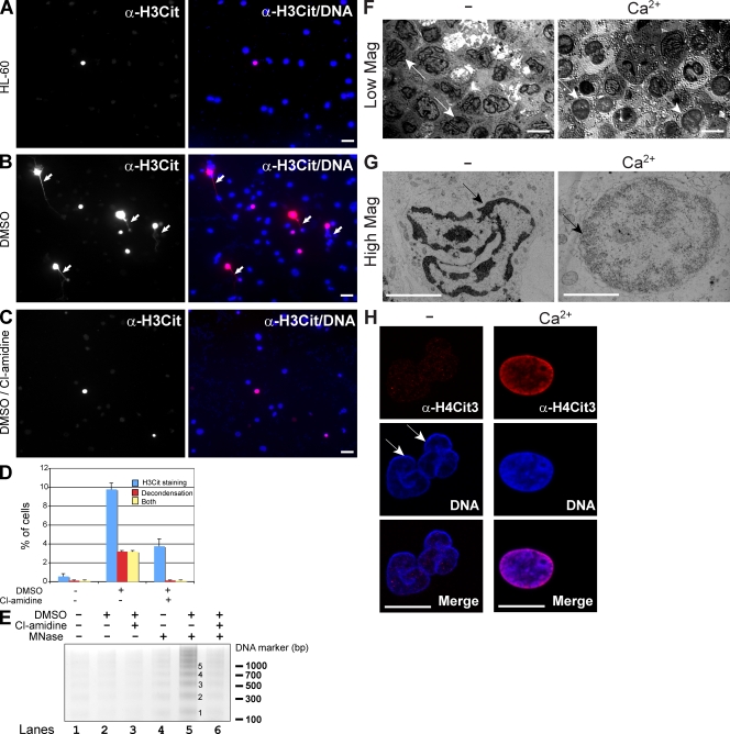
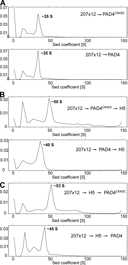
Peripheral blood neutrophils form highly decondensed chromatin structures, termed neutrophil extracellular traps (NETs), that have been implicated in innate immune response to bacterial infection. Neutrophils express high levels of peptidylarginine deiminase 4 (PAD4), which catalyzes histone citrullination. However, whether PAD4 or histone citrullination plays a role in chromatin structure in neutrophils is unclear. In this study, we show that the hypercitrullination of histones by PAD4 mediates chromatin decondensation. Histone hypercitrullination is detected on highly decondensed chromatin in HL-60 granulocytes and blood neutrophils. The inhibition of PAD4 decreases histone hypercitrullination and the formation of NET-like structures, whereas PAD4 treatment of HL-60 cells facilitates these processes. The loss of heterochromatin and multilobular nuclear structures is detected in HL-60 granulocytes after PAD4 activation. Importantly, citrullination of biochemically defined avian nucleosome arrays inhibits their compaction by the linker histone H5 to form higher order chromatin structures. Together, these results suggest that histone hypercitrullination has important functions in chromatin decondensation in granulocytes/neutrophils.


Figures

Figure 1.
Figure 1.
Chromatin decondensation induced by PAD4 activation and histone citrullination in HL-60 granulocytes. (A–C) Loss of histone H4Arg3 methylation on the decondensed chromatin (denoted by arrows) after calcium ionophore treatment. (D–F) Increase in H4Cit3 on the decondensed chromatin (denoted by arrows). (G and H) Grayscale images show an increase of H4Cit3 on decondensed chromatin (denoted by arrows). (I and J) Grayscale images show that H4K16 acetylation was not elevated on the decondensed chromatin (denoted by arrows). (K) Changes in histone H3 and H4 citrullination analyzed by Western blotting. Ponceau S staining shows the amount of histones. Bars, 20 µm.
Figure 2.
Figure 2.
Histone citrullination on highly decondensed chromatin in peripheral blood neutrophils treated with TNF-α. (A–C) H4Cit3 staining before TNF-α treatment. (D–F) Approximately 10% of cells were stained with H4Cit3 after 15-min TNF-α treatment. The white arrows denote decondensed chromatin. (G–L) Representative images of NETs stained by both DNA dye and H4Cit3 antibody. Bars, 20 µm.
Figure 3.
Figure 3.
PAD4 activity is important for the formation of highly decondensed chromatin. (A–C) H4Cit3 and DNA staining of HL-60 granulocytes without treatment (A), with calcium ionophore treatment (B), and with the PAD4 inhibitor Cl-amidine treatment before calcium ionophore treatment (C). The arrows denote decondensed chromatin stained by the H4Cit3 antibody. (D) Western blot assays of histone H3 and H4 citrullination. The histone H3 blot is a loading control. (E–G) Decondensed chromatin (denoted by arrows) was detected in HL-60 cells after Triton X-100 and GST-PAD4 treatment (E and F) but not after the GST-PAD4C645S mutant treatment (G). (H) Western blot assays of histone H3 and H4 citrullination in HL-60 cells after incubation with GST-PAD4 or the GST-PAD4C645S mutant. Ponceau S staining shows the amount of histones. (I) MNase digestion of HL-60 cells treated with GST-PAD4 or the GST-PAD4C645S mutant. Chromatin in GST-PAD4–treated cells (lane 7) is more accessible than that in GST-PAD4C645S–treated cells (lane 8). The red box highlights lanes with a clear difference in MNase digestion. Bars, 30 µm.
Figure 4.
Figure 4.
PAD4 activity is important for NET formation after cytokine and bacteria treatment and for loss of heterochromatin and multilobular nuclear structures. (A-C) Histone H3 citrullination and DNA staining in undifferentiated HL-60 cells (A), DMSO-differentiated HL-60 cells (B), and DMSO-differentiated HL-60 cells pretreated with Cl-amidine (C) after treatment with IL-8 and bacteria S. flexneri. The arrows denote decondensed chromatin stained by the H3Cit antibody. (D) Percentages of cells with positive staining of H3 citrullination and/or chromatin decondensation shown with standard deviations (error bars; n = 4; >3,000 cells counted in each experiment). (E) After 15 h of IL-8 and bacteria treatment, NET formation was measured by MNase digestion. Numbers denote mono- and polynucleosomal DNA. (F and G, left) Transmission electron microscopy analyses of HL-60 granulocytes before calcium ionophore treatment. The arrows denote nuclei with distinct heterochromatin (dark regions) underlining the nuclear envelope. (right) Transmission electron microscopy analyses after calcium ionophore treatment. The arrows denote nuclei that lost the distinct heterochromatin structure underlining the nuclear envelope. (H) H4 citrullination and DNA staining before (left column; arrows denote two nuclei) and after calcium ionophore treatment. Notice the loss of both multilobular nuclear structure and rimlike heterochromatin after H4 citrullination. Bars: (A–C) 20 µm; (F and H) 10 µm; (G) 5 µm.
Figure 5.
Figure 5.
Histone citrullination inhibits nucleosome array compaction by linker histone H5. (A) A 35-S structure was detected after treatment of the 207 × 12 array (containing 12 nucleosome core particles in an array) with the GST-PAD4C645S mutant or GST-PAD4. (B) After adding the linker histone H5, the 207 × 12 nucleosome core particle array pretreated with the PAD4C645S mutant was detected as a 50-S structure, whereas the 207 × 12 nucleosome core particle array pretreated with PAD4 was detected as a 40-S structure. (C) The H5-bound 207 × 12 array treated with the PAD4C645S mutant was detected as a 53-S structure, whereas the H5-bound 207 × 12 array treated with PAD4 was detected as a 45-S structure.

References

    1. Balint B.L., Szanto A., Madi A., Bauer U.M., Gabor P., Benko S., Puskas L.G., Davies P.J., Nagy L. 2005. Arginine methylation provides epigenetic transcription memory for retinoid-induced differentiation in myeloid cells.Mol. Cell. Biol. 25:5648–5663 - PMC - PubMed
    1. Beiter K., Wartha F., Albiger B., Normark S., Zychlinsky A., Henriques-Normark B. 2006. An endonuclease allows Streptococcus pneumoniae to escape from neutrophil extracellular traps.Curr. Biol. 16:401–407 - PubMed
    1. Brinkmann V., Reichard U., Goosmann C., Fauler B., Uhlemann Y., Weiss D.S., Weinrauch Y., Zychlinsky A. 2004. Neutrophil extracellular traps kill bacteria.Science. 303:1532–1535 - PubMed
    1. Brown P.H., Schuck P. 2006. Macromolecular size-and-shape distributions by sedimentation velocity analytical ultracentrifugation.Biophys. J. 90:4651–4661 - PMC - PubMed
    1. Buchanan J.T., Simpson A.J., Aziz R.K., Liu G.Y., Kristian S.A., Kotb M., Feramisco J., Nizet V. 2006. DNase expression allows the pathogen group A Streptococcus to escape killing in neutrophil extracellular traps.Curr. Biol. 16:396–400 - PubMed

Publication types