. 2009 Apr;83(7):3007-18.
doi: 10.1128/JVI.01505-08. Epub 2009 Jan 19.

Severe acute respiratory syndrome coronavirus nsp9 dimerization is essential for efficient viral growth

Affiliations

Severe acute respiratory syndrome coronavirus nsp9 dimerization is essential for efficient viral growth

Zachary J Miknis et al. J Virol. 2009 Apr.

Abstract

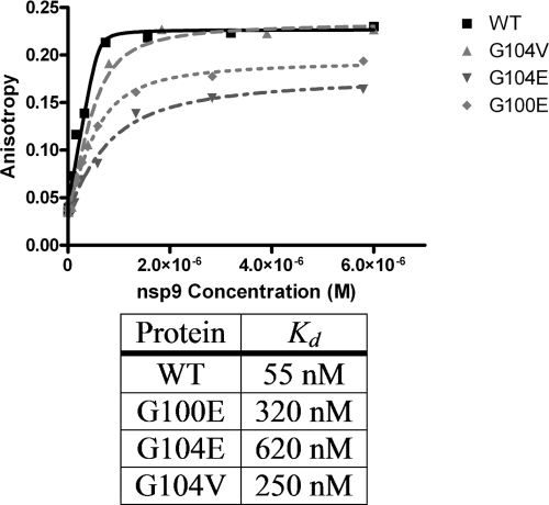
The severe acute respiratory syndrome coronavirus (SARS-CoV) devotes a significant portion of its genome to producing nonstructural proteins required for viral replication. SARS-CoV nonstructural protein 9 (nsp9) was identified as an essential protein with RNA/DNA-binding activity, and yet its biological function within the replication complex remains unknown. Nsp9 forms a dimer through the interaction of parallel alpha-helices containing the protein-protein interaction motif GXXXG. In order to study the role of the nsp9 dimer in viral reproduction, residues G100 and G104 at the helix interface were targeted for mutation. Multi-angle light scattering measurements indicated that G100E, G104E, and G104V mutants are monomeric in solution, thereby disrupting the dimer. However, electrophoretic mobility assays revealed that the mutants bound RNA with similar affinity. Further experiments using fluorescence anisotropy showed a 10-fold reduction in RNA binding in the G100E and G104E mutants, whereas the G104V mutant had only a 4-fold reduction. The structure of G104E nsp9 was determined to 2.6-A resolution, revealing significant changes at the dimer interface. The nsp9 mutations were introduced into SARS-CoV using a reverse genetics approach, and the G100E and G104E mutations were found to be lethal to the virus. The G104V mutant produced highly debilitated virus and eventually reverted back to the wild-type protein sequence through a codon transversion. Together, these data indicate that dimerization of SARS-CoV nsp9 at the GXXXG motif is not critical for RNA binding but is necessary for viral replication.


Figures

FIG. 1.
FIG. 1.
Dimer arrangements in nsp9. (A) nsp9 crystal structure (1QZ8) showing presumed biological dimer and helix-helix interface. The individual monomers are colored in blue/green and red/yellow, respectively. The positions of G100 and G104 are depicted as space-filling models for both monomers. G100E and G104E are labeled in one monomer. (B) Alternate nsp9 dimer (1UW7) stabilized through sheet regions. Each monomer is colored from the N terminus (blue) to the C terminus (red). (C) Antiparallel helix-helix dimer of HCoV-229E nsp9 stabilized by a disulfide linkage at C69. Each monomer is colored from the N terminus (blue) to the C terminus (red). (D) Multiple sequence alignment of CoV nsp9 homologs showing absolute conservation of glycines equivalent to G100 (green) and G104 (cyan) in SARS-CoV. Images were prepared by using PyMol.
FIG. 2.
FIG. 2.
Wild-type nsp9 along with mutants bind ssRNA. Lane 1, nsp9 wild type/RNA (WT); lane 2, G100E/RNA; lane 3, G104E/RNA; lane 4, G104V/RNA; lane 5, ssRNA. The positions of free probe and protein/RNA shifts are indicated. Protein-RNA complexes were resolved on a 4 to 20% TBE gel and observed after detection of the biotinylated ssRNA.
FIG. 3.
FIG. 3.
ssDNA does not prevent a ssRNA from binding to nsp9. Wild-type nsp9 was loaded into lanes 1, 4, and 7. nsp9 G100E is found in lanes 2, 5, and 8. G104E nsp9 is loaded into lanes 3, 6, and 9. Lanes 1 to 3 contain only RNA and protein, lanes 4 to 6 contain equivalent amounts of ssRNA and ssDNA, and lanes 7 to 9 contain an 10-fold excess of ssDNA over ssRNA. Lane 10 is an RNA-only loading control.
FIG. 4.
FIG. 4.
FA measurement of RNA binding by wild-type (WT) (▪), G100E (⧫), G104E (▾), or G104V (▴) nsp9. Each measurement is an average of 10 individual measurements. The data were fitted to the equation given in Materials and Methods to generate the curves depicted in the graph and derive binding equilibrium Kd values.
FIG. 5.
FIG. 5.
Tetrameric arrangement of G104E crystals. (A) G104E structure exhibiting sheet-sheet dimer (center of structure, yellow and blue monomers) and loop-sheet interface (flanking sheet-sheet dimer on either side, red/yellow or blue/green monomers) present within the asymmetric unit containing four monomers of nsp9. (B) Helix-helix dimer in the G104E structure, similar to the helix-helix dimer in 1QZ8. Alignment of the two structures is depicted with 1QZ8 in red and G104E in green. Significant deviations in both crossing angles and buried surface area arise as a result of the G104E mutation. (C) Electrostatic surface of helix-like arrangement of G104E monomers along the b axis of a unit cell. Colors: blue, negative surface; white, neutral surface; red, positive surface. (D) Interactions stabilizing the helix-helix dimer. Primary contacts are formed between G104E and L9 of a symmetry related monomer. Distances between atoms are in angstroms. The L9′ and G104E′ designations indicate residues are a from symmetry-related monomer. Images were prepared by using PyMol.

References

    1. Andrade, M. A., P. Chacon, J. J. Merelo, and F. Moran. 1993. Evaluation of secondary structure of proteins from UV circular dichroism spectra using an unsupervised learning neural network. Protein Eng. 6383-390. - PubMed
    1. Bahadur, R. P., P. Chakrabarti, F. Rodier, and J. Janin. 2003. Dissecting subunit interfaces in homodimeric proteins. Proteins 53708-719. - PubMed
    1. Bailey, S. 1994. The Ccp4 Suite: programs for protein crystallography. Acta Crystallogr. D Biol. Crystallogr. 50760-763. - PubMed
    1. Bost, A. G., R. H. Carnahan, X. T. Lu, and M. R. Denison. 2000. Four proteins processed from the replicase gene polyprotein of mouse hepatitis virus colocalize in the cell periphery and adjacent to sites of virion assembly. J. Virol. 743379-3387. - PMC - PubMed
    1. Brockway, S. M., C. T. Clay, X. T. Lu, and M. R. Denison. 2003. Characterization of the expression, intracellular localization, and replication complex association of the putative mouse hepatitis virus RNA-dependent RNA polymerase. J. Virol. 7710515-10527. - PMC - PubMed

Publication types

MeSH terms