. 2009 Apr;83(7):3268-75.
doi: 10.1128/JVI.01849-08. Epub 2009 Jan 19.

The NS4A protein of hepatitis C virus promotes RNA-coupled ATP hydrolysis by the NS3 helicase

Affiliations

The NS4A protein of hepatitis C virus promotes RNA-coupled ATP hydrolysis by the NS3 helicase

Rudolf K F Beran et al. J Virol. 2009 Apr.

Abstract

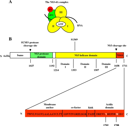
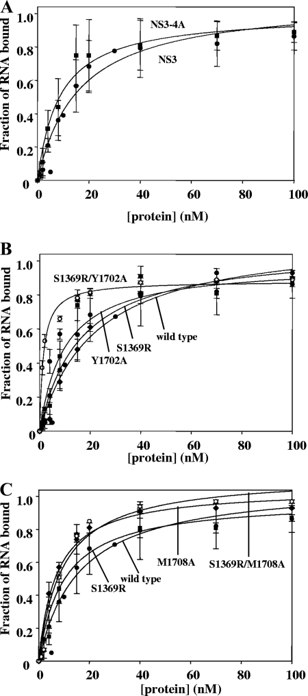
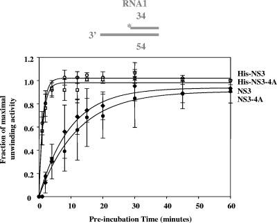
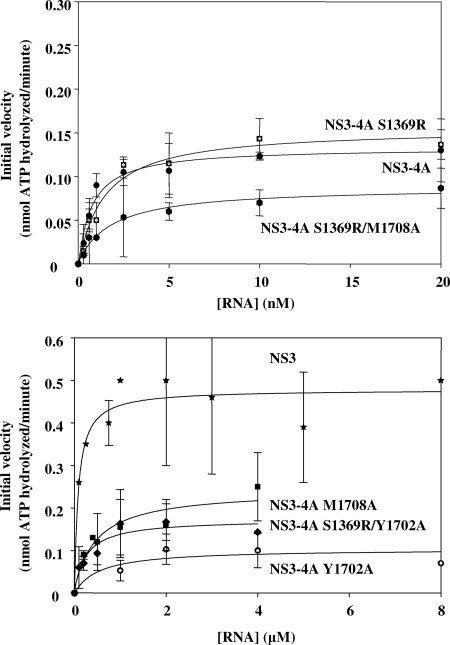
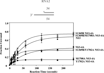
Nonstructural protein 3 (NS3) is an essential replicative component of the hepatitis C virus (HCV) and a member of the DExH/D-box family of proteins. The C-terminal region of NS3 (NS3hel) exhibits RNA-stimulated NTPase and helicase activity, while the N-terminal serine protease domain of NS3 enhances RNA binding and unwinding by NS3hel. The nonstructural protein 4A (NS4A) binds to the NS3 protease domain and serves as an obligate cofactor for NS3 serine protease activity. Given its role in stimulating protease activity, we sought to determine whether NS4A also influences the activity of NS3hel. Here we show that NS4A enhances the ability of NS3hel to bind RNA in the presence of ATP, thereby acting as a cofactor for helicase activity. This effect is mediated by amino acids in the C-terminal acidic domain of NS4A. When these residues are mutated, one observes drastic reductions in ATP-coupled RNA binding and duplex unwinding by NS3. These same mutations are lethal in HCV replicons, thereby establishing in vitro and in vivo that NS4A plays an important role in the helicase mechanism of NS3 and its function in replication.


Figures

FIG. 1.
FIG. 1.
Composition of NS3-4A and the NS3-4A expression construct. (A and B) The NS3-4A complex organization and construct design is illustrated schematically. In panel A, “pro” refers to the serine protease domain, and the Roman numerals indicate the respective NS3 helicase subdomains. The regions where ATP, RNA, and the NS4A cofactor bind are indicated as well. The protein construct expressed in E. coli is depicted in panel B. The numbers below the map refer to the HCV polyprotein numbering of the amino acids of NS3-4A. A detailed map of NS4A (i.e., HCV polyprotein residues 1658 to 1711) (18) is shown below the His-SUMO-NS3-4A expression construct. Residues Y1702 and M1708 are indicated in white font.
FIG. 2.
FIG. 2.
The NS4A cofactor does not affect direct RNA binding by NS3. The parameters for each binding curve are listed in Table 1. In all cases, the Hill equation with a cooperativity value of n = 1 provided the best-fit curve (see Materials and Methods). In panel A, the curves are NS3 (solid circles) and NS3-4A (solid squares). In panel B, the curves are NS3-4A (solid circles), NS3-4A S1369R (solid squares), NS3-4A Y1702A (solid diamonds), and NS3-4A S1369R/Y1702A (hatched circles). In panel C, the curves are NS3-4A (solid circles), NS3-4A S1369R (solid squares), and NS3-4A M1708A (solid diamonds), NS3-4A S1369R/M1708A (hatched circles). These data are the averages of three experiments, and the error represents the standard deviation.
FIG. 3.
FIG. 3.
The NS4A cofactor does not affect the rate of functional helicase formation on an RNA unwinding substrate. The rate constant for functional complex formation for NS3 was 0.05 ± 0.03 min−1 (solid diamonds); for NS3-4A, it was 0.02 ± 0.01 min−1 (solid circles); for His-NS3, it was 0.7 ± 0.1 min−1 (hatched circles); and for His-NS3-4A, it was 1.0 ± 0.3 min−1 (hatched squares). The data shown are the averages of three experiments, and the error represents the standard deviation.
FIG. 4.
FIG. 4.
RNA-stimulated ATPase activity of NS3-4A variants. The Michaelis-Menten kinetic parameters for each fit are listed in Table 2. The data plotted are NS3 (stars), NS3-4A (solid circles), NS3-4A (S1369R) (hatched squares), NS3-4A (Y1702A) (hatched circles), NS3-4A (M1708A) (solid squares), NS3-4A (S1369R/Y1702A) (solid diamonds), and NS3-4A (S1369R/M1708A) (solid hexagons). The data shown are the averages of three assays, and the error represents the standard deviation.
FIG. 5.
FIG. 5.
RNA unwinding by NS3-4A variants. The unwinding parameters of the 34-bp RNA duplex RNA2 for NS3 (solid diamonds) are A = 0.67 ± 0.02 and kobs = 0.020 ± 0.002 s−1; for NS3-4A (solid circles), they are A = 0.41 ± 0.02 and kobs = 0.020 ± 0.002 s−1; for NS3-4A Y1702A (solid squares), they are A = 0.13 ± 0.01 and kobs = 0.06 ± 0.01 s−1; for NS3-4A M1708A (crossed circles), they are A = 0.13 ± 0.01 and kobs = 0.06 ± 0.01 s−1; for NS3-4A S1369R (hatched squares), they are A = 0.74 ± 0.02 and kobs = 0.03 ± 0.01 s−1; for NS3-4A S1369R/M1708A (hatched diamonds), they are A = 0.70 ± 0.02 and kobs = 0.02 ± 0.01 s−1; and for NS3-4A S1369R/Y1702A (hatched circles), they are A = 0.35 ± 0.01 and kobs = 0.04 ± 0.01 s−1. These data are the averages of three experiments, and the error represents the standard deviation.

References

    1. Appel, N., T. Schaller, F. Penin, and R. Bartenschlager. 2006. From structure to function: new insights into hepatitis C virus RNA replication. J. Biol. Chem. 2819833-9836. - PubMed
    1. Beran, R. K., M. M. Bruno, H. A. Bowers, E. Jankowsky, and A. M. Pyle. 2006. Robust translocation along a molecular monorail: the NS3 helicase from hepatitis C virus traverses unusually large disruptions in its track. J. Mol. Biol. 358974-982. - PubMed
    1. Beran, R. K., and A. M. Pyle. 2008. Hepatitis C viral NS3-4A protease activity is enhanced by the NS3 helicase. J. Biol. Chem. 28329929-29937. - PMC - PubMed
    1. Beran, R. K., V. Serebrov, and A. M. Pyle. 2007. The serine protease domain of hepatitis C viral NS3 activates RNA helicase activity by promoting the binding of RNA substrate. J. Biol. Chem. 28234913-34920. - PubMed
    1. Campo, D. S., Z. Dimitrova, R. J. Mitchell, J. Lara, and Y. Khudyakov. 2008. Coordinated evolution of the hepatitis C virus. Proc. Natl. Acad. Sci. USA 1059685-9690. - PMC - PubMed

Publication types

MeSH terms

LinkOut - more resources