

Endothelial progenitor cells restore renal function in chronic experimental renovascular disease
- PMID: 19153272
- PMCID: PMC2758066
- DOI: 10.1161/CIRCULATIONAHA.108.788653
Endothelial progenitor cells restore renal function in chronic experimental renovascular disease
Abstract
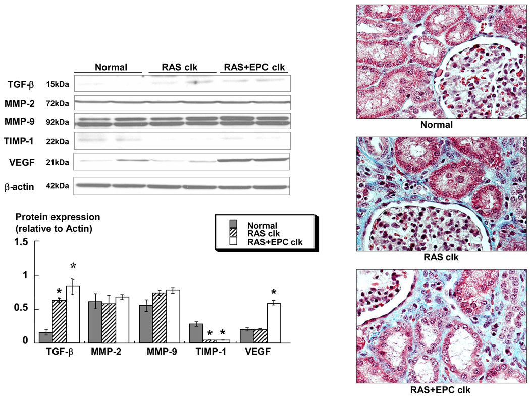
Background: Endothelial progenitor cells (EPCs) promote neovascularization and endothelial repair. Renal artery stenosis (RAS) may impair renal function by inducing intrarenal microvascular injury and remodeling. We investigated whether replenishment with EPCs would protect the renal microcirculation in chronic experimental renovascular disease.
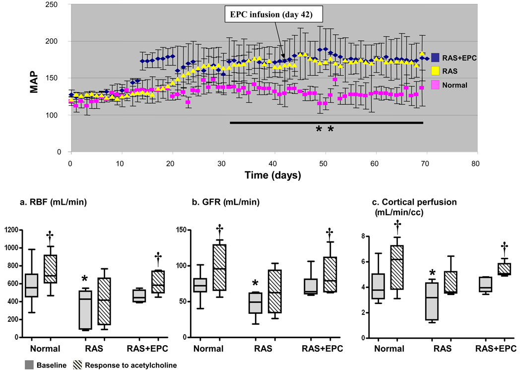
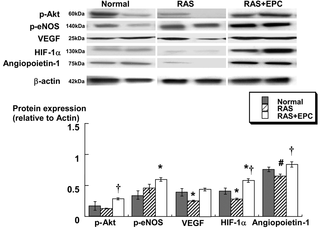
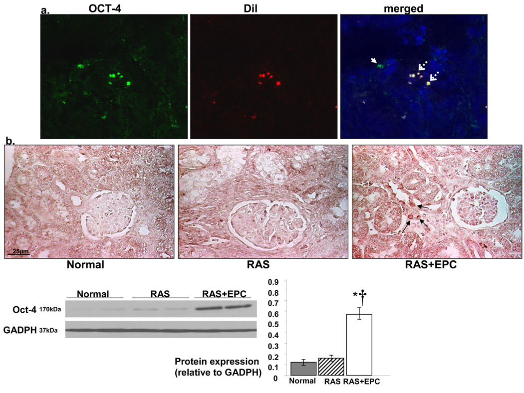
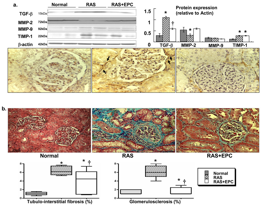
Methods and results: Single-kidney hemodynamics and function were assessed with the use of multidetector computed tomography in vivo in pigs with RAS, pigs with RAS 4 weeks after intrarenal infusion of autologous EPCs, and controls. Renal microvascular remodeling and angiogenic pathways were investigated ex vivo with the use of micro-computed tomography, histology, and Western blotting. EPCs increased renal expression of angiogenic factors, stimulated proliferation and maturation of new vessels, and attenuated renal microvascular remodeling and fibrosis in RAS. Furthermore, EPCs normalized the blunted renal microvascular and filtration function.
Conclusions: The present study shows that a single intrarenal infusion of autologous EPCs preserved microvascular architecture and function and decreased microvascular remodeling in experimental chronic RAS. It is likely that restoration of the angiogenic cascade by autologous EPCs involved not only generation of new vessels but also acceleration of their maturation and stabilization. This contributed to preserving the blood supply, hemodynamics, and function of the RAS kidney, supporting EPCs as a promising therapeutic intervention for preserving the kidney in renovascular disease.
Figures

References
-
- Rafii S, Lyden D. Therapeutic stem and progenitor cell transplantation for organ vascularization and regeneration. Nat Med. 2003;9:702–712. - PubMed
-
- Urbich C, Dimmeler S. Endothelial progenitor cells: characterization and role in vascular biology. Circ Res. 2004;95:343–353. - PubMed
-
- Kawamoto A, Murayama T, Kusano K, Ii M, Tkebuchava T, Shintani S, Iwakura A, Johnson I, von Samson P, Hanley A, Gavin M, Curry C, Silver M, Ma H, Kearney M, Losordo DW. Synergistic effect of bone marrow mobilization and vascular endothelial growth factor-2 gene therapy in myocardial ischemia. Circulation. 2004;110:1398–1405. - PubMed
-
- Napoli C, Williams-Ignarro S, de Nigris F, de Rosa G, Lerman LO, Farzati B, Matarazzo A, Sica G, Botti C, Fiore A, Byrns RE, Sumi D, Sica V, Ignarro LJ. Beneficial effects of concurrent autologous bone marrow cell therapy and metabolic intervention in ischemia-induced angiogenesis in the mouse hindlimb. Proc Natl Acad Sci U S A. 2005;102:17202–17206. - PMC - PubMed
-
- Urbich C, Heeschen C, Aicher A, Sasaki K, Bruhl T, Farhadi MR, Vajkoczy P, Hofmann WK, Peters C, Pennacchio LA, Abolmaali ND, Chavakis E, Reinheckel T, Zeiher AM, Dimmeler S. Cathepsin L is required for endothelial progenitor cell-induced neovascularization. Nat Med. 2005;11:206–213. - PubMed
Publication types
MeSH terms
Grants and funding
LinkOut - more resources
Full Text Sources
Other Literature Sources
Medical
