. 2009 Jan;65(Pt 1):24-33.
doi: 10.1107/S0907444908036524. Epub 2008 Dec 18.

Structures of the multicomponent Rieske non-heme iron toluene 2,3-dioxygenase enzyme system

Affiliations

Structures of the multicomponent Rieske non-heme iron toluene 2,3-dioxygenase enzyme system

Rosmarie Friemann et al. Acta Crystallogr D Biol Crystallogr. 2009 Jan.

Abstract

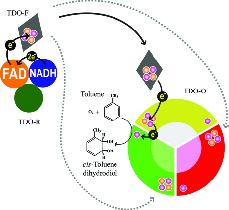
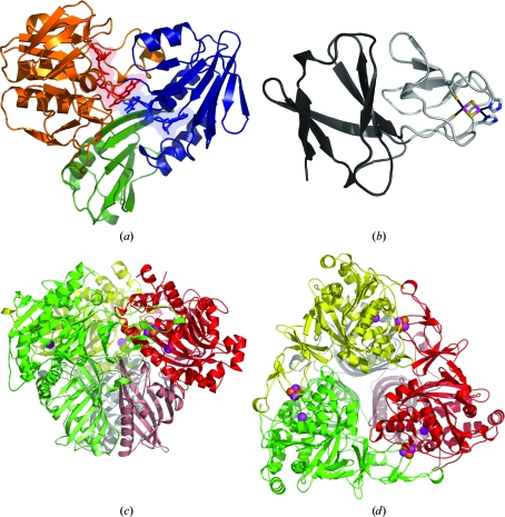
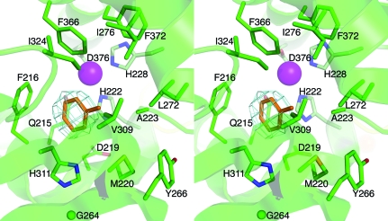
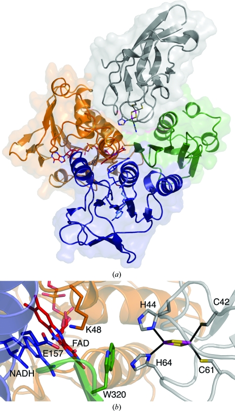
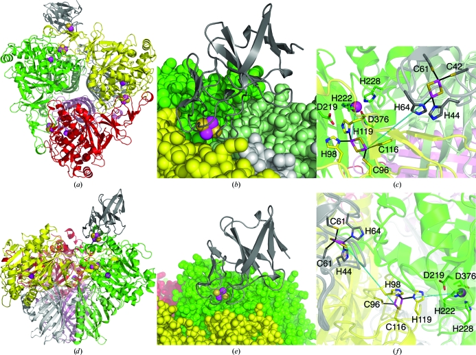
Bacterial Rieske non-heme iron oxygenases catalyze the initial hydroxylation of aromatic hydrocarbon substrates. The structures of all three components of one such system, the toluene 2,3-dioxygenase system, have now been determined. This system consists of a reductase, a ferredoxin and a terminal dioxygenase. The dioxygenase, which was cocrystallized with toluene, is a heterohexamer containing a catalytic and a structural subunit. The catalytic subunit contains a Rieske [2Fe-2S] cluster and mononuclear iron at the active site. This iron is not strongly bound and is easily removed during enzyme purification. The structures of the enzyme with and without mononuclear iron demonstrate that part of the structure is flexible in the absence of iron. The orientation of the toluene substrate in the active site is consistent with the regiospecificity of oxygen incorporation seen in the product formed. The ferredoxin is Rieske type and contains a [2Fe-2S] cluster close to the protein surface. The reductase belongs to the glutathione reductase family of flavoenzymes and consists of three domains: an FAD-binding domain, an NADH-binding domain and a C-terminal domain. A model for electron transfer from NADH via FAD in the reductase and the ferredoxin to the terminal active-site mononuclear iron of the dioxygenase is proposed.


Figures

Figure 1
Figure 1
Schematic representation of electron transfer in the TDO enzyme system. Two electrons are transferred from NADH to FAD in the reductase (TDO-R). The ferredoxin (TDO-F; shown as a grey rhombus) shuttles one electron from FAD via the dioxygenase (TDO-O) [2Fe–2S] Rieske center to the mononuclear iron in the neighboring subunit, where the catalytic reaction take place. The S and Fe atoms in the Rieske clusters are represented as orange and magenta circles, respectively.
Figure 2
Figure 2
Overall structures of the TDO enzyme system. (a) TDO-R. The FAD-binding domain, NADH-binding domain and C-terminal domain are colored blue, orange and green, respectively. FAD (red) and NADH (blue) are shown in stick representation and as a transparent surface representation. NADH was modeled in the NADH-binding pocket based on the structure of BPDO-RKKS102. (b) TDO-F. The large and cluster-binding domains are colored dark and light gray, respectively. The Rieske center and coordinating side-chain residues are shown in stick representation, with Fe, S and N atoms colored magenta, orange and blue, respectively. (c) Side view of the mushroom-shaped TDO-O α3β3 hexamer. The α-subunits are colored red, green and yellow and the β-subunits pink, light green and gray. The Rieske [2Fe–2S] cluster and the mononuclear iron are shown in CPK model representation, with Fe and S atoms colored magenta and orange, respectively. (d) Top view of TDO-O using the same color representations as in (c). This figure was produced using PyMOL (http://www.pymol.org).
Figure 3
Figure 3
Stereo representation of the active site of TDO-O with toluene bound. The F obsF calc map was computed before toluene was modeled. Residues that coordinate (light green) the mononuclear iron (magenta) and those that line the active site (green) are shown in stick representation. This figure was produced using PyMOL (http://www.pymol.org).
Figure 4
Figure 4
Modeled interactions between TDO-R and TDO-F. The FAD-binding domain, NADH-binding domain and C-terminal domain of the TDO-R are colored orange, blue and green, respectively. FAD (red) and NADH (blue) are shown in stick representation. NADH has been modeled in the NADH-binding pocket based on the structure of BPDO-RKKS102. TDO-F is colored gray and the Rieske iron–sulfur cluster with its coordinating residues is shown in stick representation. (a) Overall representation of a possible binding site for TDO-F between the FAD-binding and C-­terminal domains of TDO-R. (b) Close-up of the modeled interactions for electron transfer between TDO-R and TDO-F via FAD and Trp320 of TDO-R. Two residues (Lys48 and Glu157) that are believed to be involved in hydride transfer in GR and BPDO-RKKS102 are represented. This figure was produced using PyMOL (http://www.pymol.org).
Figure 5
Figure 5
Modeled interactions between TDO-F and TDO-O. Color representations are the same as in Fig. 2 ▶. Cys42, Cys61, His44 and His64 belong to TDO-F. Cys116, Cys96, His98 and His119 belong to the α-subunit of TDO-O, while the rest of the residues belong to a neighboring TDO-O α-subunit. (a) and (b) TDO-F is modeled in a shallow depression between two adjacent TDO-O heterodimers. (c) A line is drawn from the Rieske center of TDO-F to the Rieske center of TDO-O. (d), (e) and (f) TDO-F is modelled in accordance with carbazole 1,9a-dioxygenase and ferredoxin at the top of the cap of the mushroom-shaped α3β3 TDO-O with a possible electron-pathway route represented in (f). This figure was produced using PyMOL (http://www.pymol.org).

References

    1. Arand, M., Hällberg, B. M., Zou, J., Bergfors, T., Oesch, F., van der Werf, M. J., de Bont, J. A., Jones, T. A. & Mowbray, S. L. (2003). EMBO J.22, 2583–2592. - PMC - PubMed
    1. Ashikawa, Y., Fujimoto, Z., Noguchi, H., Habe, H., Omori, T., Yamane, H. & Nojiri, H. (2006). Structure, 14, 1779–1789. - PubMed
    1. Behrens, M. R., Mutlu, N., Chakraborty, S., Dumitru, R., Jiang, W. Z., Lavallee, B. J., Herman, P. L., Clemente, T. E. & Weeks, D. P. (2007). Science, 316, 1185–1188. - PubMed
    1. Beil, S., Mason, J. R., Timmis, K. N. & Pieper, D. H. (1998). J. Bacteriol.180, 5520–5528. - PMC - PubMed
    1. Berendsen, H. J. C., Postma, J. P. M., van Gunsteren, W. F. & Hermans, J. (1981). Intermolecular Forces, edited by B. Pullman, pp. 331–342. Dordrecht: Reidel.

Publication types

MeSH terms