. 2009 Feb 18;28(4):347-58.
doi: 10.1038/emboj.2008.294. Epub 2009 Jan 15.

Raf kinase inhibitory protein suppresses a metastasis signalling cascade involving LIN28 and let-7

Affiliations

Raf kinase inhibitory protein suppresses a metastasis signalling cascade involving LIN28 and let-7

Surabhi Dangi-Garimella et al. EMBO J. .

Abstract

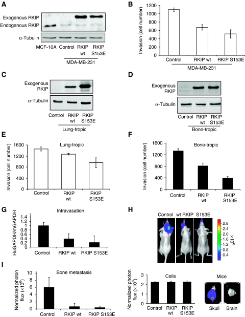
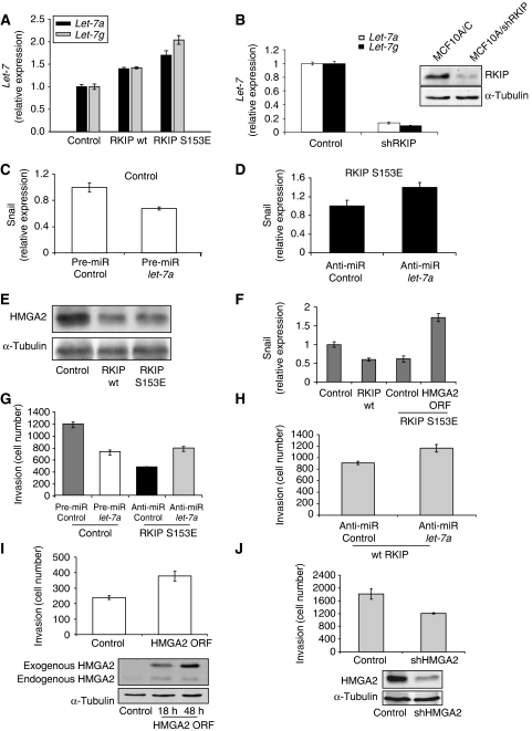
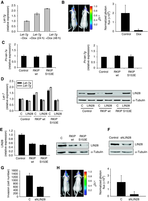
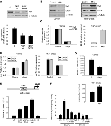
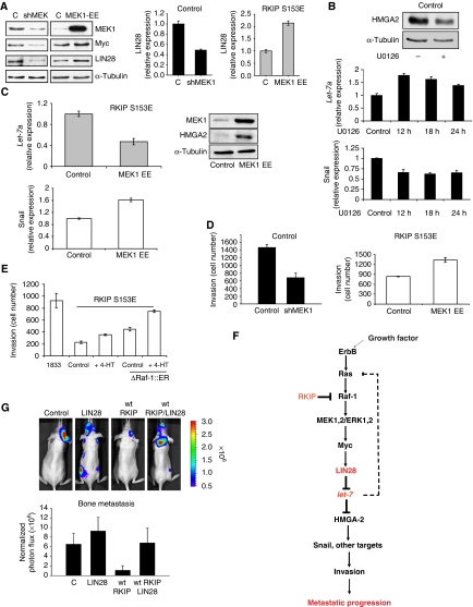
Raf kinase inhibitory protein (RKIP) negatively regulates the MAP kinase (MAPK), G protein-coupled receptor kinase-2, and NF-kappaB signalling cascades. RKIP has been implicated as a metastasis suppressor for prostate cancer, but the mechanism is not known. Here, we show that RKIP inhibits invasion by metastatic breast cancer cells and represses breast tumour cell intravasation and bone metastasis in an orthotopic murine model. The mechanism involves inhibition of MAPK, leading to decreased transcription of LIN28 by Myc. Suppression of LIN28 enables enhanced let-7 processing in breast cancer cells. Elevated let-7 expression inhibits HMGA2, a chromatin remodelling protein that activates pro-invasive and pro-metastatic genes, including Snail. LIN28 depletion and let-7 expression suppress bone metastasis, and LIN28 restores bone metastasis in mice bearing RKIP-expressing breast tumour cells. These results indicate that RKIP suppresses invasion and metastasis in part through a signalling cascade involving MAPK, Myc, LIN28, let-7, and downstream let-7 targets. RKIP regulation of two pluripotent stem cell genes, Myc and LIN28, highlights the importance of RKIP as a key metastasis suppressor and potential therapeutic agent.


Figures

Figure 1
Figure 1
RKIP regulates breast cancer invasion and metastasis. (A) RKIP is expressed in MCF10A mammary gland and depleted in metastatic MDA-MB-231 cells. MDA-MB-231 cells were stably transduced with wt or S153E RKIP and the lysates immunoblotted with anti-RKIP or anti-tubulin antibody. (B) Wt and S153E RKIP inhibit invasion of MDA-MB-231 cells. Cells were assayed for invasion as described in Materials and methods. Results represent the mean±s.e. for four independent samples (P<0.001 for wt and P=0.002 for S153E RKIP relative to Control). (C, D) Expression of wt and S153E RKIP in lung-tropic (4175) or bone-tropic (1833) breast cancer cells. Cells were stably transduced with wt or S153E RKIP and the lysates were immunoblotted with anti-RKIP or anti-tubulin antibody. All lanes in (C) come from the same gel, but one lane after sample 2 was omitted leading to a composite figure. (E, F) Wt and S153E RKIP inhibit invasion of lung-tropic (4175) or bone-tropic (1833) cells. The 1833 cells stably expressing control vector, wt RKIP, or S153E RKIP were assayed for invasion as described in Materials and methods. Results represent the mean±s.e. for four independent samples (4175) or mean±s.d. for three samples (1833) (P<0.05 for wt RKIP and P<0.05 for S153E RKIP relative to control 4175 cells; P=0.02 for wt RKIP and P<0.001 for S153E RKIP relative to control 1833 cells). (G) Wt and S153E RKIP inhibit intravasation of bone-tropic tumour cells (1833). The 1833 cells stably expressing control vector (six mice), wt RKIP (five mice), or S153E RKIP (five mice) were injected into the mammary fat pad of mice. After 3 weeks, cells isolated from the blood were analysed for GAPDH transcripts derived from human (tumour) or mouse (control). Results represent the mean±s.d. for the animals (P<0.002 for wt RKIP and P<0.001 for S153E RKIP relative to control). (H, I) Wt and S153E RKIP inhibit bone metastases. The 1833 cells expressing luciferase and control vector (six mice), wt RKIP (seven mice), or S153E RKIP (seven mice) were injected into the left ventricle of the mice, and the mice were imaged for luciferase activity after 3 weeks. Representative images show that RKIP wt and S153E greatly reduced bone metastases in skull (H, upper right panel; lower right panel). The 1833 cells stably expressing luciferase have an identical luciferase reporter activity before injection into mice (H, lower left panel). Comparable regions of the mouse skulls were optically imaged and quantified. Results (I) represent the mean±s.d. for the animals (P<0.01 for wt RKIP and P<0.01 for S153E RKIP relative to Control).
Figure 2
Figure 2
RKIP regulates let-7, HMGA2, and Snail. (A) Wt and S153E RKIP increase let-7a and let-7g expression. The 1833 cells expressing control, wt, or S153E were assayed for let-7a or let-7g by qRT–PCR. Results represent the mean±s.d. for three samples (P=0.03 for wt RKIP and P<0.01 for S153E RKIP relative to control). (B) Depletion of RKIP in MCF10A cells suppresses let-7a and let-7g expression. MCF10A cells expressing control vector or shRNA for human RKIP were assayed for let-7a or let-7g by qRT–PCR. Results represent the mean±s.d. for three samples (P<0.0001 for MCF10A/shRKIP relative to MCF10A cells). Cell lysates were immunoblotted with anti-RKIP or anti-tubulin antibodies. (C) Pre-miR let-7a decreases Snail mRNA levels. Snail mRNA isolated from 1833 cells transfected with pre-miR let-7a was quantitated by qRT–PCR. Results represent the mean±s.d. for three samples (P<0.02 for let-7a relative to Control). (D) Anti-miR let-7a increases Snail mRNA levels. Snail mRNA isolated from 1833 cells stably expressing S153E RKIP and transfected with anti-miR let-7a was quantitated by qRT–PCR. Results represent the mean±s.d. for three samples (P=0.03 for anti-miR let-7a relative to Control). (E) RKIP inhibits HMGA2 expression. The 1833 cells expressing vector, wt, or S153E RKIP were lysed and immunoblotted with anti-HMGA2 or anti-tubulin antibody. (F) Snail expression inhibited by RKIP and rescued by let-7-insensitive HMGA2. Snail mRNA was isolated from 1833 cells expressing control vector, wt RKIP, S153E RKIP, or S153E RKIP and HMGA2 lacking the 3′-untranslated region that binds let-7 (ORF). Snail mRNA was quantitated by qRT–PCR. Results represent the mean±s.e. for three independent samples (P<0.001 for wt RKIP and for S153E RKIP relative to control). (G, H) Let-7 regulates invasion. The 1833 cells were transfected with control or pre-miR let-7a, and 1833 cells expressing S153E or wt RKIP were transfected with control or anti-miR let-7a. Cells were assayed for invasion as described in Materials and methods. Results represent the mean±s.e. for four independent samples. (P=0.004 for pre-miR let-7 relative to Control, P=0.008 for anti-miR let-7 relative to Control in S153E RKIP cells, and P=0.025 for anti-miR let-7 relative to Control in wt RKIP cells). (I, J) HMGA2 regulates invasion. The 1833 cells were transfected with either HMGA2 ORF or shRNA for HMGA2 (shHMGA2). Cells were lysed at 18 and 48 h after transfection of HMGA2 ORF or 48 h after transfection of shHMGA2 and immunoblotted with anti-HMGA2 or anti-tubulin antibody. Cells were assayed for invasion as described in Materials and methods. Results represent the mean±s.e. for three independent samples (I: P=0.02 for HMGA2 relative to Control; J: P<0.04 for shHMGA2 relative to Control). Results are representative of at least three independent experiments.
Figure 3
Figure 3
RKIP induces let-7 through the inhibition of LIN28. (A) Let-7g is induced by doxycycline. The 1833 cells expressing inducible let-7g were treated with 2 μg/ml doxycycline for the indicated times. Results represent the mean±s.d. for three samples (P=0.006 for 24 h and P=0.004 for 48 h treatment relative to Control). (B) Let-7g inhibits bone metastases. The 1833 cells expressing luciferase and either control vector (7 mice) or tet-inducible let-7g (7 mice) were grown in the presence of 2 μg/ml doxycycline for 24 h. Cells were injected into the left ventricle of mice, and 2 days later, mice were administered with drinking water containing 4% sucrose only or 2 mg/ml doxycycyline and 4% sucrose. Mice were imaged for luciferase activity after 3 weeks. Representative images show let-7g greatly reduced bone metastases in skull. Results represent the mean±s.d. for the animals (P=0.001 for let-7g relative to Control). (C) RKIP does not alter primary let-7 expression. The 1833 cells expressing control vector, wt RKIP, or S153E RKIP were lysed. Primary let-7a and let-7g transcripts were analysed by qRT–PCR. Results represent the mean±s.d. for three samples. (D) LIN28 inhibits let-7 expression. The 1833 cells expressing control, wt RKIP, or S153E RKIP were stably transfected with LIN28 expression vector. Let-7a and g transcripts were analysed by qRT–PCR. Left: results represent the mean±s.d. for three samples (P=0.01 and P<0.001 for LIN28 in wt RKIP cells and S153E RKIP cells, respectively, relative to Control); Right: 1833 cells expressing control vector, wt RKIP, or S153E RKIP were lysed and immunoblotted with anti-LIN28 or anti-tubulin antibodies. Results are representative of at least three independent experiments. (E) RKIP decreased LIN28 mRNA in 1833 cells. The 1833 cells expressing control vector, wt RKIP, or S153E RKIP were lysed. LIN28 transcripts were analysed by qRT–PCR. Left: results represent the mean±s.d. for three samples (P=0.003 for wt RKIP and P=0.002 for S153E RKIP relative to Control); right: 1833 cells expressing control vector, wt RKIP, or S153E RKIP were lysed and immunoblotted with anti-LIN28 or anti-tubulin antibodies. Results are representative of at least three independent experiments. (F) shLIN28 downregulates LIN28 expression. The 1833 cells expressing control vector or shLIN28 were lysed and immunoblotted with anti-LIN28 or anti-tubulin antibodies. Results are representative of at least three independent experiments. (G) LIN28 depletion inhibits invasion of 1833 cells. The 1833 cells expressing control vector or shLIN28 were assayed for invasion as described in Materials and methods. Results represent the mean±s.d. for three independent samples (P=0.003 for shLIN28 relative to Control). (H) ShLIN28 inhibits bone metastasis. The 1833 cells expressing luciferase and vector control (seven mice) or shRNA for LIN28 (eight mice) were injected into the left ventricle of mice. Mice were imaged for luciferase activity after 3 weeks. Results represent the mean±s.d. for the animals (P=0.002 for shLIN28 relative to Control).
Figure 4
Figure 4
Myc regulates LIN28 transcription. (A) RKIP downregulates Myc expression. Blot: 1833 cells expressing control vector, wt RKIP, or S153E RKIP were lysed and immunoblotted with anti-Myc or anti-tubulin antibodies. Results are representative of at least three independent experiments. Graph: results represent the mean±s.d. for three independent samples (P=0.03 for wt RKIP and P=0.003 for S153E RKIP relative to Control). (B) Myc regulates LIN28 expression. The 1833 cells transfected with scrambled control or siRNA for Myc (left) or 1833 S153E RKIP cells transfected with a control or a Myc expression vector (right) were lysed and immunoblotted with anti-Myc, anti-LIN28, or anti-tubulin antibodies. Results are representative of at least three independent experiments. (C) Left: Myc regulates LIN28 transcript levels. The 1833 cells were transfected with a control or siRNA for Myc. LIN28 and Myc transcripts were analysed by qRT–PCR 48 h after transfection. Right: 1833 cells expressing S153E RKIP were transfected with a control or Myc expression vector. LIN28 transcripts were analysed by qRT–PCR 48 h after transfection. Results represent the mean±s.d. for three samples (P<0.0001 for siMyc and P<0.0001 for Myc relative to Control). (D) Myc regulates let-7 expression. The 1833 cells were transfected with a control or siRNA for Myc. Let-7a and g transcripts were analysed by qRT–PCR 48 h after transfection. The 1833 cells expressing S153E RKIP were transfected with a control or Myc expression vector. Let-7a and g transcripts were analysed by qRT–PCR 48 h after transfection. Results represent the mean±s.d. for three samples (P<0.01 for siMyc and P<0.01 for Myc relative to Control). (E) Myc regulates LIN28 transcription by binding to its promoter. Schematic representation of LIN28 promoter with the putative Myc-binding site. Chromatin immunoprecipitations (ChIPs) were carried out with anti-Myc antibody and anti-Jun antibody (a positive control for ChIP assay). ChIP was analysed by qRT–PCR, with primers in the LIN28, WNT5A (a positive control), CyclinD1 (a positive control for ChIP assay), and β-globin (a negative control; NC) promoters. Results represent the mean±s.d. for three samples (P<0.004 for LIN28 relative to IgG). (F) RKIP regulates Myc binding to the LIN28 promoter. Chromatin immunoprecipitation (ChIP) were carried out with anti-Myc antibody on the LIN28 promoter. The 1833 cells expressing control vector (C), wt RKIP, or S153E RKIP were treated with 2% serum or 2% serum with U0126 (10 μM) for 2 h after 48 h serum starvation. Results represent the mean±s.d. for three samples (P=0.01 and 0.001 for wt RKIP S153E RKIP, respectively, relative to untreated Control). (G) Myc expression regulates invasion of 1833 cells. Top: 1833 cells transfected with scrambled control or siRNA for Myc were assayed for invasion as described in Materials and methods. Results represent the mean±s.d. for three independent samples (P=0.004 for siMyc relative to Control). Bottom: 1833 S153E RKIP cells transfected with vector control or an expression vector for Myc were assayed for invasion as described in Materials and methods. Results represent the mean±s.d. for three independent samples (P=0.01 for Myc relative to Control).
Figure 5
Figure 5
RKIP regulates let-7 in part through the MAPK pathway, and LIN28 reverses the inhibition of bone metastasis by RKIP. (A) MEK1 regulates LIN28 and Myc in 1833 cells. Constitutively, active MEK induces LIN28 and Myc. The 1833 cells expressing S153E RKIP were transfected with control vector (C) or MEK1-EE for 48 h. LIN28 was also assayed by qRT–PCR. MEK1, Myc, lIN28, and α-tubulin expression were measured by western blot. Conversely, stable depletion of MEK by lentiviral shRNA downregulates LIN28 and Myc in 1833 cells. Results represent the mean±s.d. for three samples (P=0.005 for shMEK and P=0.001 for MEK1-EE relative to Control). Immunoblot results are representative of at least three independent experiments. (B) Inhibition of MEK by U0126 induces let-7a and inhibits HMGA2 and Snail. Blot: 1833 cells treated with or without the MEK inhibitor U0126 (10 μM, 12 h) were lysed and immuoblotted with antibodies to HMGA2 or tubulin. Graphs: 1833 cells were treated with 10 μM U0126 for the indicated times, and let-7a was assayed by qRT–PCR. Snail expression was quantitated by qRT–PCR. Results represent the mean±s.d. for three samples (Let-7: P=0.001, 0.01, and 0.03 for 12, 18, and 24 h, respectively, relative to Control; Snail: P=0.01, 0.004, and 0.01 for 12, 18, and 24 h, respectively, relative to Control). Results are representative of at least three independent experiments. (C) Constitutively active MEK inhibits let-7a expression and induces HMGA2 and Snail. The 1833 cells expressing S153E RKIP were transfected with control vector or MEK1-EE for 48 h, and let-7a and Snail were assayed by qRT–PCR. Results represent the mean±s.d. for three independent samples (Let-7a: P<0.02 for MEK1-EE relative to Control; Snail: P=0.01 for MEK1-EE relative to Control). MEK1 and HMGA2 expression were measured by western blot. Results are representative of at least three independent experiments. (D) MEK regulates invasion. Left: 1833 cells expressing either vector control or shRNA for MEK1 were assayed for invasion as described in Materials and methods. Results represent the mean±s.d. for three independent samples (P=0.004 for shMEK1 relative to Control). Right: 1833 cells expressing either vector control or an expression vector for MEK1-EE were assayed for invasion as described in Materials and methods. Results represent the mean±s.d. for three independent samples (P<0.005 for MEK1-EE relative to Control). (E) Inducible Raf kinase rescues the inhibition of invasion caused by S153E RKIP. The 1833 cells expressing S153E RKIP and transfected with control vector or ΔRaf-1:ER were untreated or treated with tamoxifen (4-HT). Results represent the mean±s.e. for three independent samples (P<0.001 for S153E RKIP+ΔRaf-1:ER+HT relative to S153E RKIP Control; P<0.001 for S153E RKIP+ΔRaf-1:ER+HT relative S153E RKIP+ΔRaf-1:ER). (F) Scheme showing the mechanism for RKIP regulation of invasion and metastasis through the MAPK/LIN28/let-7 pathway. (G) LIN28 overcomes the inhibitory effect of wt RKIP on bone metastasis. The 1833 cells expressing luciferase and either control vector or wt RKIP were stably transfected with control vector or LIN28 and injected into the left ventricle of the mice (Control, six mice; LIN28, 5 mice; wt RKIP, six mice; wt RKIP+LIN28, five mice), and mice were imaged for luciferase activity after 3 weeks. Results represent the mean±s.d. for the animals (P=0.001 for wt RKIP relative to Control, and P=0.01 for wt RKIP+LIN28 relative to wt RKIP).

Similar articles

Cited by

References

    1. Akaishi J, Onda M, Asaka S, Okamoto J, Miyamoto S, Nagahama M, Ito K, Kawanami O, Shimizu K (2006) Growth-suppressive function of phosphatidylethanolamine-binding protein in anaplastic thyroid cancer. Anticancer Res 26: 4437–4442 - PubMed
    1. Al-Mulla F, Hagan S, Behbehani AI, Bitar MS, George SS, Going JJ, Garcia JJ, Scott L, Fyfe N, Murray GI, Kolch W (2006) Raf kinase inhibitor protein expression in a survival analysis of colorectal cancer patients. J Clin Oncol 24: 5672–5679 - PubMed
    1. Beach S, Tang H, Park S, Dhillon AS, Keller ET, Kolch W, Yeung KC (2007) Snail is a repressor of RKIP transcription in metastatic prostate cancer cells. Oncogene - PMC - PubMed
    1. Burk U, Schubert J, Wellner U, Schmalhofer O, Vincan E, Spaderna S, Brabletz T (2008) A reciprocal repression between ZEB1 and members of the miR-200 family promotes EMT and invasion in cancer cells. EMBO Rep 9: 582–589 - PMC - PubMed
    1. Chang TC, Yu D, Lee YS, Wentzel EA, Arking DE, West KM, Dang CV, Thomas-Tikhonenko A, Mendell JT (2008) Widespread microRNA repression by Myc contributes to tumorigenesis. Nat Genet 40: 43–50 - PMC - PubMed

Publication types