. 2009 Feb 18;28(4):383-93.
doi: 10.1038/emboj.2008.281. Epub 2009 Jan 15.

Two-polymerase mechanisms dictate error-free and error-prone translesion DNA synthesis in mammals

Affiliations

Two-polymerase mechanisms dictate error-free and error-prone translesion DNA synthesis in mammals

Sigal Shachar et al. EMBO J. .

Erratum in

  • EMBO J. 2009 Apr 8;28(7):992

Abstract

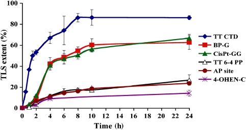
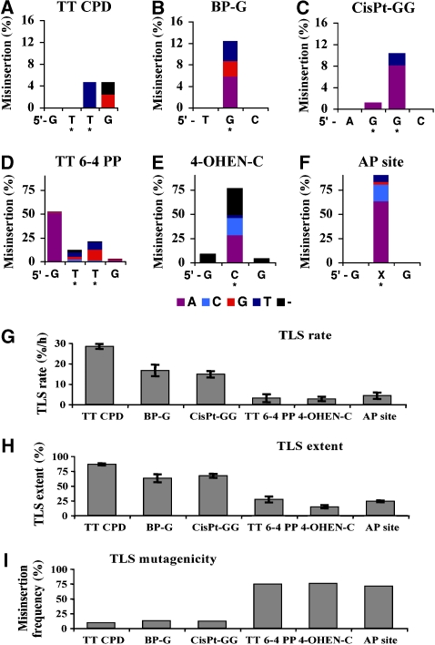
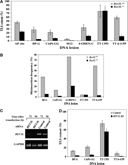
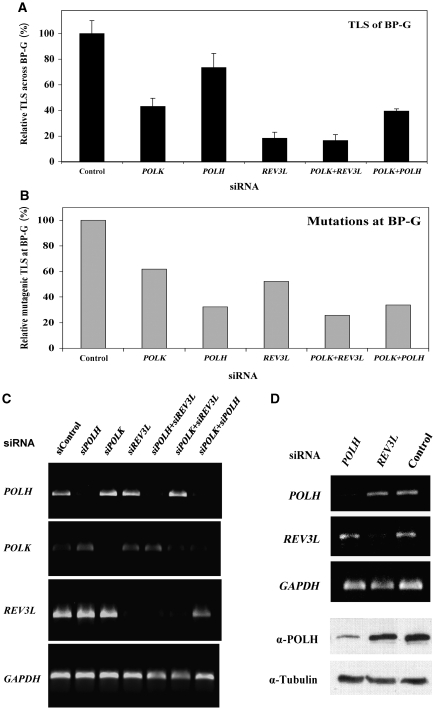
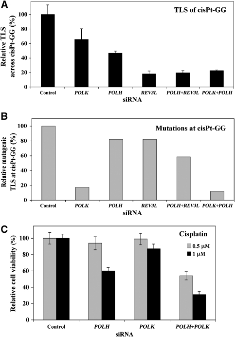
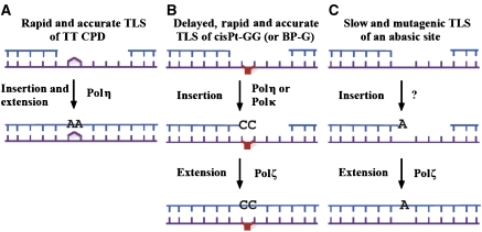
DNA replication across blocking lesions occurs by translesion DNA synthesis (TLS), involving a multitude of mutagenic DNA polymerases that operate to protect the mammalian genome. Using a quantitative TLS assay, we identified three main classes of TLS in human cells: two rapid and error-free, and the third slow and error-prone. A single gene, REV3L, encoding the catalytic subunit of DNA polymerase zeta (pol zeta), was found to have a pivotal role in TLS, being involved in TLS across all lesions examined, except for a TT cyclobutane dimer. Genetic epistasis siRNA analysis indicated that discrete two-polymerase combinations with pol zeta dictate error-prone or error-free TLS across the same lesion. These results highlight the central role of pol zeta in both error-prone and error-free TLS in mammalian cells, and show that bypass of a single lesion may involve at least three different DNA polymerases, operating in different two-polymerase combinations.


Figures

Figure 1
Figure 1
Kinetics of TLS across six different types of DNA damage in human U2OS cells. Plasmid mixtures containing the indicated gap-lesion plasmid, along with the control and the carrier plasmids, were introduced into human U2OS cells. Following incubation of 0–24 h to allow TLS, the DNA was extracted and used to transform an E. coli indicator strain. The extent of TLS was calculated as described under Materials and methods. Each of the data represents the average of four TLS experiments. The detailed data are presented in Supplementary Tables 1s–6s.
Figure 2
Figure 2
Mutagenicity of TLS across six different types of DNA damage in human U2OS cells. Individual colonies from the TLS reactions presented in Figure 1 were picked, and their plasmid content was analysed for mutations in the DNA region corresponding to the original site of the lesion. The cumulative height of each column represents the misinsertion frequency opposite the corresponding lesion, whereas the coloured column sections represent specific mutational events, colour coded as shown underneath. The DNA sequence with the damaged bases (marked by stars) is shown in the 5′ → 3′ direction. The detailed DNA sequence data are presented in Supplementary Tables 7s–12s. Results are presented for mutations formed at TT CPD (A), BP-G (B), cisPt-GG (C), TT 6-4 PP (D), 4-OHEN-C (E), and AP site (F). (G) TLS rates, calculated for the linear TLS phase of each lesion from the data presented in Figure 1, namely 0–2 h for TT CPD, TT 6-4 PP, 4-OHEN-C and the AP site, and 2–4 h for BP-G and cisP-GG, are shown. (H) Maximal values of TLS for the six lesions measured at 24 h are shown. The data were taken from Figure 1 and Supplementary Tables 1s–6s. (I) Mutagenic TLS out of total TLS events. For the AP site, insertion of A or G was taken as a mutagenic event. The data were taken from Figures 1 and 2, and Supplementary Tables 1s–12s.
Figure 3
Figure 3
Involvement of Rev3L in TLS in mammalian cells. (A) The TLS assay was performed with Rev3L+/+ or Rev3L−/− MEFs. Cells were assayed for TLS as described under Materials and methods, using the indicated gap-lesion plasmids. Average results of at least four experiments are presented. The detailed data are presented in Supplementary Table 13s. (B) Mutagenicity of the TLS reaction across different lesions in MEF Rev3L+/+ and Rev3L−/− cells. Colonies obtained in the experiments described in (A) were picked, their plasmid contents extracted, and subjected to DNA sequence analysis. The graph shows the percentage of incorrect nucleotides inserted opposite each of the lesions. The statistical significance of the differences in mutagenicity between the two cell types was calculated by the χ2 test, yielding the following P-values: BP-G, 0.0006; cisPt-GG, 0.006; M12, <0.0001; 4-OHEN-C, 0.0002; TT 6-4 PP, <0.0001. The detailed data are presented in Supplementary Tables 14s–18s. (C) RT–PCR of RNA extracted from human U2OS cells transiently transfected with siRNA for REV3L. (D) Extent of TLS in U2OS cells in which REV3L expression was knocked down using specific siRNA. Cells were assayed for TLS as described under Materials and methods, using the indicated gap-lesion plasmids. Average results of at least four experiments are presented. The detailed data are presented in Supplementary Table 21s.
Figure 4
Figure 4
Polκ and polζ operate in the same TLS pathway to bypass BP-G in human cells. (A) Relative TLS extent across BP-G in U2OS cells in which the expression of specific TLS DNA polymerases was knocked down. The experiments were performed as described in the legend of Figure 3D, and the detailed data are presented in Supplementary Table 22s. TLS extents were given as percentage relative to TLS assayed with control siRNA, which was 10.9±1.1% (see Supplementary Table 22s). (B) Relative mutagenic TLS across BP-G based on DNA sequence information of plasmids isolated from colonies obtained in the experiments described in (A). The detailed data are presented in Table I. Mutagenic TLS, namely the fraction of nucleotides other than C inserted opposite BP-G, was calculated out of all TLS events, and presented relative to the mutagenic TLS in cells transfected with control siRNA (which was 13.6%; see Table I). (C) RT–PCR of RNA extracted from human U2OS cells transiently transfected with the indicated siRNA. (D) RT–PCR of POLH and REV3L mRNAs (top) and an immunoblot with antibodies against polη (bottom) performed with RNA and protein extracts prepared from U2OS cells transfected with the indicated siRNAs.
Figure 5
Figure 5
Polζ cooperates with polη and polκ in error-free and error-prone TLS, respectively, across cisPt-GG in human cells. (A) Relative extent of TLS across cisPt-GG in U2OS cells in which the expression of specific TLS DNA polymerases was knocked down. The experiments were performed as described in the legend of Figure 4A, and the detailed data are presented in Supplementary Table 23s. (B) Relative mutagenic TLS across cisPt-GG based on DNA sequence information of plasmids isolated from colonies obtained in the experiments described in (A). The detailed data are presented in Table I. Mutagenic TLS, namely the fraction of nucleotides other than CC inserted opposite cisPt-GG, was calculated out of all TLS events, and presented relative to the mutagenic TLS in cells transfected with control siRNA (which was 18.3%; see Table I). (C) Cisplatin sensitivity of XPA cells in which the expression of POLH and POLK was knocked down. XP12RO cells were transfected with the indicated siRNAs, after which they were treated with 0.5 or 1 μM cisplatin. Results are given relative to the viability of cells transfected with control siRNA (77 and 55% for 0.5 and 1 μM cisplatin, respectively). Cell viability was measured using a luminescence-based assay for measuring ATP.
Figure 6
Figure 6
A model for three pathways of TLS in mammalian cells. TLS across a TT CPD is rapid and accurate, and occurs most likely by a single DNA polymerase, polη (A). TLS across cisPt-GG and BP-G occurs by a two-polymerase TLS pathway, which is rapid and accurate but delayed, and involves a combination of polζ with either polη or polκ (B). TLS across an AP site occurs by a slow and highly mutagenic TLS pathway, which involves polζ, and most likely an additional DNA polymerase (C). See text for details.

Comment in

  • Bypass specialists operate together.
    Takata K, Wood RD. Takata K, et al. EMBO J. 2009 Feb 18;28(4):313-4. doi: 10.1038/emboj.2008.303. EMBO J. 2009. PMID: 19225445 Free PMC article. Review.

References

    1. Adar S, Livneh Z (2006) Translesion DNA synthesis across non-DNA segments in cultured human cells. DNA Repair (Amst) 5: 479–490 - PubMed
    1. Alt A, Lammens K, Chiocchini C, Lammens A, Pieck JC, Kuch D, Hopfner KP, Carell T (2007) Bypass of DNA lesions generated during anticancer treatment with cisplatin by DNA polymerase eta. Science 318: 967–970 - PubMed
    1. Avkin S, Adar S, Blander G, Livneh Z (2002) Quantitative measurement of translesion replication in human cells: evidence for bypass of abasic sites by a replicative DNA polymerase. Proc Natl Acad Sci USA 99: 3764–3769 - PMC - PubMed
    1. Avkin S, Goldsmith M, Velasco-Miguel S, Geacintov N, Friedberg EC, Livneh Z (2004) Quantitative analysis of translesion DNA synthesis across a benzo[a]pyrene-guanine adduct in mammalian cells. The role of DNA polymerase κ. J Biol Chem 279: 53298–53305 - PubMed
    1. Avkin S, Sevilya Z, Toube L, Geacintov NE, Chaney SG, Oren M, Livneh Z (2006) p53 and p21 regulate error-prone DNA repair to yield a lower mutation load. Mol Cell 22: 407–413 - PubMed

Publication types