. 2009 Feb 4;28(3):183-92.
doi: 10.1038/emboj.2008.267. Epub 2009 Jan 15.

Ciliary targeting motif VxPx directs assembly of a trafficking module through Arf4

Affiliations

Ciliary targeting motif VxPx directs assembly of a trafficking module through Arf4

Jana Mazelova et al. EMBO J. .

Abstract

Dysfunctions of primary cilia and cilia-derived sensory organelles underlie a multitude of human disorders, including retinal degeneration, yet membrane targeting to the cilium remains poorly understood. Here, we show that the newly identified ciliary targeting VxPx motif present in rhodopsin binds the small GTPase Arf4 and regulates its association with the trans-Golgi network (TGN), which is the site of assembly and function of a ciliary targeting complex. This complex is comprised of two small GTPases, Arf4 and Rab11, the Rab11/Arf effector FIP3, and the Arf GTPase-activating protein ASAP1. ASAP1 mediates GTP hydrolysis on Arf4 and functions as an Arf4 effector that regulates budding of post-TGN carriers, along with FIP3 and Rab11. The Arf4 mutant I46D, impaired in ASAP1-mediated GTP hydrolysis, causes aberrant rhodopsin trafficking and cytoskeletal and morphological defects resulting in retinal degeneration in transgenic animals. As the VxPx motif is present in other ciliary membrane proteins, the Arf4-based targeting complex is most likely a part of conserved machinery involved in the selection and packaging of the cargo destined for delivery to the cilium.


Figures
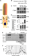
Figure 1
Figure 1
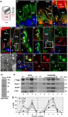
Rhodopsin VxPx motif engages Arf4 at the TGN to regulate RTC budding. (A) Diagram of the photoreceptor cell. RTCs travel from the Golgi/TGN (arrow) to the base of the cilium (C). BB, basal body; M, mitochondria; N, nucleus; Sy, synapse; AJ, adherens junction. (B) adRP mutations in the rhodopsin VxPx targeting motif are indicated with asterisks. (C) Frog retinas were pulse-labelled for 60 min and retinal PNS was incubated for 30 min with 50 μM peptides, as indicated in the panel, prior to a 2 h cell-free chase. Radiolabelled membrane proteins from two retinas were fractionated into Golgi/TGN and RTCs and analysed by SDS–PAGE and autoradiography ([35S]Rh). (D) Immunoblots of a duplicate gel were probed successively with anti-Arf3 and anti-Arf4, which specifically recognizes an ∼20-kDa protein (left panel) and quantified (lower right panels). (E) Golgi/TGN and RTCs were separated on sucrose density gradients, and distribution of rhodopsin ([35S]Rh) was visualized as in (C). Duplicate gels were blotted and probed with antibodies, as indicated in the panel (partial loss of the Rab11 band in RTC fractions is due to a cut to allow Arf3 detection on the same immunoblot). The differential distribution of calnexin, galactosyltransferase (Gal T) and sialyltransferase (Sial T), markers for the ER, trans-Golgi and TGN, respectively, in membranes from identically run sucrose gradients, as reported (Deretic and Papermaster, 1991, 1993; Deretic et al, 2004). (F) Quantification of autoradiograms and immunoblots from (E).
Figure 2
Figure 2
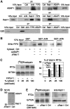
ASAP1 is a GAP for Arf4 and regulates RTC budding from the TGN. (A) Membrane proteins from one retina were separated by SDS–PAGE and immunoblotted with anti-ASAP1, which recognized an ∼120-kDa protein. Upon fractionation (lower panel), ASAP1 was detected in the G/TGN and the cytosol, with a small portion associated with RTCs. (B) Schematic representation of the ASAP1 BAR-PZA construct. (C) ASAP1 BAR-PZA was incubated with recombinant Arfs 1, 4, 5 or 6, and the hydrolysis of [α32P]GTP bound to Arf was measured. The data are presented as the means±s.d. of three separate experiments. (D) Radiolabelled retinal PNS was separated into membrane and cytosolic fractions, which were reconstituted as indicated in the panel. ASAP1-BAR-PZA (1 μM) was added to the assay in limited (25%) cytosol during the cell-free chase, and incorporation of rhodopsin into RTCs was determined ([35S]Rh). The quantification data are presented as the means±s.e. of three separate experiments (*P=0.01). (EG) Negatively stained in vitro budded RTCs (∼250 nm) examined by EM. (H) Following in vitro budding, Golgi and RTCs were immunoblotted with anti-GM130. (I) Cytosol was immunodepleted with anti-ASAP1 and protein A beads, reducing ASAP1 to ∼10% of control. (J) After in vitro budding in ASAP1-immunodepleted cytosol, membrane proteins from two retinas were separated by SDS–PAGE and autoradiographed ([35S]rhodopsin, left panels). Duplicate gels were immunoblotted with ASAP1 (right panels).
Figure 3
Figure 3
ASAP1 is associated with both the Golgi/TGN and actin cytoskeleton, and colocalizes with Rab11 and FIP3 on nascent RTCs at the TGN. (A) A diagram representing the microfilaments (red) and calycal processes (CPs) that evaginate from the RIS and surround the base of the ROS (modified from Deretic et al, 1995). (B) A confocal optical section (0.7 μm) of frog retina. ASAP1 (green) is detected in the RIS cytoplasm, in punctate structures around the Golgi (G, arrow), and in axially aligned structures (arrowheads), resembling microfilaments. N, nucleus (TO-PRO-3, blue). (C) ASAP1 (green) and phalloidin (red), colocalize (yellow, arrowheads) along microfilaments, including the CPs (arrow). Microfilaments are anchored at the adherens junctions (AJs). (D) Rhodopsin C-terminal mAb 11D5 (red) labels the ROS and the Golgi (G), where ASAP1-positive puncta (yellow, arrows) line up with regular periodicity. ASAP1 is also detected in CPs. (E) Rab11-positive puncta (yellow, arrows) aligned with rhodopsin-laden Golgi. Rab11 is also present on RTCs (arrowheads). (F) ASAP1 (blue) and Rab11 (red) colocalize in the bud-like profiles at the tips of the trans-Golgi (Rab6, green) (boxed area magnified in G). (G) Magnified trans-Golgi area from (F), with ASAP1- and Rab11-positive buds (arrows). (H) Rab11 (red) and ASAP1 (blue) are shown separately. (I) FIP3 (green, rabbit Ab; Wilson et al, 2005), overlaps with Rab11 (red) and ASAP1 (blue) in the punctate structures within the Golgi region (boxed area magnified in J). (J) FIP3, Rab11 and ASAP1 are shown in separate channels to better visualize their colocalization (arrows). (K) An EM image showing a greatly enlarged nascent bud at the TGN (arrowhead) and enlarged RTCs (arrows) in propranolol-treated retina (modified from Deretic et al, 2004). (L) Rab11 (red) and ASAP1 (green) colocalize (arrows, yellow) in propranolol-enlarged RTCs and buds lining Rab6-postive trans-Golgi (blue). (M, N) Rab11 and ASAP1 shown individually. Insets demonstrate identical distribution of Rab11 and ASAP1. (O) Goat anti-FIP3 detects an ∼80-kDa protein on isolated RTCs. (P) Following in vitro budding and subcellular fractionation, the distribution of rhodopsin was determined by autoradiography ([35S]Rh). Duplicate gels were immunoblotted as indicated in the panel. (Q) Quantification of autoradiograms and immunoblots. Bar, 4 μm in (B, C), 3 μm in (D–F, I), 2 μm in (L–N), 1 μm in (H, J), 0.7 μm in (G, K) and insets in (L–N).
Figure 4
Figure 4
Arf4, Rab11, FIP3 and ASAP1 form a complex and regulate RTC budding. (A) Specific antibodies were incubated with retinal PNS and proteins bound to protein-A beads were separated by SDS–PAGE and analysed by immunoblotting, as indicated. Left panels, pAb IPs; right panels, mAb IPs. (B) Arf4 and Arf6 were expressed as GST fusion proteins and incubated with 6His–FIP3. Proteins bound to glutathione beads were separated by SDS–PAGE and analysed by immunoblotting with anti-FIP3. (C) FIP3-F1 C-terminal fragment (aa 441–756) (1 μM) was added to the assay in limited (25%) cytosol during the cell-free chase, and rhodopsin incorporation into RTCs was determined ([35S]rhodopsin). Autoradiograms and immunoblots of duplicate gels probed for Rab11 and ASAP1 were quantified (right panel). (D) Cytosol was immunodepleted with anti-Rab11, reducing it to nearly undetectable levels. (E) Following in vitro budding in Rab11-immunodepleted cytosol, membrane proteins from two retinas were separated by SDS–PAGE and autoradiographed ([35S]rhodopsin, left panels). Rab11 immunoblots of duplicate gels (right panels).
Figure 5
Figure 5
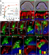
Expression of [I46D]Arf4 deficient in ASAP1-mediated GTP hydrolysis disrupts rhodopsin trafficking and causes retinal degeneration in transgenic X. laevis. (A) Arf4 or [I46D]Arf4 was incubated with ASAP1 BAR-PZA, or with AGAP1, and the hydrolysis of [α32P]GTP bound to Arf was measured. The data are presented as the means±s.d. of three separate experiments. (B) Transgenic expression of Arf4–GFP (green) in retinal photoreceptors. WGA (red) detects glycosylated membrane proteins. Nuclei are stained with DAPI (blue). (C) Expression of [I46D]Arf4–GFP (green) causes photoreceptor loss and retinal degeneration. (D) Arf4–GFP colocalizes with WGA-labelled membranes in the RIS. ONL, outer nuclear layer. (E) RIS is constricted (arrows) in [I46D]Arf4 mutants. (F) Arf4–GFP is present in the Golgi (G). Microfilaments (phalloidin, red), span the RIS (large bracket) from the outer limiting membrane (OLM, dotted line) to the CPs. N, nucleus. (G, H) In [I46D]Arf4 retinas, microfilaments (phalloidin, red) do not span the RIS (indicated by large brackets). The OLM (dotted line) is displaced and RIS is constricted (arrow in H). (I) Rhodopsin (red) colocalizes with Arf4–GFP in the Golgi (G, arrows). N, nucleus (TO-PRO-3, blue). (J) [I46D]Arf4–GFP and rhodopsin (red) colocalize (arrow) at the site of RIS constriction. (K) Rhodopsin-rich membranes (red, arrow) are elaborating from the [I46D]Arf4 Golgi (G), towards the nucleus (N). (LN) Arf4–GFP (green, L) and rhodopsin (red, M) extensively colocalize in the Golgi (G, arrows, yellow in N). Inset in N: surface density of Golgi membranes (SvG) containing Arf4, rhodopsin or both, determined within the RIS area occupied by the Golgi, indicated by a large bracket in (N). The data from a representative transgenesis experiment are presented as the means±s.e., n=11. (OQ) [I46D]Arf4 (green, O) and rhodopsin (red, P) partition into distinct membranes. Arrows in (O–Q) indicate atypical rhodopsin-rich membranes juxtaposed to the Golgi/TGN-containing Arf4. Inset in Q: surface density of Golgi membranes (SvG) in photoreceptors expressing [I46D]Arf4–GFP. The data are presented as in the inset in (N), n=11. Bar, 500 μm (B, C), 20 μm (D, E), 5 μm (F–H, J), 4 μm in (I, K) and 3 μm in (L–Q).

Similar articles

Cited by

References

    1. Beronja S, Laprise P, Papoulas O, Pellikka M, Sisson J, Tepass U (2005) Essential function of Drosophila Sec6 in apical exocytosis of epithelial photoreceptor cells. J Cell Biol 169: 635–646 - PMC - PubMed
    1. Berson EL, Rosner B, Weigel-DiFranco C, Dryja TP, Sandberg MA (2002) Disease progression in patients with dominant retinitis pigmentosa and rhodopsin mutations. Invest Ophthalmol Vis Sci 43: 3027–3036 - PubMed
    1. Brown MT, Andrade J, Radhakrishna H, Donaldson JG, Cooper JA, Randazzo PA (1998) ASAP1, a phospholipid-dependent arf GTPase-activating protein that associates with and is phosphorylated by Src. Mol Cell Biol 18: 7038–7051 - PMC - PubMed
    1. Burguete AS, Fenn TD, Brunger AT, Pfeffer SR (2008) Rab and Arl GTPase family members cooperate in the localization of the Golgin GCC185. Cell 132: 286–298 - PMC - PubMed
    1. Chen W, Feng Y, Chen D, Wandinger-Ness A (1998) Rab11 is required for trans-Golgi network-to-plasma membrane transport and a preferential target for GDP dissociation inhibitor. Mol Biol Cell 9: 3241–3257 - PMC - PubMed

Publication types

MeSH terms