Review
. 2009;48(8):1378-98.
doi: 10.1002/anie.200802092.

RNA interference: from basic research to therapeutic applications

Affiliations
Review

RNA interference: from basic research to therapeutic applications

Jens Kurreck. Angew Chem Int Ed Engl. 2009.

Abstract

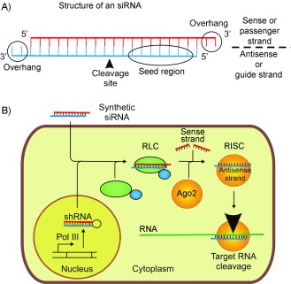
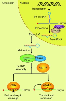
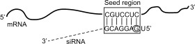
An efficient mechanism for the sequence-specific inhibition of gene expression is RNA interference. In this process, double-stranded RNA molecules induce cleavage of a selected target RNA (see picture). This technique has in recent years developed into a standard method of molecular biology. Successful applications in animal models have already led to the initiation of RNAi-based clinical trials as a new therapeutic option.Only ten years ago Andrew Fire and Craig Mello were able to show that double-stranded RNA molecules could inhibit the expression of homologous genes in eukaryotes. This process, termed RNA interference, has developed into a standard method of molecular biology. This Review provides an overview of the molecular processes involved, with a particular focus on the posttranscriptional inhibition of gene expression in mammalian cells, the possible applications in research, and the results of the first clinical studies.


Figures

Figure 1
Figure 1
A) Structure of an siRNA. The two strands of the siRNA form an approximately 19 nucleotide duplex. Two nucleotides hang over from each 3′ end. Deoxythymidine is often used as the overhangs in chemically synthesized siRNAs. The position at which the complementary target RNA is cleaved is indicated with an arrow, and the seed region, through which the interaction with the target RNA begins, is indicated. B) Simplified model of the RNAi mechanism in mammalian cells. After uptake of the chemically synthesized siRNAs into the cells, they are loaded onto the RISC by the RLC, in the course of which the sense strand is removed. The antisense strand guides the RISC to the complementary target RNA, which is cleaved by the Ago2 protein. A longer term inhibition of gene expression can be accomplished when an shRNA is expressed intracellularly instead of by the exogenous application of an siRNA. (Figure adapted from Ref. 9.)
Figure 2
Figure 2
miRNA pathways in mammalian cells. RNAs are transcribed in the nucleus in the form of a precursor (pri‐miRNA), which is processed by the RNase III Drosha to pre‐miRNA. In this process, the Drosha complexes with the DGCR8 protein. The pre‐miRNA is exported out of the nucleus and into the cytoplasm by Exportin‐5 and cleaved there by Dicer (complexed with TRBP) to form the functional miRNA, which in turn combines with an Argonaut protein (Ago) to form an miRNA–ribonucleoprotein (miRNP) complex. The miRNA can either cause endonucleolytic cleavage of the target mRNA through Ago2 or block translation in the case of partial complementarity. (Figure adapted from Ref. 27.)
Figure 3
Figure 3
Two‐step model to explain the efficiency of siRNA (s: sense strand, as: antisense strand): 1) Depending on the relative stability of the two ends of an siRNA, one of the two strands is preferentially assembled into the RISC. The retention of the strand complementary to the target RNA can be achieved through the selection of a suitable sequence. 2) An antisense strand assembled into the RISC can, however, be unsuitable for silencing when the complementary sequence of the target RNA is inaccessible. The local structure of the target region thus also influences silencing significantly. (Figure adapted from Ref. 48 with permission from Elsevier.)
Figure 4
Figure 4
Selected modified nucleotides which can be employed to stabilize siRNAs.
Figure 5
Figure 5
Base pairing between nucleotides 2–8 of the siRNA (seed region) and mRNAs can lead to off‐target effects in RNAi applications. These undesired side effects can be significantly reduced by a 2′‐O‐methyl substitution of the second nucleotide (circled).
Figure 6
Figure 6
Toll‐like Receptors (TLR). A) Signaling pathway of the TLRs. B) Cellular localization and ligands which activate the various TLRs. LPS: Lipopolysaccharide; CpG: cytidine‐phosphate‐guanosine; MyD88: myeloid differentiation primary response protein 88; NF‐ϰB: nuclear factor kappa beta; MAPK: mitogen‐activated protein kinases; IRF: interferon regulatory factor. (Figure modified from Ref. 104.)
Figure 7
Figure 7
Nonviral delivery of siRNAs. A) Lipoplex: cationic lipids (gray) form complexes with the negatively charged siRNAs (red). PEG (yellow) is frequently attached to improve the pharmacokinetic characteristics. B) Liposomes in which the cationic lipids enclose the siRNA. C) siRNA coupled to cholesterol to increase its lipophilicity. D) Specific delivery by coupling of siRNAs on the antigen‐binding fragment of an antibody through positively charged protamine. E) Direct coupling of an siRNA to an aptamer for tumor‐cell‐specific delivery. F) Neuronal delivery by a peptide of the rabies virus glycoprotein (RVG) with an arginine nonamer (9R) at its carboxy terminus to bind the siRNA. G) Receptor‐mediated delivery by coupling of a ligand (F: Folate) to a DNA oligonucleotide (blue), that hybridizes with siRNA (sense strand: green, antisense strand: red). Further details are described in the text.
Figure 8
Figure 8
Creation of replication‐deficient viral vectors. For gene transfer, the central protein‐coding genes of the viral genome are removed and replaced with the transgene or an shRNA expression cassette. The vector genome is packaged in a packaging cell line expressing the viral genes, which in most cases are spread over several plasmids. The resulting virus vector only includes the expression cassette for the transgene, while the essential virus genes are missing, so that further replication is impossible.
Figure 9
Figure 9
Function of the tumor suppressor CYLD, which was identified by means of an RNAi screen. CYLD works as an inhibitor in the NF‐κB signaling pathway. The loss of the CYLD function leads to uncontrolled growth. The pathway can also be inhibited by using sodium salicylate or prostaglandin‐1 (PGA1). TRAF: TNF‐receptor‐associated factor; IKK: IκB kinase complex. Scheme adapted from Ref. 146.

Similar articles

Cited by

References

    1. Napoli C., Lemieux C., Jorgensen R., Plant Cell 1990, 2, 279–289. - PMC - PubMed
    1. Fire A., Xu S., Montgomery M. K., Kostas S. A., Driver S. E., Mello C. C., Nature 1998, 391, 806–811. - PubMed
    1. Mello C. C., Angew. Chem. 2007, 119, 7114–7124;
    2. Angew. Chem. Int. Ed. 2007, 46, 6985–6994. - PMC - PubMed
    1. Fire A. Z., Angew. Chem. 2007, 119, 7094–7113;
    2. Angew. Chem. Int. Ed. 2007, 46, 6966–6984. - PubMed
    1. Taroncher‐Oldenburg G., Marshall A., Nat. Biotechnol. 2007, 25, 961. - PubMed

Publication types

LinkOut - more resources