. 2009 Feb;15(2):156-64.
doi: 10.1111/j.1469-0691.2008.02648.x. Epub 2008 Dec 22.

The Panton-Valentine leukocidin vaccine protects mice against lung and skin infections caused by Staphylococcus aureus USA300

Affiliations

The Panton-Valentine leukocidin vaccine protects mice against lung and skin infections caused by Staphylococcus aureus USA300

E L Brown et al. Clin Microbiol Infect. 2009 Feb.

Abstract

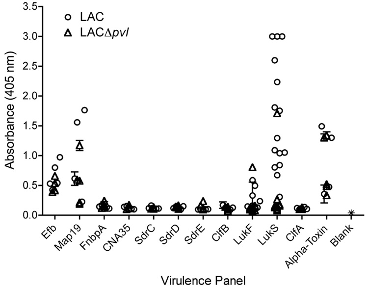
Methicillin-resistant Staphylococcus aureus is increasingly responsible for staphylococcal infections in the community. A large percentage of the community-acquired methicillin-resistant (CA-MRSA) strains in the USA produce Panton-Valentine leukocidin (PVL), which is associated with severe infections. The virulence of the clinical CA-MRSA strain USA300 was compared to that of its isogenic pvl-deleted mutant, and it was shown that PVL contributes to lung and muscle tissue destruction, respectively, in murine necrotizing pneumonia and skin infection models. Mice infected with the USA300 strain developed a dominant anti-PVL response. The PVL subunits were therefore tested as vaccinogens against this isolate, and their vaccine efficacy correlated with both the route of vaccination and infection. These data suggest that PVL is a virulence factor in murine CA-MRSA infections.


Figures

Fig. 1
Fig. 1. Pneumonia survival curve
Balb/c mice were intranasally inoculated with a suspension of 1–3 × 10⁸ CFUs of either Staphylococcus aureus USA300 LAC (●) or S. aureus LACΔpvl (■). Mortality was recorded at 24 and 48 h post-infection, and animals were monitored for up to 7 days.
Fig. 2
Fig. 2. Panton–Valentine leukocidin-deleted mutants cause less severe infections
(a–e) Mice were infected intranasally with 3–5 × 10⁷ CFUs of each Staphylococcus aureus strain, LAC (●) or LACΔpvl (○). (a) Weight loss in grams over time (days). (b, c) Gross lung morphology 2 days post-infection (b, LAC; c, LACΔpvl). (d, e) Five-micrometre lung sections were stained with haematoxylin and eosin (H&E), ×20 magnification. (f–l) Mice were infected intradermally with 1 × 10⁷ CFUs of each S. aureus strain, LAC (■) or LACΔpvl (□). (f) Weight loss in grams over time (days). (g, h) Gross superficial skin morphology. Intradermally infected animals were killed and photographed 7 days post-inoculation (g, LAC; h, LACΔpvl). (i, j) Gross morphology of tissues underlying the infection site (i, LAC; j, LACΔpvl). (k, l) Five-micrometre tissue sections stained with H&E, 10× magnification.
Fig. 3
Fig. 3. Antibody response to virulence factors
Mice were infected intradermally with 1 × 10⁷ CFUs of either Staphylococcus aureus LAC or S. aureus LACΔpvl. Twenty-eight days later, sera from five mice were collected, and the reactivity of antibodies towards selected recombinant S. aureus virulence factors was tested. CNA was used as a negative control, as it is not encoded in the S. aureus USA300 genome. The presence of antibodies from groups of five animals infected with LAC (●), infected with LACΔpvl (▽) or uninfected (*), that were reactive to the S. aureus virulence factors was tested using ELISA as previously described. Each symbol represents the response of each animal. The data are expressed as the mean ± SD of three wells.
Fig. 4
Fig. 4. Subcutaneous immunizations
(a) Isotype profile analysis following vaccination. Twenty-eight days following a subcutaneous immunization regimen with either LukF or LukS (or LukF and LukS in one group of subcutaneously vaccinated mice), mice were bled and their sera were analysed for isotype-specific (IgG1, IgG2a, IgG2b, IgG3, IgE, IgA, or IgM) anti-LukF or anti-LukS responses. Only IgG1-, IgG2a-, IgG2b- and IgA-specific antibodies were detected. The IgG3 response is representative of negative responses, i.e. IgE and IgM. The number of mice used in each treatment group is indicated in parentheses. Each symbol represents the mean ± SE response within each group. †p <0.05, IgG2a anti-LukS response as compared to the IgG2a anti-LukF response in subcutaneously vaccinated mice; unpaired t-test with Welch correction. (b) Delayed-type hypersensitivity response to LukF and LukS following vaccination. Thirty-four days post-vaccination, mice from all groups were challenged on the right and left footpads with 2.5 µg of LukF and LukS, respectively. Footpads were measured at 0 and 24 h after challenge, and the response to LukS (black) and to LukF (white) is expressed as the mean ± SE of six mice/group (*p <0.0001 as compared to the contralateral footpad or to the challenge-only control; unpaired t-test with Welch correction). (c) Percent survival following intranasal inoculation with Staphylococcus aureus USA300. Balb/c mice (total numbers used/group indicated in parentheses) were vaccinated subcutaneously with recombinant LukF, LukS, control proteins or adjuvant alone prior to infection with 5 × 10⁷ CFUs of S. aureus in a volume of 20 µL. Mouse survival was monitored for up to 7 days post-infection. (d) Weight change in vaccinated mice following intradermal infection. Vaccinated mice were intradermally infected with S. aureus USA300 and weighed before and after infection. Symbols represent the weight loss in mice vaccinated with LukS (●), LukF (■), CFA only (▼), or unvaccinated (○). †p <0.01 LukF- and LukS-vaccinated mice as compared to CFA-only-vaccinated groups of mice; unpaired t-test with Welch correction.
Fig. 5
Fig. 5. Mucosal immunizations
(a) Isotype profile analysis following vaccination. Twenty-eight days following a intranasal immunization regimen with either LukF or LukS (or control proteins), mice were bled and their sera were analysed for isotype-specific (IgG1, IgG2a, IgG2b, IgG3, IgE, IgA, or IgM) anti-LukF or anti-LukS responses. Only IgG1-, IgG2a-, IgG2b- and IgA-specific antibodies were detected. The IgG3 response is representative of negative responses, i.e. IgE and IgM. The number of mice used in each treatment group is indicated in parentheses. Each symbol represents the mean ± SE response within each group. ‡p <0.005, IgG2b and IgA anti-LukS response as compared to the IgG2b and IgA anti-LukF response in intranasally vaccinated mice; unpaired t-test with Welch correction. (b) Delayed-type hypersensitivity response to LukF and LukS following vaccination. Thirty-four days post-vaccination, mice from all groups were challenged on the right and left footpads with 2.5 µg of LukF and LukS, respectively. Footpads were measured at 0 and 24 h after challenge, and the response to LukS (black) and to LukF (white) is expressed as the mean ± SE of six mice/group (*p <0.0001 as compared to the contralateral footpad or to the challenge-only control; unpaired t-test with Welch correction). (c) Percent survival following intranasal inoculation with Staphylococcus aureus USA300. Balb/c mice (total numbers used/group indicated in parentheses) were vaccinated subcutaneously with recombinant LukF, LukS, control proteins or adjuvant alone prior to infection with 5 × 10⁷ CFUs of S. aureus in a volume of 20 µL. Mouse survival was monitored for up to 7 days post-infection. The percent survival of mice immunized intranasally with LukS was significantly different when compared to LukF-, DbpA- and alpha toxin-immunized mice (‡p <0.05) or to non-immunized mice (†p <0.0001; Fisher’s exact test). (d) Weight change in vaccinated mice following intradermal infection. Vaccinated mice were intradermally infected with S. aureus USA300 and weighed before and after infection. Symbols represent the weight loss in mice vaccinated with LukS (●), LukF (■), cholera toxin (CT) only (▲), or unvaccinated (○).

References

    1. Lowy FD. Staphylococcus aureus infections. N Engl J Med. 1998;339:520–532. - PubMed
    1. Klevens RM, Edwards JR, Richards CL, Jr, et al. Estimating health care-associated infections and deaths in U.S. hospitals, 2002. Public Health Rep. 2007;122:160–166. - PMC - PubMed
    1. Klevens RM, Morrison MA, Nadle J, et al. Invasive methicillin-resistant Staphylococcus aureus infections in the United States. JAMA. 2007;298:1763–1771. - PubMed
    1. Davis SL, Perri MB, Donabedian SM, et al. Epidemiology and outcomes of community-associated methicillin-resistant Staphylococcus aureus infection. J Clin Microbiol. 2007;45:1705–1711. - PMC - PubMed
    1. Gonzalez BE, Hulten KG, Dishop MK, et al. Pulmonary manifestations in children with invasive community-acquired Staphylococcus aureus infection. Clin Infect Dis. 2005;41:583–590. - PubMed

Publication types

MeSH terms