Review
. 2009 Jan;16(1):21-34.
doi: 10.1016/j.devcel.2008.12.012.

The Jekyll and Hyde functions of caspases

Affiliations
Review

The Jekyll and Hyde functions of caspases

Caroline H Yi et al. Dev Cell. 2009 Jan.

Abstract

Apoptosis is an ancient form of regulated cell death that functions under pathological and nonpathological contexts in all metazoans. More than a decade of intense research has led to extensive characterization of the core molecular mechanisms for apoptotic cell death. This includes the identification of a family of cysteine proteases, caspases, which are critical for the execution of apoptosis. Whereas completion of the proteolytic caspase cascade leads to elimination of a cell by apoptosis, caspase activation, when finely tuned, directs alternative cellular functions independent of cell death. Exciting recent developments have focused on uncovering nonapoptotic roles of caspases ranging from immune regulation to spermatogenesis, in highly specialized cellular frameworks.


Figures
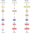
Figure 1
Figure 1. The canonical apoptotic pathway is well conserved in C. elegans, Drosophila, and mammals
Functional orthologs are color-coded. In C. elegans inhibition of the anti-apoptotic Bcl-2 family member, CED-9, by the pro-apoptotic BH3-only protein, EGL-1, releases CED-4 to promote activation of the caspase, CED-3. In Drosophila, the pro-apoptotic multi-domain Bcl-2 family members, Debcl and Buffy, promote Dark-mediated activation of Dronc followed by induction of the caspase cascade. Similarly, mammalian pro-apoptotic BH3-only proteins inhibit the anti-apoptotic Bcl-2 family members, Bcl-2 and Bcl-xL, to promote oligomerization of the multi-domain Bcl-2 family members, Bax and Bak. Adaptor proteins, such as Apaf-1, promote caspase activation, leading to apoptotic cell death. Note that in mammals, the caspase family has expanded substantially (see text). Bcl, B-cell lymphoma; CED, cell death abnormal; BH3, Bcl-2 homology 3; EGL, egg-laying defective; Dark, Drosophila Apaf-1 related killer.
Figure 2
Figure 2. Inflammatory caspases mediate an innate immune response to microbial invasion
Cellular recognition of foreign invaders leads to formation of inflammatory caspase-containing protein complexes, called the inflammasomes. The best characterized is the NALP1 inflammasome, which consists of a NLR family member, an adaptor protein called ASC, and caspase-1/5. Inflammasome assembly leads to activation of caspase-1 and maturation of IL-1β. A recent study has implicated caspase-1 in regulating non-conventional protein secretion, which may be intimately involved in the inflammatory response (Keller et al., 2008). However the exact mechanism for caspase-1 in mediating protein secretion remains to be determined.
Figure 3
Figure 3. The role of caspase-8 in maintaining the survival of activated T cells
Caspase-8 deficient T cells undergo necroptosis in response to antigen stimulation, which can be inhibited by necrostatin-1 (Nec-1). Autocrine secretion of TNFα is hypothesized to mediate necroptosis in activated caspase-8 deficient T cells.
Figure 4
Figure 4. Local caspase activation influences dendritic pruning during Drosophila metamorphosis
Extensive severing of neuronal dendrites occurs in the class IV dendritic arborization (C4da) neurons within the fly peripheral nervous system during metamorphosis (Kuo et al., 2006). Local activation of caspases is restricted to dendrites targeted for degradation at 4hr after pupae formation (APF) as indicated by antibody staining with anti-activated caspase-3. Activated caspase-3 is not observed at early metamorphosis or white pupae stage (WP). Images courtesy of Yuh-Nung Jan.
Figure 5
Figure 5. Non-apoptotic function of caspases in Drosophila neural development
Low-level caspase activity, which would otherwise fall below the threshold of detection in some assays, was measured using a fluorescence resonance energy transfer (FRET)-based probe containing enhanced cyan fluorescence protein (ECFP) as the FRET donor and a variant of enhanced yellow fluorescence protein, Venus, as the FRET acceptor. A caspase cleavage site was designed within the peptide linker that connects ECFP and Venus (Takemoto et al., 2003). In situ confocal imaging of the FRET emission ratio (Venus/ECFP) reveals caspase activity in wing imaginal discs (A), including the specific subset of cells that have the potential to give rise to scutellar sensory organ precursors (B, upper panel). This level of caspase activation is dependent on DmIKKε-activated degradation of DIAP1 and is insufficient to induce apoptosis. Rather, it is required for proper regulation of sensory organ precursor cell number (Kuranaga et al., 2006). Suppression of caspase activation via loss of DmIKKεresults in the eventual formation of an extra sensory bristle, which co-stains with a neuronal marker, anti-senseless (B, lower panels). Images courtesy of Masayuki Miura.

References

    1. Aballay A, Ausubel FM. Programmed cell death mediated by ced-3 and ced-4 protects Caenorhabditis elegans from Salmonella typhimurium-mediated killing. Proc Natl Acad Sci U S A. 2001;98:2735–2739. - PMC - PubMed
    1. Allan LA, Clarke PR. Phosphorylation of caspase-9 by CDK1/cyclin B1 protects mitotic cells against apoptosis. Mol Cell. 2007;26:301–310. - PubMed
    1. Allan LA, Morrice N, Brady S, Magee G, Pathak S, Clarke PR. Inhibition of caspase-9 through phosphorylation at Thr 125 by ERK MAPK. Nat Cell Biol. 2003;5:647–654. - PubMed
    1. Amer A, Franchi L, Kanneganti TD, Body-Malapel M, Ozoren N, Brady G, Meshinchi S, Jagirdar R, Gewirtz A, Akira S, Nunez G. Regulation of Legionella phagosome maturation and infection through flagellin and host Ipaf. J Biol Chem. 2006;281:35217–35223. - PubMed
    1. Arama E, Agapite J, Steller H. Caspase activity and a specific cytochrome C are required for sperm differentiation in Drosophila. Dev Cell. 2003;4:687–697. - PubMed

Publication types

LinkOut - more resources