. 2009 Jan;16(1):58-69.
doi: 10.1016/j.devcel.2008.11.008.

Hey2 regulation by FGF provides a Notch-independent mechanism for maintaining pillar cell fate in the organ of Corti

Affiliations

Hey2 regulation by FGF provides a Notch-independent mechanism for maintaining pillar cell fate in the organ of Corti

Angelika Doetzlhofer et al. Dev Cell. 2009 Jan.

Abstract

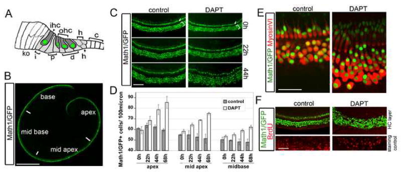
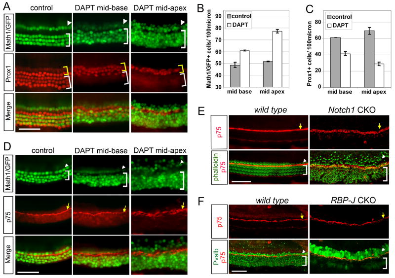
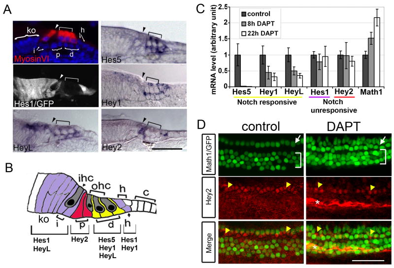
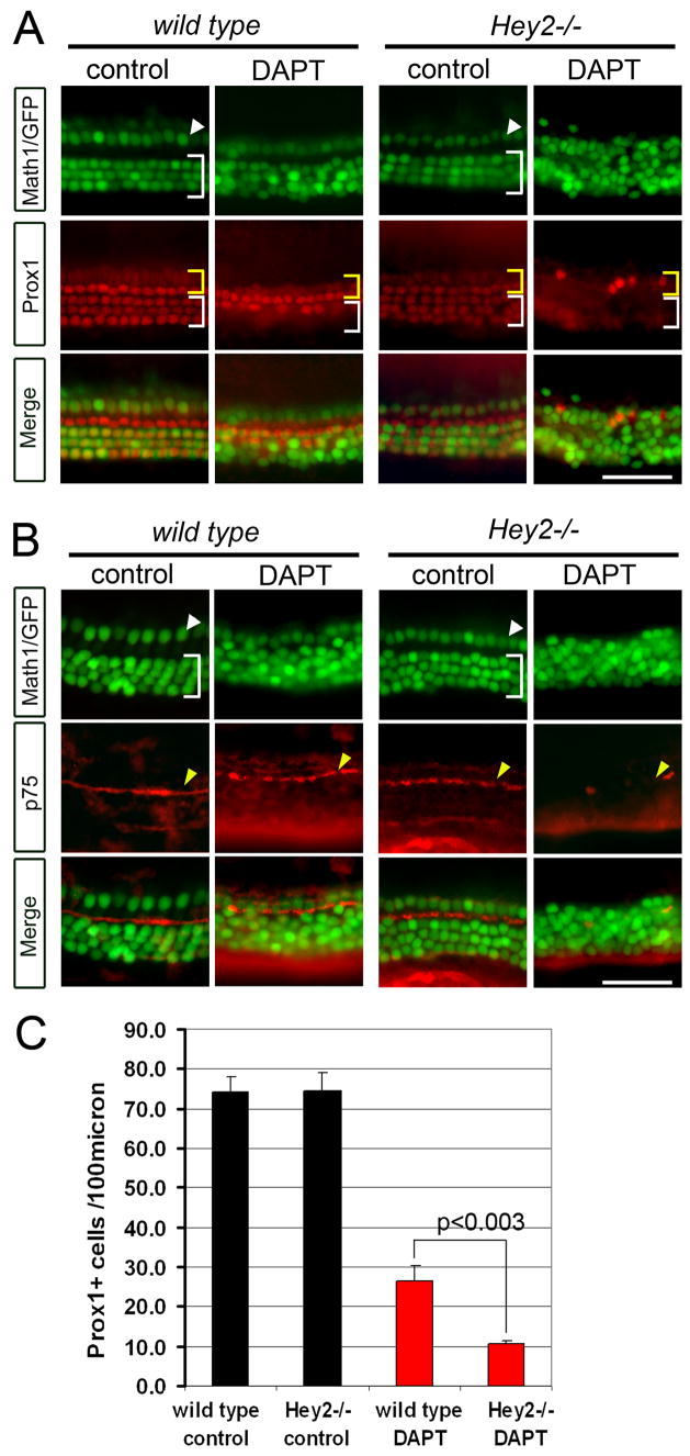
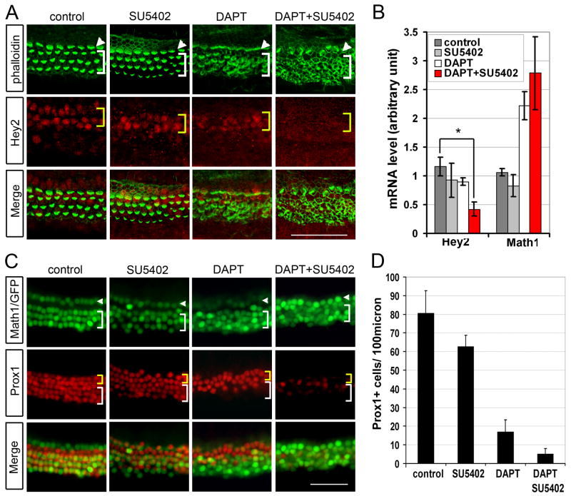
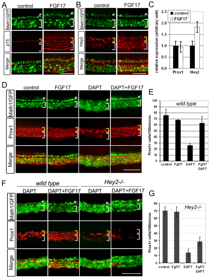
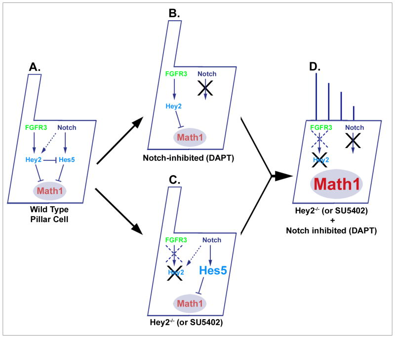
The organ of Corti, the auditory organ of the inner ear, contains two types of sensory hair cells and at least seven types of supporting cells. Most of these supporting cell types rely on Notch-dependent expression of Hes/Hey transcription factors to maintain the supporting cell fate. Here, we show that Notch signaling is not necessary for the differentiation and maintenance of pillar cell fate, that pillar cells are distinguished by Hey2 expression, and that-unlike other Hes/Hey factors-Hey2 expression is Notch independent. Hey2 is activated by FGF and blocks hair cell differentiation, whereas mutation of Hey2 leaves pillar cells sensitive to the loss of Notch signaling and allows them to differentiate as hair cells. We speculate that co-option of FGF signaling to render Hey2 Notch independent also liberated pillar cells from the need for direct contact with surrounding hair cells, and enabled evolutionary remodeling of the complex cellular mosaic of the inner ear.


Figures

Fig. 1
Fig. 1. Treatment of neonatal organ of Corti explants with the gamma-secretase inhibitor DAPT induces ectopic hair cells
A: Schematic of the organ of Corti at postnatal day 0 (P0). Inner (ihc) and outer (ohc) hair cells (green nuclei) are surrounded by supporting cell subtypes: Inner phalangeal cells (i), inner and outer pillar cells (p), Deiters’ cells (d) and Hensen cells (h). B: Whole mount of a neonatal Math1/GFP transgenic organ of Corti. Math1/GFP expression labels hair cells (green). C: Time course of ectopic hair cell production in neonatal cochlear organ cultures in response to DAPT. Arrow marks inner and bracket marks outer hair cells (Math1/GFP, green). D: Quantification of C. Similar results were obtained in two independent preparations (error bar ± s.e.m.). E: Ectopic Math1/GFP+ cells (green) in DAPT express MyosinVI (red). F: No BrdU incorporation (red) is seen in Math1/GFP+ hair cells (green) in control or DAPT treated cochlear organs. The mesenchymal layer of cochlear organs (taken from a confocal plane below the basal lamina of the sensory epithelium) was used as BrdU staining control. Scale bar: 0.5mm in A; 50μm in C, E, F.
Fig. 2
Fig. 2. Deiters’ cells, but not pillar cells, trans-differentiate into hair cells in the absence of Notch signaling
A–D: P0 Math1/GFP transgenic cochlear explants cultured for 72 hours in the presence of DAPT or DMSO (control). A: Ectopic hair cells (green) in DAPT are accompanied by a loss of Prox1+ cells (red) in the Deiters’ cell region (white bracket). B–C: Quantification of Math1/GFP+ hair cells (B) and Prox1+ supporting cells (C) after 72 hours with and without DAPT treatment. The number of Math1/GFP+ hair cells and Prox1+ supporting cells of mid-apical and mid-basal regions of cochlear explants was normalized to 100μm (n=5; error bar ± s.e.m.). D: Pillar cells persist in the absence of Notch signaling: p75 antibody staining (red) marks pillar cells in control (DMSO treated) and DAPT treated cochlear explants (yellow arrowhead). E-: Pillar cells are maintained in P0 Notch1 mutant organ of Corti: Phalloidin staining (green) labels the actin rich hair cell bundles (stereocilia). p75 antibody staining (red) labels the apical tips of pillar cells (yellow arrow). F: Pillar cell differentiation is unaffected in the RBP-J mutant organ of Corti. E13 RBP-J mutant and wild type cochlear explants were cultured for 4 days. p75 antibody staining labels pillar cells (red, yellow arrow). Parvalbumin staining labels inner (green, arrowhead) and outer hair cells (green, bracket). Scale bar: 50μm in A, D, E, F.
Fig. 3
Fig. 3. Notch signaling is not necessary for Hey2 expression in pillar cells
A: Expression of Hes5, Hey1, Hey2 and HeyL transcripts and Hes1/GFP transgene in the neonatal (P0-P2) organ of Corti. Hair cells are visualized by MyosinVI staining (red). Arrowheads point to inner hair cells and bracket marks outer hair cells. Two brackets point to nuclei (blue) of pillar cells (p) and Deiters’ cells (d); arrows point to Hensen (h) and inner phalangeal cells (iph). Large bracket marks Köllikers organ (ko). B: Schematic of the organ of Corti indicating the organization of expression patterns of Hes and Hey genes. C: Hey2 and Hes1 mRNA levels are unchanged in the presence of DAPT. Relative expression levels (QPCR) of Math1 and Hey2, Hes1, HeyL, Hey1, Hes5 mRNA in stage P1 cochlear organs exposed for 8h DAPT (gray bar), 22h DAPT (white bar) and 22h DMSO vehicle control (black bar; n=3; error bars ± s.e.m.). D: Hey2 protein expression is maintained in pillar cells in the absence of Notch signaling. Math1/GFP transgenic P1 cochlear organs were cultured for 48 hours in the presence of DAPT or DMSO (control) and stained with Hey2 antibody (red). Math1/GFP expression (green) labels inner (white arrow) and outer hair cells (white bracket). Yellow arrowhead marks pillar cells. * Non-specific binding of Hey2 antibody to extracellular matrix. Scale bar: 50μm in A, D.
Fig. 4
Fig. 4. Hey2 is required to maintain pillar cell fate in the absence of Notch signaling
A–C: P0 Math1/GFP+ wild type and mutant Hey2 (Hey2−/−) cochlear explants were cultured for 72 hours in DAPT or DMSO (control). A: DAPT treated Hey2−/− cochlear explants have strongly reduced numbers of Prox1+ cells (red) in pillar cell region (yellow bracket) and no Prox1+ cells in Deiters’ cell region (white bracket). B: Pillar cell specific p75 staining (red, yellow arrow) confirms the severe loss of pillar cells in Hey2−/− cochlear explants in DAPT. C: Quantification of Prox1+ cells/100 μm in control wild type and Hey2 mutant cultures (black bars) and DAPT treated cultures (red bars). For each condition a minimum of three cochlear cultures from three independent experiments were analyzed (error bars ± s.e.m.). Scale bar: 50μm in A, B.
Fig. 5
Fig. 5. FGF and Notch signaling prevent pillar cells from trans-differentiating into hair cells
A: Inhibition of Notch and FGF signaling results in a loss of pillar cell specific Hey2 protein expression (red) and an increase in phalloidin+ hair cells (green). P0 cochlear explants were stained with Hey2 antibody (red) and phalloidin (green) after being cultured for 48 hours in DMSO (control) or in the presence of FGF inhibitor SU5402, DAPT or both. Note: As previously reported (Kiernan et al., 2005), loss of Notch signaling results in disorganization of phalloidin labeled hair cell bundles (DAPT, DAPT+SU5402, green) (see Fig. 2E). B: Culture of cochlear explants in DAPT and SU5402 (red bar) for 22 hours results in a significant decrease in Hey2 mRNA levels. Bars represent mean of three independent experiments performed (error bars ± s.e.m.) (*<0.01). C: P0 Math1/GFP transgenic cochlear explants were stained with Prox1 antibody (red) after being cultured for 72 hours in the presence or absence of DAPT and SU5402. D: Quantification of Prox1+cells as in C. A minimum of three cochlear cultures were analyzed for each condition (error bars ± s.e.m.). Scale bar: 50μm in A, C.
Fig. 6
Fig. 6. Up-regulation of Hey2 by FGF17 prevents Deiters’ cell conversion in the absence of Notch signaling
A–C: P0 cochlear explants were cultured for 48 hours in presence of FGF17. A–B: Expansion of pillar cell specific p75 and Hey2 expression in the presence of FGF17. C: Up-regulation of Hey2 mRNA expression in the presence of FGF17. Bars represent mean of three independent experiments performed (error bars ± s.e.m.). D: FGF17 prevents Deiters’ cell trans-differentiation in the absence of Notch signaling. E: Quantification of Prox1+ cells in D. Three cochlear cultures were analyzed for each condition and black bar represents mean of Prox1+ cell/100μm (error bars ± s.e.m.). F: FGF17 does not block the effect of DAPT in Hey2 mutant cochlear explants. G: Quantification of Prox1+ cells in F. A minimum of three cochlear cultures were analyzed for each condition (error bars ± s.e.m.). Top panel (A, B, D, F): Math1/GFP (green) labels inner (arrow) and outer (bracket) hair cells. Middle panel shows p75 (A), Hey2 (B) and Prox1 (D, F) antibody staining in red. Yellow bracket marks pillar cell domain, white bracket marks Deiters’ cell domain. Scale bar: 50μm in A, B, D, F.
Fig. 7
Fig. 7. FGF and Notch signaling act redundantly to prevent pillar cells from transdifferentiating into hair cells
A: Signaling diagram representing FGF and Notch-mediated effects on Math1 expression and maintenance of the pillar cell phenotype. Math1 is both necessary and sufficient for hair cell differentiation in the context of the inner ear (Bermingham et al., 1999; Zheng and Gao, 2000), and either Hey2 or Hes5 can inhibit Math1 expression and thus prevent pillar cell transdifferentiation into hair cells. B: Hey2 is expressed under the control of FGF signaling and is largely independent of changes in Notch signaling. C: In the absence of Hey2, Hes5 is upregulated in pillar cells (see Fig. S5), leading to a continued Notch-dependent block to transdifferentiation, and suggesting that Hey2 normally inhibits Hes5 expression. D: If both FGF and Notch signaling are blocked, neither Hey2 nor Hes5 is expressed leading to Math1 de-repression and trans-differentiation of pillar cells into hair cells.

References

    1. Adam J, Myat A, Le Roux I, Eddison M, Henrique D, Ish-Horowicz D, Lewis J. Cell fate choices and the expression of Notch, Delta and Serrate homologues in the chick inner ear: parallels with Drosophila sense-organ development. Development (Cambridge, England) 1998;125:4645–4654. - PubMed
    1. Artavanis-Tsakonas S, Rand MD, Lake RJ. Notch signaling: cell fate control and signal integration in development. Science. 1999;284:770–776. - PubMed
    1. Barald KF, Kelley MW. From placode to polarization: new tunes in inner ear development. Development (Cambridge, England) 2004;131:4119–4130. - PubMed
    1. Bermingham NA, Hassan BA, Price SD, Vollrath MA, Ben-Arie N, Eatock RA, Bellen HJ, Lysakowski A, Zoghbi HY. Math1: an essential gene for the generation of inner ear hair cells. Science. 1999;284:1837–1841. - PubMed
    1. Bermingham-McDonogh O, Oesterle EC, Stone JS, Hume CR, Huynh HM, Hayashi T. Expression of Prox1 during mouse cochlear development. The Journal of comparative neurology. 2006;496:172–186. - PMC - PubMed

Publication types

MeSH terms