. 2009 Jan;16(1):118-31.
doi: 10.1016/j.devcel.2008.11.004.

BubR1 N terminus acts as a soluble inhibitor of cyclin B degradation by APC/C(Cdc20) in interphase

Affiliations

BubR1 N terminus acts as a soluble inhibitor of cyclin B degradation by APC/C(Cdc20) in interphase

Liviu A Malureanu et al. Dev Cell. 2009 Jan.

Abstract

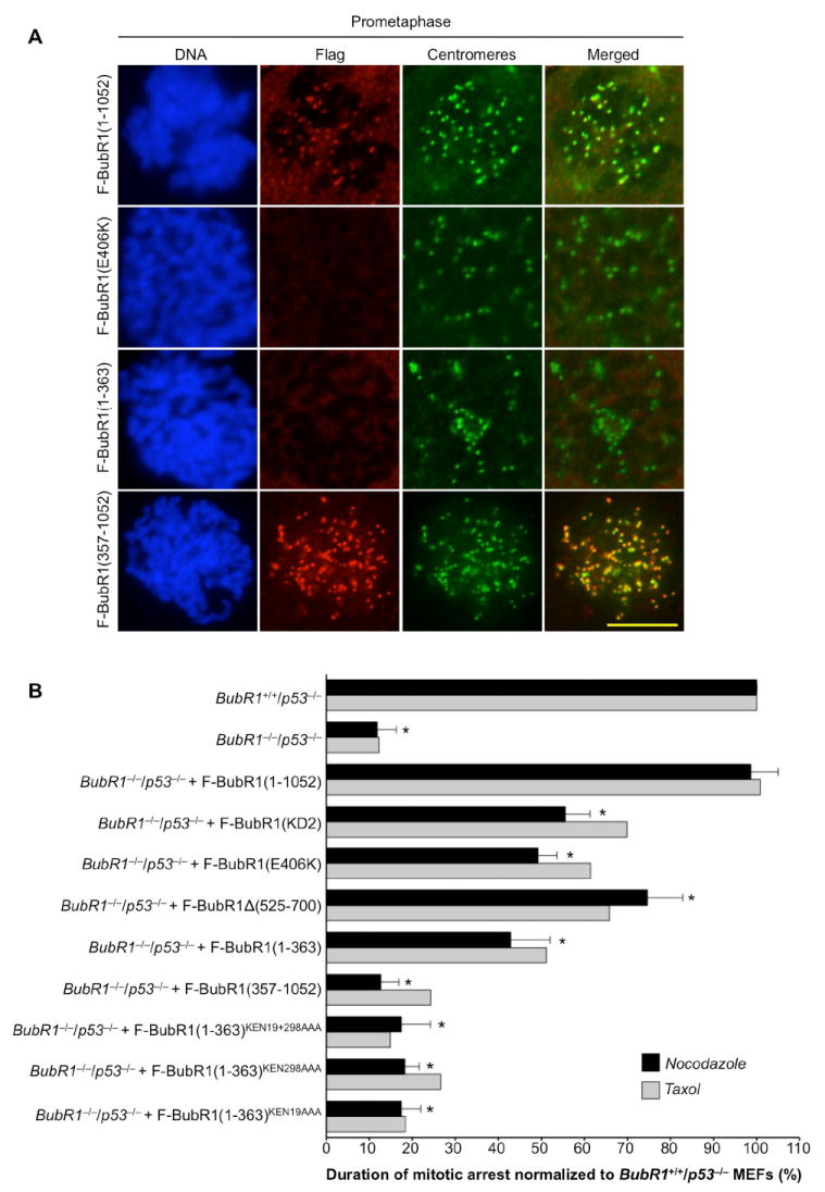
BubR1 is an essential mitotic checkpoint protein with multiple functional domains. It has been implicated in mitotic checkpoint control, as an active kinase at unattached kinetochores, and as a cytosolic inhibitor of APC/C(Cdc20) activity, as well as in mitotic timing and stable chromosome-spindle attachment. Using BubR1-conditional knockout cells and BubR1 domain mutants, we demonstrate that the N-terminal Cdc20 binding domain of BubR1 is essential for all of these functions, whereas its C-terminal Cdc20-binding domain, Bub3-binding domain, and kinase domain are not. We find that the BubR1 N terminus binds to Cdc20 in a KEN box-dependent manner to inhibit APC/C activity in interphase, thereby allowing accumulation of cyclin B in G(2) phase prior to mitosis onset. Together, our results suggest that kinetochore-bound BubR1 is nonessential and that soluble BubR1 functions as a pseudosubstrate inhibitor of APC/C(Cdc20) during interphase to prevent unscheduled degradation of specific APC/C substrates.


Figures

Figure 1
Figure 1. Cell Growth and Survival Requires the N Terminus of BubR1
(A) Schematic overview of wild-type and mutant BubR1 proteins. (B) Images of BubR1H/H/p53−/− MEFs containing indicated expression constructs taken 6 days after infection with Cre retrovirus. (C) Viability of BubR1H/H/p53−/− MEFs ectopically expressing the indicated mutant BubR1 proteins in the presence (− Cre) or absence (+ Cre) of endogenous BubR1. (D) Southern blot of BubR1H/H/p53−/−MEFs expressing the indicated mutant BubR1 proteins subjected to Cre-mediated disruption of endogenous BubR1. DNA was digested with BamH1 and blots were probed with a 3’ BubR1 genomic probe (Baker et al., 2004). (E) Western blot analysis of MEFs carrying the indicated BubR1 expression constructs. Blots were probed with the indicated antibodies. Panels labeled αBubR1: lanes 1-6 were probed with antibodies against hBubR1(1-350) and lanes 7 and 8 with antibodies against mBubR1(382-420). Actin was used as loading control. The genotype of MEFs in lanes 3-6 and 8 was BubR1−/−/p53−/−, that of MEFs in lane 7 was BubR1H/H/p53−/−.
Figure 2
Figure 2. Cdc20 Binding to BubR1 N Terminus Is Sufficient for Cell Viability
(A) Schematic overview of wild-type and mutant BubR1 proteins. K, KEN box. (B and C) Same as legends to Fig. 1B and 1C, respectively. (D) Western blot analysis of MEFs carrying the indicated BubR1 expression constructs. Blots were probed with the indicated antibodies. Actin was used as loading control. The genotype of cells in lanes 1 and 5 was BubR1−/−/p53−/−, whereas that of cells in lanes 2-4 and 6-8 was BubR1H/H/p53−/−. (E) Immunoblots of mitotic extracts of MEFs carrying the indicated BubR1 expression constructs subjected to immunoprecipitation with Cdc20 or Flag antibody, and probed with the indicated antibodies.
Figure 3
Figure 3. The Mitotic Checkpoint Has Different Requirements for Cytosolic and Kinetochore-Bound BubR1
(A) Immunolocalization of Flag-tagged BubR1 proteins in MEFs during prometaphase. F-BubR1(1-1052), F-BubR1(E406K) and F-BubR1(1-363) were expressed in BubR1−/−/p53−/− MEFs and F-BubR1(357-1052) in BubR1H/H/p53−/− MEFs. Flag-tagged proteins were visualized with mouse anti-FLAG antibody and centromeres with ACA antibody. DNA was stained with Hoechst. Bar = 10 μm. (B) Analysis of mitotic checkpoint activity of MEFs challenged with nocodazole or taxol. Three independent MEF lines per genotype were challenged with nocodazole. Error bars represent the SD. *p < 0.0001 versus BubR1+/+/p53−/− MEFs (Log rank test). One line per genotype was challenged with taxol.
Figure 4
Figure 4. The BubR1 N Terminus Regulates Chromosome-Spindle Attachment and Mitotic Timing
(A) Cdc20 binding to the BubR1 N terminus is essential for chromosome alignment. MEFs were treated with 10 μM MG132 for 2 h, fixed and immunostained for α–tubulin and centromeres. The second and third image from the left represent BubR1−/−/p53−/− metaphases with minor and major misalignment phenotypes, respectively. Bar = 10 μm. (B) Quantification of chromosome misalignment defects of MEFs shown in (A). MEF genotypes and BubR1 expression constructs are as indicated in (C). Shaded and non-shaded areas represent major and minor misalignments, respectively. (C) Quantification of misalignment defects in MG132-treated MEFs by live-cell imaging. Shaded and non-shaded areas represent major and minor misalignments, respectively. (D) Examples of metaphase-arrested MEFs displaying proper chromosome alignment (top), minor misalignment (middle) or major misalignment (bottom). Cells were MG132 treated. Arrows designate misaligned chromosomes. Bar = 10 μm. (E) Analysis of chromosome misalignment and cell death by live-cell imaging. We note that nearly all cells dying within 8 h after mitosis had major alignment defects. (F) Timing of anaphase onset. Three MEF lines were used per genotype. Error bars represent the SD. *p < 0.0001 versus BubR1+/+/p53−/− MEFs (Mann-Whitney test).
Figure 5
Figure 5. Binding of BubR1 N Terminus to Cdc20 Inhibits APC/C activity
(A) Prophases stained for cyclin B, phosphohistone H3Ser10 (P-H3) and DNA (Hoechst). (B) Quantification of cyclin B levels in prophase. (C) Representative prophases treated with 10 μM MG132 1 h prior to staining for cyclin B, P-H3 and DNA (Hoechst). (D) Quantification of cyclin B levels in prophase after MG132 treatment. (E) Incidence of PMSCS in MEFs expressing the indicated BubR1 mutants.
Figure 6
Figure 6. BubR1 Stabilizes Cyclin B in Interphase by Inhibiting APC/CCdc20
(A) BubR1−/−/p53−/− MEFs have low cyclin B-GFP levels in G2 and mitosis. Cyclin B-GFP fluorescence of MEF cells of the indicated genotypes was measured and the average fluorescence intensity in G2, prophase (P), prometaphase (PM), metaphase (M) and anaphase (A) plotted (n > 10 cells per genotype). Error bars represent the SD. *p < 0.0001 versus BubR1+/+/p53−/− MEFs at the same cell cycle stage (Mann-Whitney test). Bar = 10 μm. (B) MG132 treatment of BubR1−/−/p53−/− MEFs restores normal cyclin B-GFP expression during G2 and mitosis. Error bars represent the SD. *p < 0.0001 versus BubR1+/+/p53−/− MEFs at the same cell cycle stage (Mann-Whitney test). Bar = 10 μm. (C) BubR1−/−/p53−/− MEFs express normal levels of non-degradable cyclin BR42A-GFP G2 and mitosis. Error bars represent the SD. (D) Expression of F-BubR1(1-363) in BubR1−/−/p53−/− MEFs restores normal cyclin B-cerulean levels in G2 and mitosis, whereas F-BubR1(1-363) lacking its KEN boxes does not. Error bars represent the SD. *p < 0.0001 versus BubR1+/+/p53−/− MEFs at the same cell cycle stage (Mann-Whitney test). Bar = 10 μm.
Figure 7
Figure 7. BubR1 Interacts with APC/CCdc20 in Interphase Cells
(A) Percentage of mitotic cells present in G2 and mitotic BubR1+/+/p53−/− MEF extracts used in coimmunoprecipitation experiments shown in (C). (B) Western blot analysis of G2 and mitotic BubR1+/+/p53−/− MEF extracts for actin. 5 μl of extracts used in (C) was loaded in each lane. (C) Cdc20 binds to BubR1 in both interphase and mitosis. G2 and mitotic MEF extracts prepared from equal amounts of cells were subjected to immunoprecipitation with anti-BubR1 antibody and probed with the indicated antibodies. (D) As (C) but with G2 extracts from BubR1−/−/p53−/− MEFs expressing BubR1(1-363). (E) Western blot analysis showing that BubR1+/+/p53−/− MEFs transduced with pTRIPZ lentivirus expressing mMad2-shRNAmir clone V2MM_6980 contain ~10-20% of normal Mad2 levels after 3 days of culture in medium with 1 μg/ml DOX. (F) Mad2 depletion in BubR1+/+/p53−/− MEFs has no impact on cyclin B-GFP levels in G2 and mitosis. DOX was added 72 h prior to analysis of cyclin B-GFP stability. Error bars represent the SD. Bar = 10 μm. (G) Model for BubR1 function in G2 phase and mitosis. BubR1 binds to Cdc20 or Cdc20 already bound to APC/C (probably in combination with Mad2 and Bub3) in a KEN-box-dependent manner in G2, thereby blocking access of cyclin B to the APC/C, allowing this cyclin to accumulate and perform its mitotic functions. In mitosis, mitotic checkpoint proteins accumulating at unattached kinetochores catalyze binding of Mad2 and BubR1-Bub3 to Cdc20 that might emerge due to degradation of APC/CCdc20 inhibitors, such as Emi1 (Miller et al., 2006), and/or activation of mechanisms that displace Mad2 and BubR1 from Cdc20 in mitosis, such as UbcH10-mediated ubiquitination of Cdc20. BubR1 kinase activity is critical for APC/C inhibition under circumstances where spindle damage persist for prolonged periods of time.

Comment in

References

    1. Bailly E, Pines J, Hunter T, Bornens M. Cytoplasmic accumulation of cyclin B1 in human cells: association with a detergent-resistant compartment and with the centrosome. J Cell Sci. 1992;101(Pt 3):529–545. - PubMed
    1. Baker DJ, Chen J, van Deursen JM. The mitotic checkpoint in cancer and aging: what have mice taught us? Curr Opin Cell Biol. 2005;17:583–589. - PubMed
    1. Baker DJ, Jeganathan KB, Cameron JD, Thompson M, Juneja S, Kopecka A, Kumar R, Jenkins RB, de Groen PC, Roche P, van Deursen JM. BubR1 insufficiency causes early onset of aging-associated phenotypes and infertility in mice. Nat Genet. 2004;36:744–749. - PubMed
    1. Buffin E, Emre D, Karess RE. Flies without a spindle checkpoint. Nat Cell Biol. 2007;9:565–572. - PubMed
    1. Burton JL, Solomon MJ. Mad3p, a pseudosubstrate inhibitor of APCCdc20 in the spindle assembly checkpoint. Genes & development. 2007;21:655–667. - PMC - PubMed

Publication types

MeSH terms