. 2009 Jan 22;5(1):35-46.
doi: 10.1016/j.chom.2008.12.002.

RP105 facilitates macrophage activation by Mycobacterium tuberculosis lipoproteins

Affiliations

RP105 facilitates macrophage activation by Mycobacterium tuberculosis lipoproteins

Antje Blumenthal et al. Cell Host Microbe. .

Abstract

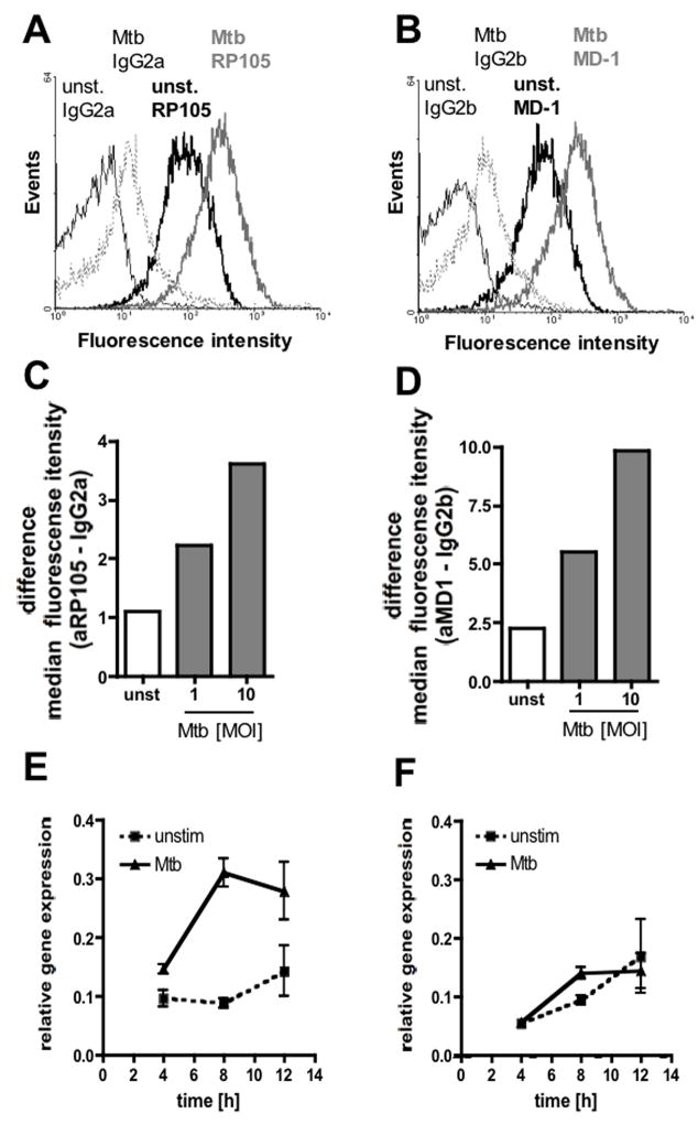
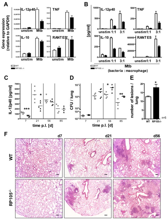
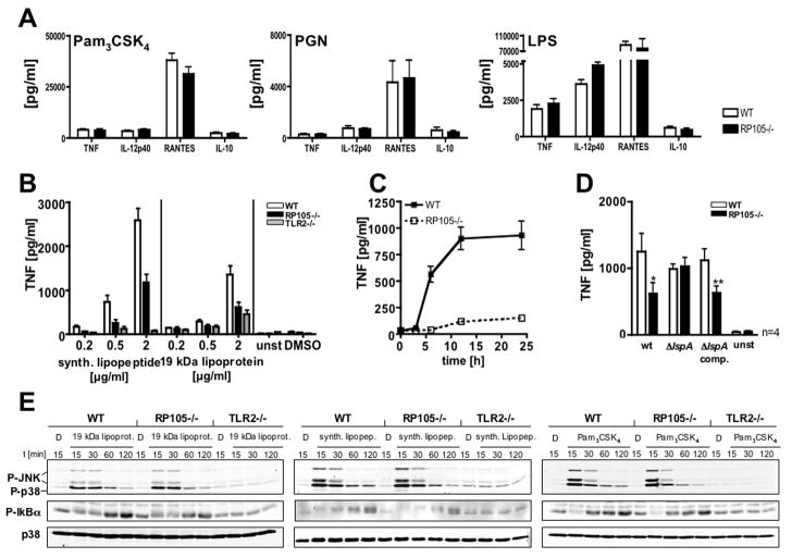
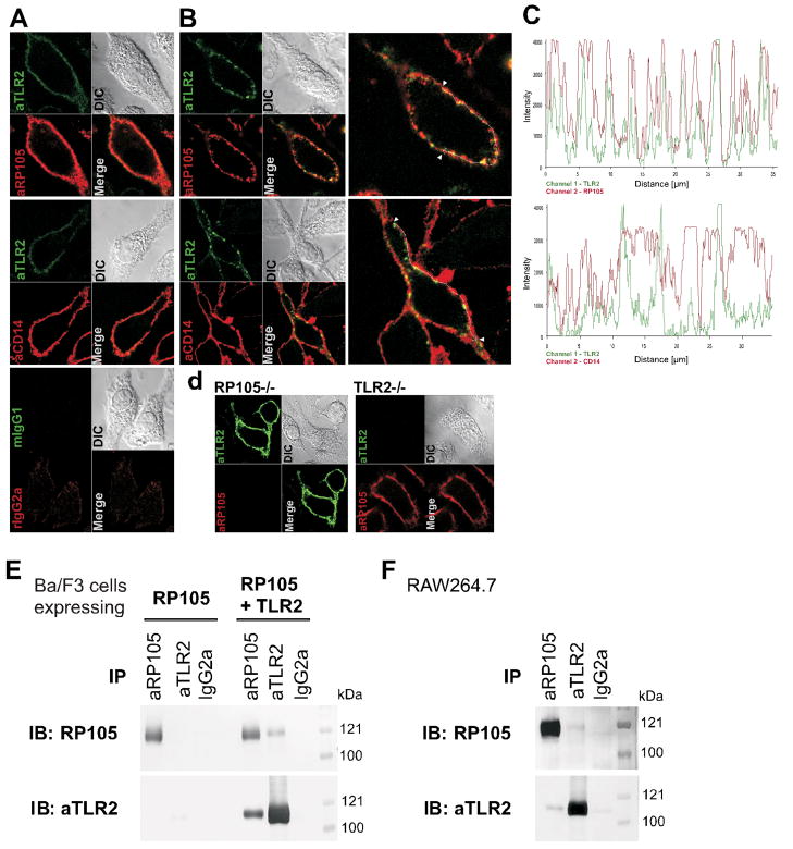
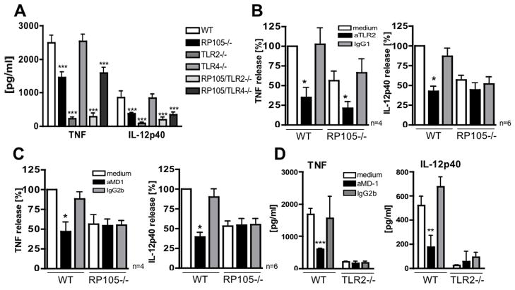
RP105, phylogenetically related to Toll-like receptor (TLR)-4, is reported to facilitate B cell activation by the TLR4-agonist lipopolysaccharide (LPS)--but to limit LPS-induced cytokine production by antigen-presenting cells. Here, we show that the role of RP105 extends beyond LPS recognition and that RP105 positively regulates macrophage responses to Mycobacterium tuberculosis (Mtb) lipoproteins. Mtb-infected RP105(-/-) mice exhibited impaired proinflammatory cytokine responses associated with enhanced bacterial burden and increased lung pathology. The Mtb 19 kDa lipoprotein induced release of tumor necrosis factor in a manner dependent on both TLR2 and RP105, and macrophage activation by Mtb lacking mature lipoproteins was not RP105 dependent. Thus, mycobacterial lipoproteins are RP105 agonists. RP105 physically interacted with TLR2, and both RP105 and TLR2 were required for optimal macrophage activation by Mtb. Our data identify RP105 as an accessory molecule for TLR2, forming part of the receptor complex for innate immune recognition of mycobacterial lipoproteins.


Figures

Figure 1
Figure 1
Mtb-induced up-regulation of RP105 and MD-1 expression by macrophages. Expression of RP105 (A) and MD-1 (B) by BMM, unstimulated (unst.) or infected with Mtb (MOI 10; 18 h). Data are from 1 experiment representative of 3. Mtb-induced increase in surface expression of RP105 (C) and MD-1 (D) by macrophages is dose dependent. Median fluorescence intensities (MFI) for isotype control stainings were subtracted from MFI for specific antibody stainings. One representative experiment of 2 is depicted. RP105 (E) and MD-1 (F) mRNA levels normalized to GAPDH from Mtb-stimulated and unstimulated BMM at indicated times. Data represent means of triplicates ± sd. Results from 1 experiment out of 2 are shown.
Figure 2
Figure 2
RP105 is required for optimal macrophage activation in response to Mtb infection and participates in control of Mtb infection in vivo. (A) mRNA levels in BMM stimulated with Mtb (MOI 3, 4 h) or unstimulated. Data represent means of triplicates ± sd. Comparison of raw data of three independent wells by unpaired t test showed a significant difference in IL-12p40 mRNA expression (*P=0.0032), but no differences for TNF, IL-10, and RANTES. One representative experiment out of 2 is shown. (B) Cytokine and chemokine release by Mtb-stimulated WT and RP105−/− macrophages (MOI 1 or 3, 24 h). Data represent means ± sd of 3 parallel cultures in a single experiment and are representative of 3 independent experiments. (C)-(F) Infection of WT and RP105−/− mice with a low dose of Mtb (175 ± 98 CFU/lung). (C) IL-12p40 concentrations in serum at indicated times post infection (p.i.). Means are indicated. Groups were compared by unpaired t test. *P<0.05, **P<0.002, ***P ≤ 0.0005. Results of 1 experiment out of 2 are shown. (D) Colony forming units (CFU) in lungs monitored over time. Means are indicated. Groups were compared by unpaired t test. *P<0.05, **P<0.002. Results of 1 experiment out of 2 are shown. (E) Number of lesions per lung section on day 56. Means of the number of lesions in one lung section of 5 mice per group ± se are shown. Groups were compared by unpaired t test. *P=0.0239. (F) Exacerbated pathology in lungs of Mtb infected RP105−/− mice. Tissue sections were stained with hematoxylin and eosin. Representative sections from 1 mouse per genotype out of 5 are shown; 2 independent experiments were performed. (Bar = 100 μm).
Figure 3
Figure 3
Mtb lipoproteins activate macrophages in an RP105-dependent fashion. (A) Cytokine responses of WT and RP105−/− macrophages after 24 h stimulation with Pam3CSK4 (10 ng/ml), peptidoglycan (PGN, 10 μg/ml), or LPS (10 ng/ml), respectively. Means ± sd of triplicate wells of 1 representative out of 3 (Pam3CSK4, LPS) or 2 (PGN) experiments are shown. Of note, the minor enhancement of cytokine production seen in RP105−/− cells, might be due to a low amount of TLR2-dependent agonists in the LPS preparation; heterologous TLR stimulation has been shown to overcome RP105-mediated inhibition of TLR4 stimulation (Divanovic et al., 2005). (B) TNF concentrations in supernatants of WT, RP105−/−, and TLR2−/−BMM after stimulation with the 19 kDa lipoprotein of Mtb or the synthetic lipopeptide (resembling the 16 N-terminal amino acids of the 19 kDa lipoprotein) for 24 h. Raw data of 3 independent wells were compared by unpaired t test. Data are means ± sd of triplicate wells of 1 experiment representative of 4. (C) TNF concentrations in culture supernatants of WT and RP105−/− after stimulation with the synthetic lipopeptide (1 μg/ml). Data are means ± sd of 5 independent wells of one experiment representative of 2. (D) TNF produced by WT and RP105−/− macrophages infected with wild type Mtb, ΔlspA or ΔlspA comp. (MOI 3) was measured after 24 h. Data represent means ± se of 4 independent experiments (performed in triplicates). Paired t test was used for comparison. *P<0.05; **P<0.005; ***P<0.0001. (E) Immunoblot analyses of p38-, JNK-, and IκB-α-phosphorylation (P) on lysates of WT, RP105−/−, and TLR2−/− BMM after stimulation with 19 kDa lipoprotein, synthetic lipopeptide (both 1 μg/ml), Pam3CSK4 (10 ng/ml), or treatment with DMSO (D) for indicated times. Total amounts of p38 were detected as control for equal loading. Data represent 1 out of 3 independent experiments.
Figure 4
Figure 4
RP105 and TLR2 co-localize and associate in the cell membrane. (A) WT BMM were incubated with heat killed Mtb for 2 h and stained with anti-TLR2 or mIgG1 isotype control antibodies that were specifically labeled with Alexa488-conjugated Fab fragments. After PFA fixation cells were incubated with anti-RP105 or anti-CD14 antibody or matched isotype control (rIgG2a) followed by an Alexa547 conjugated anti-rat secondary antibody. Samples were analyzed by confocal microscopy (DIC = differential interference contrast in transmitted light images; Merge = overlay of images acquired in different fluorescence channels). (B) WT macrophages were incubated with heat killed Mtb for 2 h and stained with anti-TLR2 or IgG1 isotype control antibodies that were specifically labeled with Alexa488-conjugated Fab fragments. Patching was achieved by crosslinking of the anti-TLR2 or mouse IgG1 antibodies with an anti-mouse antibody. After fixation cells were co-stained for RP105 or CD14 expression as described under (A). Data shown are representative for 4 (RP105) and 2 (CD14) independent experiments. Enlarged overlay images are depicted on the right. White arrowheads mark start and end points of line scans for acquisition of fluorescence profiles depicted in panel (C). (C) Fluorescence profiles for TLR2 (green) and RP105 or CD14 (red) specific staining acquired along the periphery of the cell (approximately 35 μm) as indicated by the thin white line (arrowheads mark start and end point in enlargement in panel (B)). (D) RP105−/− and TLR2−/− BMM were stained for RP105 or TLR2 as described for WT cells (A) and subject to confocal microscopy. (E, F) Immunoprecipitation (IP) and immunoblot (IB) of lysates of (E) Ba/F3 cells stably transfected with RP105/MD-1 or RP105/MD-1 and TLR2 together and (F) RAW264.7 macrophages. Co-IP demonstrating association of RP105 and TLR2 was assessed by IB using anti-RP105 and anti-TLR2 antibodies. An isotype-matched rat IgG2a antibody and Ba/F3 cells transfected with RP105/MD-1 but not TLR2 were used as controls. One representative of 3 independent experiments is shown.
Figure 5
Figure 5
RP105/MD-1 cooperates with TLR2. (A) WT, RP105−/−, TLR2−/−, TLR4−/−, RP105/TLR2−/− and RP105/TLR4−/− BMM were infected with Mtb (MOI 3). Cytokine concentrations in culture supernatants were determined 24 h post infection. Data are means ± sd from 2 independent experiments each performed in triplicates. Raw data of independent wells of knock-out cells were compared to WT cells by unpaired t test. ***P<0.0001 (B) WT and RP105−/− BMM were pre-incubated with an anti-TLR2 antibody (1–2 μg/ml) or isotype-matched control antibody (IgG1) for 1 h and infected with Mtb (MOI 3). Cytokine release was measured after 24 h (TNF) and 48 h (IL-12p40). Four (TNF) and 6 (IL-12p40) independent experiments performed in triplicates were compared by normalizing to control conditions in WT macrophages (no antibody added), means ± se are shown. Raw data were compared by paired t-test. *P<0.05. (C) Concentrations of IL-12p40 and TNF in culture supernatants of WT and RP105−/− BMM that were infected with Mtb (MOI 3) after pre-incubation (1 h) with an inhibitory anti-MD-1 antibody (1–2 μg/ml) or isotype-matched control antibody (IgG2b). Four (TNF) and 6 (IL-12p40) independent experiments, each performed in triplicates were compared by normalizing to control conditions in WT BMM (no antibody added), means ± se are shown. Raw data were compared by paired t test. *P<0.05. (D) Concentrations of IL-12p40 and TNF in supernatants of WT and TLR2−/− BMM, pre-incubated with an anti-MD-1 or matched isotype control (IgG2b) antibody, after infection with Mtb (MOI 3). Raw data of 3 independent wells were compared to control conditions (medium) by unpaired t test. Data points are means ± sd of triplicate wells of 1 experiment representative of 3. **P=0.0088, ***P=0.0005. Cytokine levels in unstimulated macrophage cultures were below the detection limit and were not significantly influenced in the presence of antibody (not shown).

Similar articles

Cited by

References

    1. Andersson U, Wang H, Palmblad K, Aveberger AC, Bloom O, Erlandsson-Harris H, Janson A, Kokkola R, Zhang M, Yang H, Tracey KJ. High mobility group 1 protein (HMG-1) stimulates proinflammatory cytokine synthesis in human monocytes. J Exp Med. 2000;192:565–570. - PMC - PubMed
    1. Bafica A, Scanga CA, Feng CG, Leifer C, Cheever A, Sher A. TLR9 regulates Th1 responses and cooperates with TLR2 in mediating optimal resistance to Mycobacterium tuberculosis. J Exp Med. 2005;202:1715–1724. - PMC - PubMed
    1. Banaiee N, Kincaid EZ, Buchwald U, Jacobs WR, Jr, Ernst JD. Potent inhibition of macrophage responses to IFN-gamma by live virulent Mycobacterium tuberculosis is independent of mature mycobacterial lipoproteins but dependent on TLR2. J Immunol. 2006;176:3019–3027. - PubMed
    1. Black RA, Rauch CT, Kozlosky CJ, Peschon JJ, Slack JL, Wolfson MF, Castner BJ, Stocking KL, Reddy P, Srinivasan S, et al. A metalloproteinase disintegrin that releases tumour-necrosis factor-alpha from cells. Nature. 1997;385:729–733. - PubMed
    1. Blumenthal A, Lauber J, Hoffmann R, Ernst M, Keller C, Buer J, Ehlers S, Reiling N. Common and unique gene expression signatures of human macrophages in response to four strains of Mycobacterium avium that differ in their growth and persistence characteristics. Infect Immun. 2005;73:3330–3341. - PMC - PubMed

Publication types

LinkOut - more resources