. 2009 Jan 22;5(1):59-71.
doi: 10.1016/j.chom.2008.12.001.

Junctional adhesion molecule-A is required for hematogenous dissemination of reovirus

Affiliations

Junctional adhesion molecule-A is required for hematogenous dissemination of reovirus

Annukka A R Antar et al. Cell Host Microbe. .

Abstract

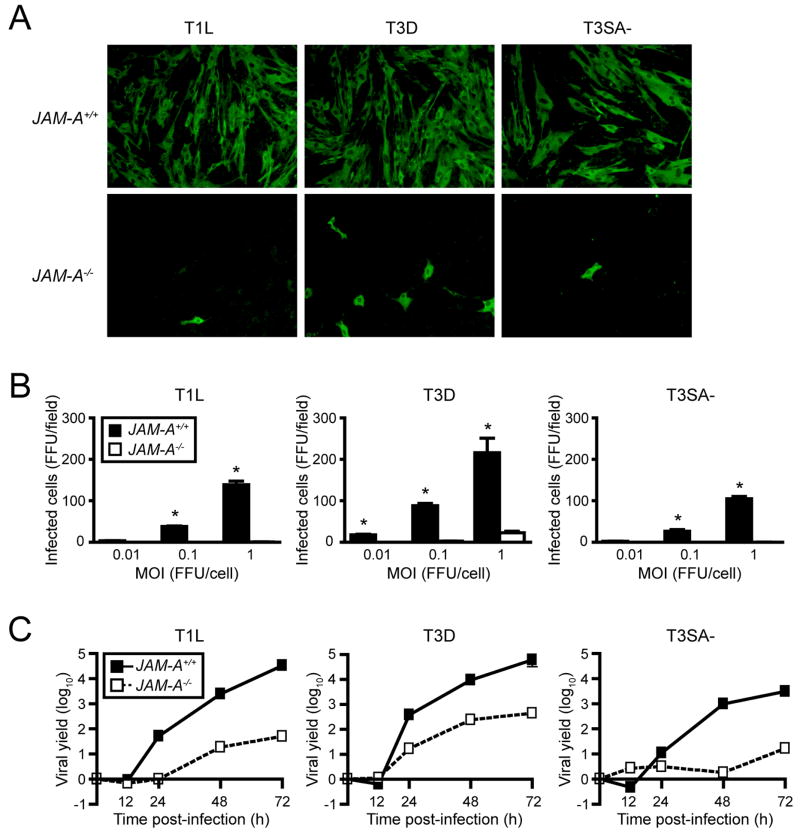
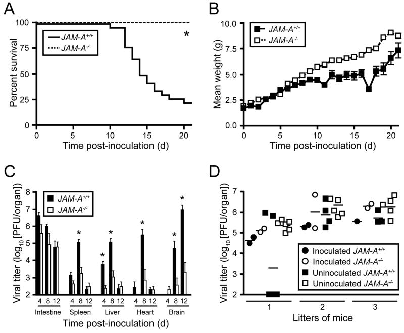
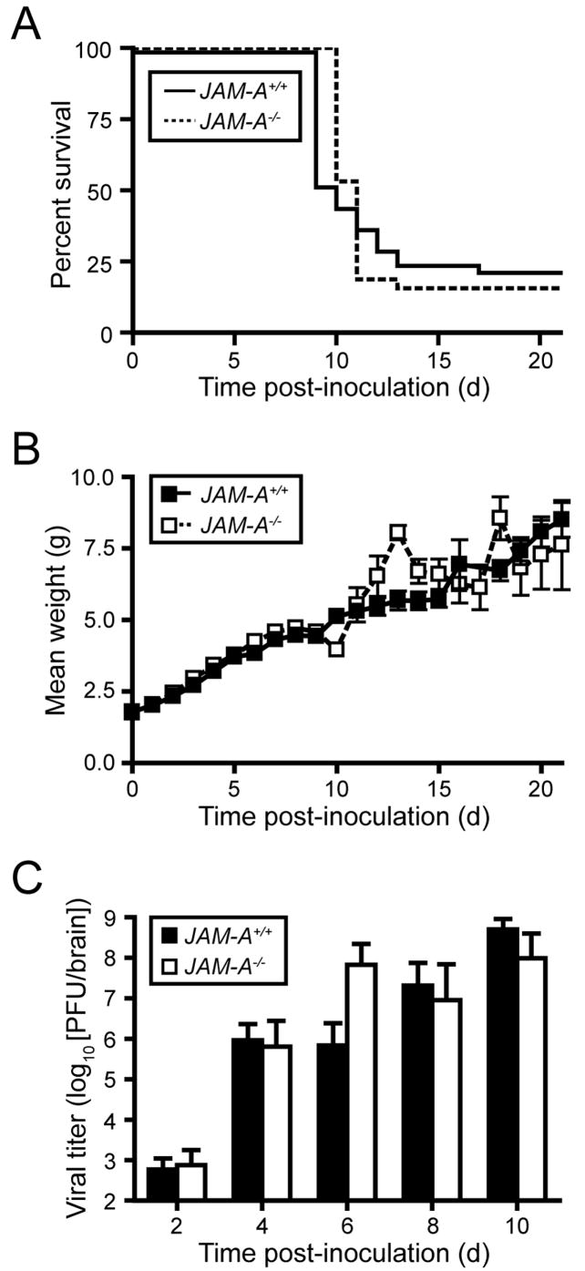
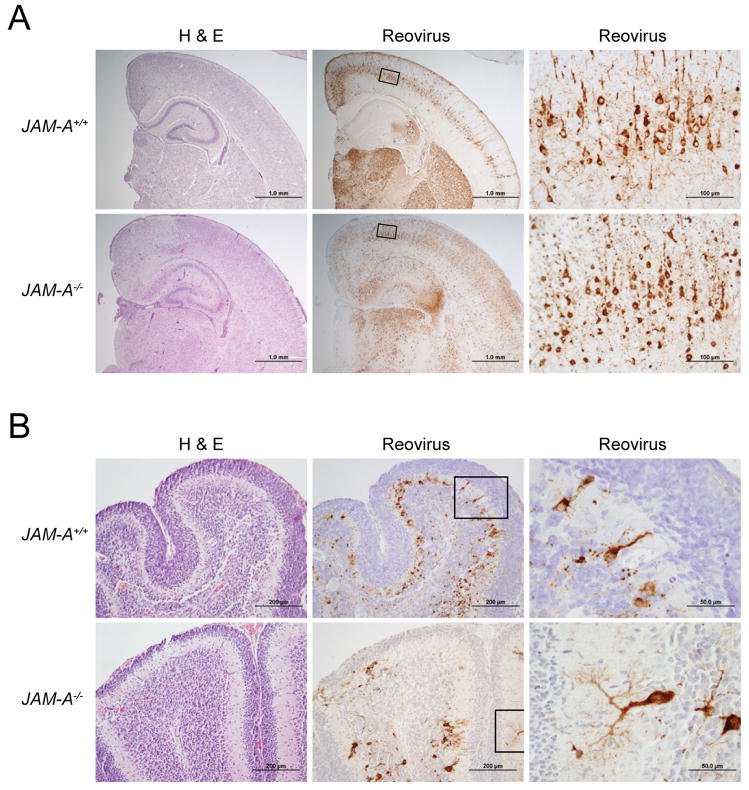
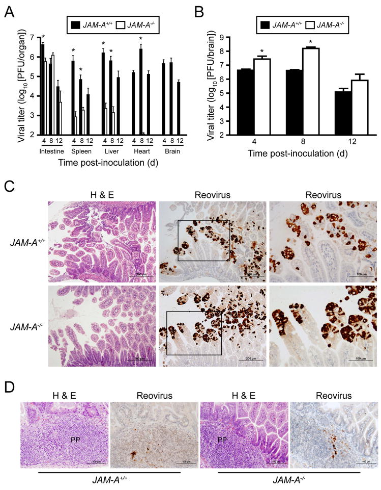
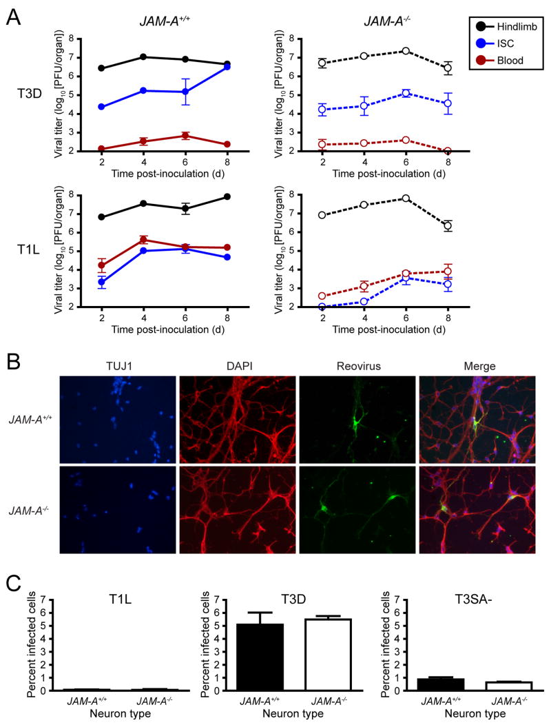
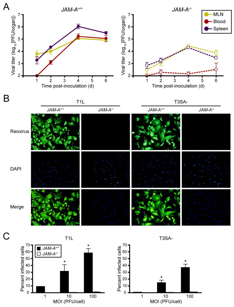
Diverse families of viruses bind immunoglobulin superfamily (IgSF) proteins located in tight junctions (TJs) and adherens junctions of epithelium and endothelium. However, little is known about the roles of these receptors in the pathogenesis of viral disease. Junctional adhesion molecule-A (JAM-A) is an IgSF protein that localizes to TJs and serves as a receptor for mammalian reovirus. We inoculated wild-type (WT) and isogenic JAM-A(-/-) mice perorally with reovirus and found that JAM-A is dispensable for viral replication in the intestine but required for systemic dissemination. Reovirus replication in the brain and tropism for discrete neural regions are equivalent in WT and JAM-A(-/-) mice following intracranial inoculation, suggesting a function for JAM-A in reovirus spread to extraintestinal sites. JAM-A promotes reovirus infection of endothelial cells, providing a conduit for the virus into the bloodstream. These findings indicate that a broadly expressed IgSF viral receptor specifically mediates hematogenous dissemination in the host.


Figures

Figure 1
Figure 1. JAM-A Is Required for Efficient Reovirus Infection of MEFs
(A) Primary MEFs generated from JAM-A+/+ and JAM-A−/− embryos were adsorbed with reovirus T1L, T3D, or T3SA- at MOIs of 0.01, 0.1, and 1 fluorescent focus unit (FFU)/cell and incubated for 20 hr. Reovirus antigen was detected by indirect immunofluorescence. Representative wells after adsorption with 1 FFU/cell are shown. (B) Infected cells were quantified in five fields of 200X view for triplicate samples. Results are expressed as the mean FFU/field for triplicate experiments. Error bars indicate SD. *, P < 0.01 as determined by Student’s t test. (C) Confluent monolayers of JAM-A+/+ and JAM-A−/− MEFs were adsorbed with reovirus T1L, T3D, or T3SA- at an MOI of 2 PFU/cell and incubated for the times shown. Viral titers were determined by plaque assay. Results are expressed as mean viral yields (tx/t0) for triplicate experiments. Error bars indicate SD.
Figure 2
Figure 2. Reovirus T3SA- Is Attenuated following Peroral Inoculation of JAM-A−/− Mice
(A–B) Newborn JAM-A+/+ and JAM-A−/− mice were inoculated perorally with 109 PFU of reovirus T3SA-. Mice (n = 26 JAM-A+/+ and n = 16 JAM-A−/−) were monitored for survival (A) and weight gain (B). *, P < 0.0001 as determined by log rank test. (C) Newborn JAM-A+/+ and JAM-A−/− mice were inoculated perorally with 104 PFU T3SA-. At days 4, 8, and 12 after inoculation, mice were euthanized, organs were resected, and viral titers were determined by plaque assay. Results are expressed as mean viral titers for 6–13 animals for each time point. Error bars indicate SD. *, P < 0.05 by Student’s t test. (D) One-two newborn JAM-A+/+ and two newborn JAM-A−/− mice from litters of 4 to 8 animals were inoculated perorally with 104 PFU T3SA- and reunited with uninoculated littermates. At day 12 after inoculation, mice were euthanized, intestines were resected, and viral titers were determined by plaque assay. Each data point represents one animal. Horizontal bars indicate the arithmetic mean of log-transformed data.
Figure 3
Figure 3. Reovirus T3SA- Is Fully Virulent following Intracranial Inoculation of JAM-A−/− Mice
(A–B) Newborn JAM-A+/+ and JAM-A−/− mice were inoculated intracranially with 100 PFU of reovirus T3SA-. Mice (n = 40 JAM-A+/+ and n = 32 JAM-A−/−) were monitored for survival (A) and weight gain (B). (C) Newborn JAM-A+/+ and JAM-A−/− mice were inoculated intracranially with 100 PFU T3SA-. At days 2, 4, 6, 8, and 10 after inoculation, mice were euthanized, brains were resected, and viral titers were determined by plaque assay. Results are expressed as mean viral titers for 5–13 animals for each time point. Error bars indicate SD.
Figure 4
Figure 4. Histopathology of Reovirus Infection following Intracranial Inoculation
Newborn JAM-A+/+ and JAM-A−/− mice were inoculated intracranially with 104 PFU T3SA-. Eight days after inoculation, brains of infected mice were resected and bisected sagittally. The left hemisphere was prepared for viral titer determination by plaque assay, and the right hemisphere was processed for histopathology. Consecutive coronal sections were stained with H&E or polyclonal reovirus antiserum. Representative sections of brain hemisphere, matched for hippocampal depth (A), and cerebellum (B) are shown. Boxes indicate areas of enlargement in the panels on the right and show cortical neurons (A) and cerebellar Purkinje neurons (B). JAM-A+/+ brain sections are from brains with left hemisphere viral titers of 4.1 × 10⁹ PFU (A) and 3.0 × 10⁹ PFU (B). JAM-A−/− brain sections are from brains with left hemisphere viral titers of 3.4 × 10⁹ PFU (A) and 1.6 × 10⁹ PFU (B).
Figure 5
Figure 5. Reovirus T1L Is Incapable of Dissemination following Peroral Inoculation of JAM-A−/− Mice
(A) Newborn JAM-A+/+ and JAM-A−/− mice were inoculated perorally with 106 PFU T1L. At days 4, 8, and 12 after inoculation, mice were euthanized, organs were resected, and viral titers were determined by plaque assay. Results are expressed as mean viral titers for 6 animals for each time point. Error bars indicate SD. *, P < 0.005 by Student’s t test. When all values are less than the limit of detection (spleen, liver, heart, and brain in JAM-A−/− mice), a Student’s t test P value cannot be calculated. (B) Newborn JAM-A+/+ and JAM-A−/− mice were inoculated intracranially with 103 PFU T1L. At days 4, 8, and 12 after inoculation, mice were euthanized, brains were resected, and viral titers were determined by plaque assay. Results are expressed as mean viral titers for 6–11 animals for each time point. Error bars indicate SD. *, P < 0.005 by Student’s t test. (C–D) Newborn JAM-A+/+ and JAM-A−/− mice were inoculated perorally with 108 PFU T1L. At days 2 and 4 after inoculation, intestines of infected mice were resected and processed for histopathology. Consecutive sections were stained with H&E or polyclonal reovirus antiserum. Representative sections of intestinal villi at day 2 (C) and Peyer’s patches (PP) at day 4 (D) are shown. Boxes indicate areas of enlargement in the panels on the right and show villus epithelial cells.
Figure 6
Figure 6. JAM-A Is Required for Hematogenous Spread of Reovirus
(A) Newborn JAM-A+/+ and JAM-A−/− mice were inoculated into the left hindlimb with 106 PFU of either T3D or T1L. At days 2, 4, 6, and 8 after inoculation, mice were euthanized, left hindlimb, blood, and inferior spinal cord (ISC), including the thoracic and lumbosacral cord segments, were resected, and viral titers were determined by plaque assay. Results are expressed as mean viral titers for 6 animals for each time point. Error bars indicate SD. (B) Primary cortical cultures generated from E15 JAM-A+/+ and JAM-A−/− embryos were cultured in vitro for 5 to 7 days, adsorbed with T3 reovirus at an MOI of 1000 PFU/cell, and incubated for 20 hr. Cells were stained with TUJ1 neural-specific marker to detect neurons (red), 4′,6-diamidino-2-phenylindole (DAPI) to detect nuclei (blue), and polyclonal reovirus antiserum to detect reovirus antigen (green) and visualized using indirect immunofluorescence microscopy. Representative wells from triplicate experiments are shown. (C) JAM-A+/+ and JAM-A−/− cortical cultures were cultured in vitro for 5 to 7 days and adsorbed with T1L, T3D, or T3SA- at an MOI of 1000 PFU/cell and incubated for 20 hr. The percentage of infected cells was quantified by dividing the number of neurons exhibiting reovirus staining by the total number of cell nuclei exhibiting DAPI staining in three fields of 400X view for triplicate experiments. Fields of view contained between 200 and 600 nuclei. Error bars indicate SD.
Figure 7
Figure 7. JAM-A Is Required for Viremia and Efficient Reovirus Infection of Endothelial Cells
(A) Newborn JAM-A+/+ and JAM-A−/− mice were inoculated perorally with 108 PFU of T1L. At days 1, 2, 4, and 6 after inoculation, mice were euthanized, mesenteric lymph node (MLN), blood, and spleen were collected, and viral titers were determined by plaque assay. Results are expressed as mean viral titers for 3–8 animals for each time point. Error bars indicate SD. (B) Primary endothelial cells generated from JAM-A+/+ and JAM-A−/− mice were adsorbed with either reovirus T1L or T3SA- at an MOI of 100 PFU/cell and incubated for 20 hr. Cells were stained with polyclonal reovirus antiserum to detect reovirus antigen (green) and 4′,6-diamidino-2-phenylindole (DAPI) to detect nuclei (blue) and visualized using indirect immunofluorescence microscopy. Representative wells from triplicate experiments are shown. (C) JAM-A+/+ and JAM-A−/− primary endothelial cells were adsorbed with T1L or T3SA- at MOIs of 1, 10, and 100 PFU/cell and incubated for 20 hr. The percentage of infected cells was quantified by dividing the number of cells exhibiting reovirus staining by the total number of cell nuclei exhibiting DAPI staining in entire wells of 96-well plates for triplicate experiments. Wells contained between 200 and 1600 nuclei. Error bars indicate SD. *, P < 0.05 as determined by Student’s t test.

Comment in

References

    1. Barton ES, Connolly JL, Forrest JC, Chappell JD, Dermody TS. Utilization of sialic acid as a coreceptor enhances reovirus attachment by multistep adhesion strengthening. J Biol Chem. 2001a;276:2200–2211. - PubMed
    1. Barton ES, Forrest JC, Connolly JL, Chappell JD, Liu Y, Schnell F, Nusrat A, Parkos CA, Dermody TS. Junction adhesion molecule is a receptor for reovirus. Cell. 2001b;104:441–451. - PubMed
    1. Barton ES, Youree BE, Ebert DH, Forrest JC, Connolly JL, Valyi-Nagy T, Washington K, Wetzel JD, Dermody TS. Utilization of sialic acid as a coreceptor is required for reovirus-induced biliary disease. J Clin Invest. 2003;111:1823–1833. - PMC - PubMed
    1. Bass DM, Trier JS, Dambrauskas R, Wolf JL. Reovirus type 1 infection of small intestinal epithelium in suckling mice and its effect on M cells. Lab Invest. 1988;58:226–235. - PubMed
    1. Becker MM, Peters TR, Dermody TS. Reovirus σNS and μNS proteins form cytoplasmic inclusion structures in the absence of viral infection. J Virol. 2003;77:5948–5963. - PMC - PubMed

Publication types

MeSH terms