. 2009 Mar 27;284(13):8963-72.
doi: 10.1074/jbc.M809395200. Epub 2009 Jan 20.

The Electron Transfer Pathway of the Na+-pumping NADH:Quinone Oxidoreductase from Vibrio cholerae

Affiliations

The Electron Transfer Pathway of the Na+-pumping NADH:Quinone Oxidoreductase from Vibrio cholerae

Oscar Juárez et al. J Biol Chem. .

Abstract

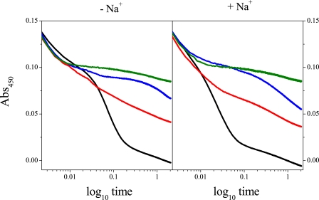
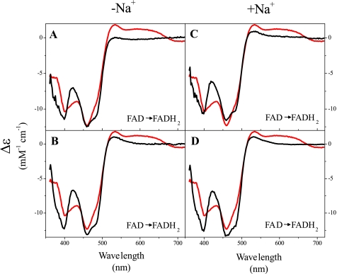
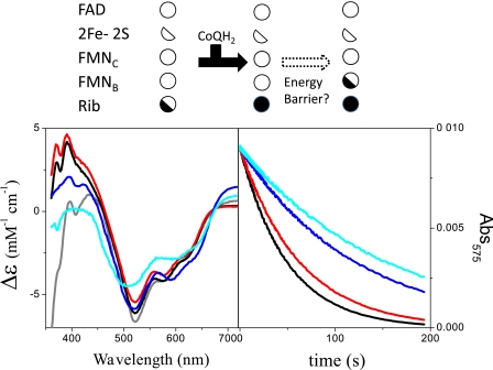
The Na(+)-pumping NADH:quinone oxidoreductase (Na(+)-NQR) is the only respiratory enzyme that operates as a Na(+) pump. This redox-driven Na(+) pump is amenable to experimental approaches not available for H(+) pumps, providing an excellent system for mechanistic studies of ion translocation. An understanding of the internal electron transfer steps and their Na(+) dependence is an essential prerequisite for such studies. To this end, we analyzed the reduction kinetics of the wild type Na(+)-NQR, as well as site-directed mutants of the enzyme, which lack specific cofactors. NADH and ubiquinol were used as reductants in separate experiments, and a full spectrum UV-visible stopped flow kinetic method was employed. The results make it possible to define the complete sequence of redox carriers in the electrons transfer pathway through the enzyme. Electrons flow from NADH to quinone through the FAD in subunit F, the 2Fe-2S center, the FMN in subunit C, the FMN in subunit B, and finally riboflavin. The reduction of the FMN(C) to its anionic flavosemiquinone state is the first Na(+)-dependent process, suggesting that reduction of this site is linked to Na(+) uptake. During the reduction reaction, two FMNs are transformed to their anionic flavosemiquinone in a single kinetic step. Subsequently, FMN(C) is converted to the flavohydroquinone, accounting for the single anionic flavosemiquinone radical in the fully reduced enzyme. A model of the electron transfer steps in the catalytic cycle of Na(+)-NQR is presented to account for the kinetic and spectroscopic data.


Figures

FIGURE 1.
FIGURE 1.
Top panel, scheme representing Na+-NQR. The six subunits (NqrA-F) are depicted together with the redox cofactors and the proposed pathway of electron flow through the enzyme (see “Discussion”). Bottom panel, scheme illustrating the results presented in Figs. 2, 3, 4, 5 and 6. The scheme depicts the redox states of the cofactors after each kinetic phase in the reaction with NADH (without sodium), in the wild type enzyme, and in the cofactor mutants. State 0 is the initial state. At the top of the scheme, the redox cofactors, FAD, 2Fe-2S center, FMNC, FMNB, and riboflavin (RIB) are shown in the order of electron flow through the enzyme. The top box represents the wild type enzyme. The mutants that lack specific cofactors are depicted in the boxes below. For each mutant, the name of the mutant (for example, NqrC-T225Y) is shown in place of the missing cofactor, representing a missing step in the sequence of electron transfer carriers and thus a blockage in the pathway. Each complete circle represents sites for two electrons; single electron reduction is shown using semi-circles. For each step the newly reduced sites are shown with black fill, whereas sites that remain reduced from the previous step are shown with gray fill.
FIGURE 2.
FIGURE 2.
Time courses at the absorbance maximum (450 nm) for flavins showing the reduction of the wild type enzyme (black line), NqrB-T236Y mutant (red line), NqrC-T225Y (blue line), and the double mutant, NqrB-T236Y/NqrC-T225Y mutant (green line).
FIGURE 3.
FIGURE 3.
Reduction of the wild type Na+-NQR from V. cholerae by 300 μm NADH; spectral components from global fit (see “Experimental Procedures”) together with reference spectra. Left panels, reactions in the absence of Na+. A, first kinetic phase (black line) compared with the F → FH2 transition of the Na+-NQR from V. harveyi (13) (red line). B, second phase (black line) compared with the FH. → FH2 transition of the Na+-NQR from V. harveyi (13) (red line). C, third phase (black line) compared with an F → formula image transition involving one equivalent of flavin (red line). D, fourth phase (black line), compared with the formula image → FH2 transition (18) (red line) and the difference spectrum of the reduction of the 2Fe-2S center from the isolated NqrF subunit (19) (blue line). Right panels, reactions in the presence of 100 mm NaCl. E, first phase (black line), compared with the F → FH2 transition (red line), the formula image → FH2 transition (blue line) and the sum of the spectra for the F → FH2 and formula image → FH2 (green line). F and G show the second and third kinetic phases (black line) compared with the F → formula image and formula image → FH2 transitions (red line), respectively. For rate constants see Table 1.
FIGURE 4.
FIGURE 4.
Reduction of the NqrB-T236Y mutant by 300 μm NADH; spectral components from global fit (see “Experimental Procedures”) together with reference spectra. Left panels, reactions in the absence of Na+. A, first kinetic phase (black line) compared with the F → FH2 transition (red line). B, second reduction phase (blue line) compared with an F → formula image transition involving one equivalent of flavin (red line), reduction of the 2Fe-2S center (green line), and the sum of the 2Fe-2S center and 0.3 equivalents of the F → formula image transition (black line). C, third phase (black line) compared with the formula image → FH2 transition (red line). Right panels, reactions in the presence of 100 mm NaCl. D, first phase (black line), compared with spectra of the F → FH2 (red line), and F → formula image (green line) transition involving one equivalent of each anionic flavosemiquinone, and the sum of the F → FH2 and F → formula image spectra (blue line). E, second reduction phase (black line) compared with the formula image → FH2 transition (red line). For rate constants see Table 1.
FIGURE 5.
FIGURE 5.
Reduction of the NqrC-T225Y mutant by 300 μm NADH; spectral components from global fit (see “Experimental Procedures”) together with reference spectra. Left panels, reactions in the absence of Na+. A, first kinetic phase (black line) compared with the F → FH2 transition (red line). B, second phase (black line) compared with the FH. → FH2 transition (red line), the reduction of the 2Fe-2S center (green line) and the sum of the FH. → FH2 transition and the reduction of the 2Fe-2S center (blue line). Right panels, reactions in the presence of 100 mm NaCl. For rate constants see Table 1.
FIGURE 6.
FIGURE 6.
Reduction of the double mutant NqrB-T236Y/NqrC-T225Y (A and C) and the NqrF-C76A mutant (B and D); spectral components from global fit (see “Experimental Procedures”) together with reference spectra. Left panels, reactions in the absence of Na+. Right panels, reactions in the presence of 100 mm NaCl. In all cases the reduction process (black lines) is compared with the F → FH2 transition (red lines). For rate constants see Table 1.
FIGURE 7.
FIGURE 7.
Reverse reduction of the wild type Na+-NQR, NqrB-T236Y, NqrC-T225Y and NqrB-T236Y/NqrC-T225Y mutants by ubiquinol; spectral components from global fit (see “Experimental Procedures”) together with reference spectra. Left panel, difference spectra of wild type (black line), NqrB-T236Y (blue line), NqrC-T225Y (red line), NqrB-T236Y/NqrC-T225Y (cyan line) compared with the FH. → FH2 transition (gray line). Right panel, time courses at the absorbance maximum of the neutral flavin radical (575 nm). Top panel, schematic model of this reaction (see Fig. 8 caption for legend).
FIGURE 8.
FIGURE 8.
Schematic model of the internal electron transfer pathway of Na+-NQR from V. cholerae. The linear sequence represents reduction of the enzyme by NADH; the cycle represents catalytic turnover with NADH and quinone. Full circles indicate two-electron acceptors; half-circles are used to indicate one-electron acceptors: half-white, oxidized; half-black, reduced. Not all states are shown. Vertical arrows indicate possible Na+ uptake and release steps (see text).

Similar articles

Cited by

References

    1. Barquera, B., Hellwig, P., Zhou, W., Morgan, J. E., Hase, C. C., Gosink, K. K., Nilges, M., Bruesehoff, P. J., Roth, A., Lancaster, C. R., and Gennis, R. B. (2002) Biochemistry 41 3781-3789 - PubMed
    1. Dibrov, P. A., Kostryko, V. A., Lazarova, R. L., Skulachev, V. P., and Smirnova, I. A. (1986) Biochim. Biophys. Acta 850 449-457 - PubMed
    1. Bogachev, A. V., and Verkhovsky, M. I. (2005) Biochemistry (Mosc.) 70 143-149 - PubMed
    1. Bogachev, A. V., Murtazina, R. A., and Skulachev, V. P. (1997) FEBS Lett. 409 475-477 - PubMed
    1. Stolpe, S., and Friedrich, T. (2004) J. Biol. Chem. 279 18377-18383 - PubMed

Publication types

MeSH terms

LinkOut - more resources