. 2009 Mar 20;284(12):7495-504.
doi: 10.1074/jbc.M808643200. Epub 2009 Jan 20.

Surfactant protein A modulates cell surface expression of CR3 on alveolar macrophages and enhances CR3-mediated phagocytosis

Affiliations

Surfactant protein A modulates cell surface expression of CR3 on alveolar macrophages and enhances CR3-mediated phagocytosis

Malgorzata Gil et al. J Biol Chem. .

Abstract

Pulmonary surfactant protein A (SP-A), a member of the collectin family, plays an important role in innate immune defense of the lung. In this study, we examined the role of SP-A in modulating complement receptor-mediated phagocytosis. Complement receptors (CR), CR3 (CD11b), and CR4 (CD11c) were expressed at reduced levels on the surface of alveolar macrophages from Sp-a(-/-) compared with Sp-a(+/+) mice. Administration of intratracheal SP-A to Sp-a(-/-) mice induced the translocation of CR3 from alveolar macrophage intracellular pools to the cell surface. Intratracheal challenge with Haemophilus influenza enhanced CR3 expression on the surface of alveolar macrophages from Sp-a(-/-) and Sp-a(+/+) mice, but relative expression remained lower in the Sp-a(-/-) mice at all time points post-inoculation. The effects of SP-A on macrophage and neutrophil CR3 redistribution between intracellular and cell surface pools were restricted to cells isolated from the lung. SP-A augmented CR3-mediated phagocytosis in a manner that was attenuated by N-glycanase or collagenase treatment of SP-A, implicating the N-linked sugar and collagen-like domains in that function. The binding of CR3 to SP-A was calcium dependent and mediated by the I-domain of CR3 and to a lesser extent by the CR3 lectin domain. Mapping of the domains of SP-A that were required for optimal binding to CR3 revealed that the N-linked sugars were more critical than the collagen-like domain or the extent of oligomeric assembly. We conclude that SP-A modulates the cell surface expression of CR3 on alveolar macrophages, binds to CR3, and enhances CR3-mediated phagocytosis.


Figures

FIGURE 1.
FIGURE 1.
SP-A-modulated cell surface expression of complement receptors on alveolar macrophages. Alveolar macrophages were recovered by bronchoalveolar lavage, incubated with the indicated fluorescently labeled anti-complement receptor antibody, and analyzed by flow cytometry. Decreased surface expression of CR3 (panel A) and CR4 (panel B) was observed on alveolar macrophages from Sp-a–/– (solid bar) compared with Sp-a+/+ (hatched bar) mice in uninfected, GBS-infected, and H. influenzae (H FLU)-infected mice. CR3 expression on alveolar macrophages was assessed at intervals for 24 h following H. influenzae infection (104 cfu). CR3 expression on alveolar macrophages fell slightly over the first 2 h post-infection and then increased through 6 h. CR3 expression on AM from Sp-a–/– mice (circle) reached a plateau at 6–24 h, but was significantly less at every time point following H. influenzae infection compared with Sp-a+/+ (triangle) mice (panel C). Total CR3 was similar for Sp-a–/– (solid bar) and Sp-a+/+ (hatched bar) alveolar macrophages and the internal pools of CR3 were increased in alveolar macrophages from Sp-a–/– mice (panel D). Data are mean ± S.E. with n = 8 mice per group, *, p < 0.05 compared with Sp-a+/+ mice for all panels.
FIGURE 2.
FIGURE 2.
SP-A-modulated cell surface expression of CR3 but not CR4 on alveolar neutrophils. Six hours following H. influenzae infection, neutrophils were isolated from BAL and CR3 and CR4 expression was determined by flow cytometry. Sp-a–/– BAL neutrophils (solid bar) expressed much less CR3 on the cell surface than Sp-a+/+ BAL neutrophils (hatched bar). BAL neutrophil CR4 expression was similar for Sp-a–/– and Sp-a+/+ mice (panel A). In contrast, blood neutrophils obtained synchronously with BAL neutrophils 6 h following H. influenzae infection expressed similar CR3 levels in Sp-a–/– and Sp-a+/+ mice (panel B). Data are mean ± S.E. with n = 6 mice per group, *, p < 0.05 compared with Sp-a+/+ mice for all panels.
FIGURE 3.
FIGURE 3.
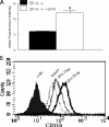
Exogenous SP-A enhanced CR3 expression on alveolar macrophages from Sp-a–/– mice. CR3 expression on alveolar macrophages was determined by flow cytometry 24 h after intratracheal injection of 100 μg of SP-A. SP-A treatment significantly enhanced expression of CR3 on alveolar macrophages from Sp-a–/– mice (open bar) compared with untreated Sp-a–/– (solid bar) mice (panel A). Data are mean ± S.E. with n = 8 mice per group, *, p < 0.05 compared with untreated Sp-a–/– mice. RAW cells, a mouse macrophage cell line, were treated with SP-A and CR3 surface expression determined by flow cytometry. SP-A enhanced surface expression of CR3 in a dose-dependent manner (panel B).
FIGURE 4.
FIGURE 4.
SP-A enhanced CR3-mediated phagocytosis. The effects of SP-A and iC3b on the phagocytosis of FITC-labeled H. influenzae were determined in CHO-WT cells and CHO-CR3 cells. In the absence of opsonins, the phagocytosis of H. influenzae was greater in CHO-CR3 cells (solid bar) compared with CHO-WT cells. iC3b, a known opsonin and ligand for CR3, enhanced CR3-mediated phagocytosis of H. influenzae in cells that expressed CR3 (open bar, panel A). SP-A enhanced CR3-mediated phagocytosis in CR3 expressing cells (hatched bar, panel A). There was no additive or synergistic effect of SP-A and iC3b on phagocytosis, the combination enhanced CR3-mediated phagocytosis to a similar extent as SP-A or iC3b alone (cross-hatched bar, panel A). SP-A-mediated enhancement of phagocytosis of H. influenzae into CR3 expressing cells was reduced by pre-treatment of SP-A with collagenase (colSPA) or N-glycanase (degSPA), suggesting roles for oligomeric assembly, the collagen like domain and N-linked carbohydrate in this effect (panel B). Data are mean ± S.E. with n = 4 experiments per group, *, p < 0.05 compared with phagocytosis by CHO-CR3 cells.
FIGURE 5.
FIGURE 5.
Binding of SP-A to CR-3 requires conversion to the active conformation. The interaction of SP-A with CR3 was determined by co-immunoprecipitation. A, in the first lane is an immunoblot of peritoneal cell lysates, the second lane is a control in which SP-A was omitted from incubation with the peritoneal lysate, the third lane shows preimmune serum rather than an anti-SP-A antibody was coupled to the gel suspension that was used for immunoprecipitation, and the fourth lane is an anti-SP-A antibody coupled to a gel suspension shown to co-immunoprecipitate CR3, detected by immunoblot analysis. Binding of SP-A to CR3 was determined in a whole cell assay, in which bound cells are detected by cell lysis and the quantification of associated alkaline phosphatase. Concentrations of PMA from 0.1 to 1 μg were required for sufficient activation of CR3 to bind SP-A (B). Binding of SP-A to CR3 was also enhanced by 1–2 mm Mn2+ (C). iC3b was used as a control. Data are mean ± S.E. of the total number of cells bound with n = 4 experiments per group, *, p < 0.05 compared with binding to non-stimulated CHO-CR3 cells.
FIGURE 6.
FIGURE 6.
Multiple domains of SP-A contribute to binding to CR3. The interaction of fluorescein-labeled SP-A with CR3 was determined by flow cytometry. The structure of the monomeric subunit of wild-type (WT) recombinant SP-A is shown in panel A. Note the branching N-linked carbohydrates attached to the N terminus and the CRD of the protein. In panel B, an E195A mutation that incapacitates the lectin function (1) of the CRD greatly reduced binding of SP-A to CR3. In panel C, tandem N1T and N187S mutations of SP-A, which prevent attachment of N-linked carbohydrates (2) also markedly reduced binding of SP-A to CR3. In panel D, truncated SP-A proteins lacking the N-terminal domain, the N-terminal N-linked carbohydrate attachment site, and first half of the collagen domain but possessing an intact Asn187 carbohydrate attachment (3), lacking the first half of the collagen domain but possessing the N-terminal segment, and both N-linked carbohydrate moieties (4), or lacking the collagen domain but possessing the N-terminal segment, and both carbohydrate moieties (5) retained significant CR3 binding activity. Mutations that resulted in a deletion of the N-terminal segment, the collagen-like domain, and the N-terminal (N1T) carbohydrate moiety (6) reduced binding of SP-A to CR3, and the binding activity that was further diminished by introduction of an additional N187S mutation prevented attachment of the CRD (7). These data implicate the N-linked carbohydrates, the CRD, and perhaps the neck domain in the interaction of SP-A with CR3. Data are mean ± S.E. with n = 4 experiments per group, *, p < 0.05 compared with binding of rat recombinant SP-A.
FIGURE 7.
FIGURE 7.
SP-A interacted with the I-domain of CR3. Binding of SP-A to CR3 was determined as outlined in the legend to Fig. 5. β-Glucan (binds the lectin domain of CR3) reduced the binding to SP-A (panel A). Antibodies to the I-domain of CR3 (M1/70 and CBRM1/5) almost completely blocked binding of SP-A to CR3 (panel A). Collagenase treatment of SP-A (colSPA) reduced the binding to CR3 compared with native SP-A. A further reduction in binding of collagenase-treated SP-A to CR3 was observed in the presence of β-glucan and M1/70 antibody (panel B) suggesting a role for the both the I-domain and lectin domain in CR3/SP-A interactions. Deglycosylation of SPA (degSPA) also reduced binding to CR3 compared with native SP-A and this binding was not inhibited in the presence of β-glucan. In contrast, the addition of the M1/70 antibody to the I-domain reduced binding of deglycosylated SP-A to CR3 (panel C), which implicates the collagen domain in the interaction with the I-domain. Data are mean ± S.E. with n = 4 experiments per group, *, p < 0.05 compared with binding of iC3b; #, p < 0.05 compared with human SP-A (panel A). *, p < 0.05 compared with human SPA; #, p < 0.05 compared with collagenase SPA or deglycosylated SPA (panels B and C).

Similar articles

Cited by

References

    1. LeVine, A. M., and Whitsett, J. A. (2001) Microbes Infect. 3 161–166 - PubMed
    1. Korfhagen, T. R. (2001) Am. J. Respir. Cell Mol. Biol. 25 668–672 - PubMed
    1. Khoor, A., Gray, M. E., Hull, W. M., Whitsett, J. A., and Stahlman, M. T. (1993) J. Histochem. Cytochem. 41 1311–1319 - PubMed
    1. Manz-Keinke, H., Plattner, H., and Schlepper-Schafer, J. (1992) Eur. J. Cell Biol. 57 95–100 - PubMed
    1. Ohmer-Schrock, D., Schlatterer, C., Plattner, H., and Schlepper-Schafer, J. (1995) J. Cell Sci. 108, 3695–3702 - PubMed

Publication types

MeSH terms