. 2009 Mar 27;284(13):8548-56.
doi: 10.1074/jbc.M805638200. Epub 2009 Jan 20.

FBP17 Mediates a Common Molecular Step in the Formation of Podosomes and Phagocytic Cups in Macrophages

Affiliations

FBP17 Mediates a Common Molecular Step in the Formation of Podosomes and Phagocytic Cups in Macrophages

Shigeru Tsuboi et al. J Biol Chem. .

Abstract

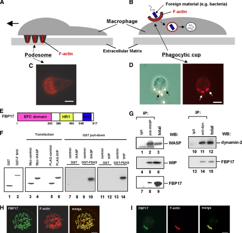
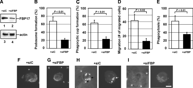
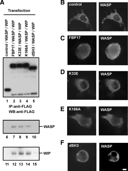
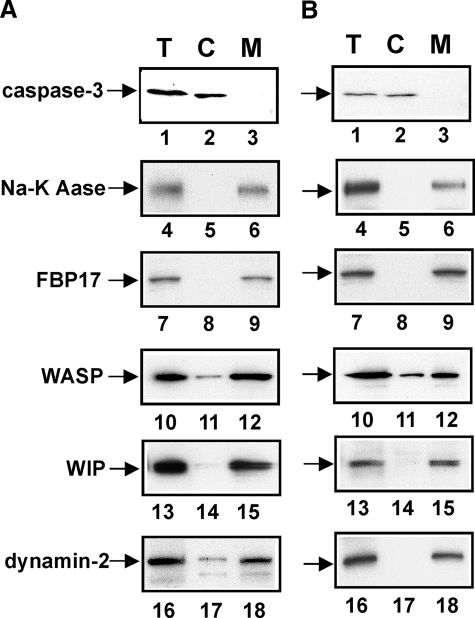
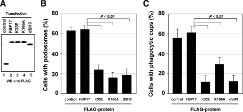
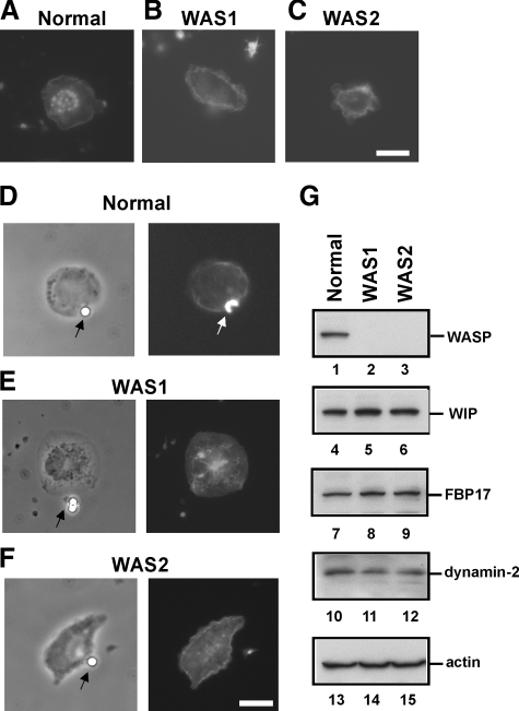
Macrophages act to protect the body against inflammation and infection by engaging in chemotaxis and phagocytosis. In chemotaxis, macrophages use an actin-based membrane structure, the podosome, to migrate to inflamed tissues. In phagocytosis, macrophages form another type of actin-based membrane structure, the phagocytic cup, to ingest foreign materials such as bacteria. The formation of these membrane structures is severely affected in macrophages from patients with Wiskott-Aldrich syndrome (WAS), an X chromosome-linked immunodeficiency disorder. WAS patients lack WAS protein (WASP), suggesting that WASP is required for the formation of podosomes and phagocytic cups. Here we have demonstrated that formin-binding protein 17 (FBP17) recruits WASP, WASP-interacting protein (WIP), and dynamin-2 to the plasma membrane and that this recruitment is necessary for the formation of podosomes and phagocytic cups. The N-terminal EFC (extended FER-CIP4 homology)/F-BAR (FER-CIP4 homology and Bin-amphiphysin-Rvs) domain of FBP17 was previously shown to have membrane binding and deformation activities. Our results suggest that FBP17 facilitates membrane deformation and actin polymerization to occur simultaneously at the same membrane sites, which mediates a common molecular step in the formation of podosomes and phagocytic cups. These results provide a potential mechanism underlying the recurrent infections in WAS patients.


Figures

FIGURE 1.
FIGURE 1.
FBP17 is a component of podosomes and phagocytic cups. A and B, schematic drawings of podosomes (A) and a phagocytic cup (B) in macrophages. C, podosomes in macrophages were visualized by F-actin staining using Alexa Fluor 568-phalloidin. D, macrophages incubated with IgG-opsonized latex beads formed phagocytic cups to ingest the beads. A phase contrast image of a macrophage forming phagocytic cups (left panel). Black arrows indicate the latex beads ingested by the macrophage. Phagocytic cups were visualized by F-actin staining using Alexa Fluor 568-phalloidin (right panel). White arrows indicate the phagocytic cups. The bar is 10 μm. E, the domain organization of FBP17. HR1, protein kinase C-related kinase homology region 1. F, FBP17 interacts directly with WASP and WIP via its SH3 domain. GST and the GST-FBP17 SH3 domain fusion protein (GST-FSH3) were purified from bacteria extracts. Purified proteins were subjected toSDS-PAGE and stained with Coomassie Brilliant Blue (lanes 1 and 2). HEK293 cells were transfected with the cDNAs of Myc-tagged control protein (Myc-PDZ-GEF), Myc-WASP, FLAG-PDZ-GEF, or FLAG-WIP, and the expression of those proteins were analyzed by immunoblotting (lanes 3–6). Lysates from the HEK293 transfected cells were incubated with the affinity matrices of GST alone or GST-FSH3. Pull-down samples were analyzed by immunoblotting using anti-Myc antibody (lanes 7–10) and anti-FLAG antibody (lanes 11–14). G, FBP17 binds WASP, WIP, and dynamin-2. WASP was immunoprecipitated (IP) from the lysates of PMA-differentiated THP-1 cells with anti-WASP or a control IgG (left panel, lanes 1–9). The WASP immunoprecipitates and total lysates were analyzed by immunoblotting (WB) for WASP (lanes 1–3), WIP (lanes 4–6), and FBP17 (lanes 7–9). Dynamin was also immunoprecipitated from the THP-1 cell lysates with an anti-dynamin polyclonal antibody. The dynamin immunoprecipitates and total lysates were analyzed by immunoblotting for dynamin-2 (lanes 10–12) and FBP17 (lanes 13–15). H and I, confocal laser scanning micrographs of PMA-differentiated THP-1 cells. H, THP-1 cells transfected with FLAG-tagged FBP17 cDNA (FBP17) were double-stained with an anti-FLAG monoclonal antibody (left panel) and phalloidin (center panel) to visualize the F-actin in podosomes. Yellow indicates co-localization of FBP17 (green) and F-actin, podosomes (red) (right panel). I, THP-1 cells transfected with FLAG-FBP17 cDNA were incubated with IgG-opsonized latex beads and double-stained with anti-FLAG antibody and phalloidin. Phagocytic cups were visualized by F-actin staining (center panel). Yellow indicates co-localization of FBP17 (green) and F-actin, phagocytic cups (red) (right panel). The bar is 10 μm.
FIGURE 2.
FIGURE 2.
The importance of FBP17 in the formation of podosomes and phagocytic cups. A, expression of FBP17 was reduced by transfection of siRNA. THP-1 cells were transfected with siRNA for FBP17 (siFBP; lanes 2 and 4) or its scrambled control siRNA (siC; lanes 1 and 3). Lysates prepared from total transfected cells were analyzed by immunoblotting for FBP17 (lanes 1 and 2) and β-actin (lanes 3 and 4). B and C, effects of FBP17 siRNA on the formation of podosomes and phagocytic cups in macrophages. Human primary monocytes were co-transfected with siFBP (closed bars) or siC (open bars) and an FITC-conjugated control siRNA and then differentiated into macrophages with M-CSF-1. FITC-positive transfected cells were examined for the formation of podosomes (B) or phagocytic cups (C), and the percentage of cells with podosomes or phagocytic cups was scored. D and E, effects of FBP17 siRNA on the functions of podosomes and phagocytic cups. Macrophages co-transfected with siFBP (closed bars) or siC (open bars) and the FITC-conjugated control siRNA were assayed for macrophage migration (D) or phagocytosis of IgG-opsonized latex beads (E). Data represent the mean ± S.D. of triplicate experiments. F–I, immunofluorescence micrographs of a representative cell from each experiment. Cells transfected with siC (F) and siFBP (G) were stained with Alexa Fluor 568-phalloidin. Cells transfected with siC (H) or siFBP (I) were incubated with IgG-opsonized latex beads and then stained with phalloidin. The left and right panels are phase contrast and immunofluorescence micrographs, respectively. The bar is 10 μm.
FIGURE 3.
FIGURE 3.
FBP17 recruits WASP, WIP, and dynamin-2 to the plasma membrane. A, HEK293 cells were co-transfected with cDNAs of the indicated FLAG-tagged proteins, Myc-tagged WASP, and HA-tagged WIP. The FLAG-tagged proteins were immunoprecipitated (IP) from lysates of the transfected cells with an anti-FLAG antibody followed by immunoblotting (WB) using antibodies to FLAG (lanes 1–5), WASP (lanes 6–10), and WIP (lanes 11–15). B–F, transfected HEK293 cells expressing FLAG-tagged proteins, Myc-WASP, and HA-WIP were double-stained with an anti-FLAG antibody and anti-WASP antibody. B–F, cells expressing FLAG-PDZ-GEF (B), FLAG-FBP17 (C), the FLAG-tagged FBP17 mutant with the K33E missense mutation (D), K166A (E), and the SH3-deleted FBP17 mutant dSH3 (F). The bar is 10 μm.
FIGURE 4.
FIGURE 4.
Subcellular localization of FBP17, WASP, WIP, and dynamin-2 in macrophages. A, macrophages forming podosomes. B, macrophages forming phagocytic cups. Total lysates (T), the cytosolic fraction (C), and the membrane fraction (M) prepared from macrophages forming podosomes (A) or phagocytic cups (B) were analyzed by immunoblotting for caspase-3 (lanes 1–3), sodium potassium ATPase (Na-K Aase; lanes 4–6), FBP17 (lanes 7–9), WASP (lanes 10–12), WIP (lanes 13–15), and dynamin-2 (lanes 16–18). Caspase-3 and sodium potassium ATPase (Na-K ATPase) are markers for the cytosol and plasma membrane, respectively.
FIGURE 5.
FIGURE 5.
The role of the EFC and SH3 domains of FBP17 in the formation of podosomes and phagocytic cups. A, expression of FLAG-tagged proteins in transfected THP-1 cells. Total lysates prepared from transfected THP-1 cells were analyzed by immunoblotting (WB) using an anti-FLAG antibody. All of the FLAG-tagged proteins, FLAG-PDZ-GEF (control, lane 1), FLAG-FBP17 (lane 2), and the FBP17 mutants, K33E, K166A, and dSH3 (lanes 3–5) were expressed in THP-1 cells at similar levels. B and C, THP-1 cells co-transfected with cDNAs for the FLAG-tagged proteins and pmaxGFP were differentiated with PMA and then assayed for the formation of podosomes (B) and phagocytic cups (C). The percentage of cells with podosomes or phagocytic cups among all GFP-positive cells was scored. Data represent the mean ± S.D. of triplicate experiments.
FIGURE 6.
FIGURE 6.
Defective formation of podosomes and phagocytic cups in macrophages from WAS patients. A–F, macrophages from a normal control and two genetically independent WAS patients (WAS1 and WAS2) were examined for the formation of podosomes (A–C) and phagocytic cups (D–F). The patients, WAS1 and WAS2, have the deletion mutations 211delT and 41–45delG, respectively, in their genomic DNAs. The bars are 10 μm. G, expression levels of WASP, WIP, FBP17, dynamin-2, and β-actin in WAS patients. Lysates prepared from macrophages from a normal control and two WAS patients (WAS1 and WAS2) were subjected to immunoblotting. WASP was not detected in the lysates from these WAS patients (lanes 2 and 3). Podosomes were completely absent (A–C) and phagocytic cup formation was severely impaired (D–F) in macrophages from both WAS patients, although FBP17, WIP, and dynamin-2 were expressed at the same level in patients as in normal individuals (G) (lanes 4–12).

Similar articles

Cited by

References

    1. Linder, S., and Aepfelbacher, M. (2003) Trends Cell Biol. 13 376-385 - PubMed
    1. Linder, S. (2007) Trends Cell Biol. 17 107-117 - PubMed
    1. Linder, S., Nelson, D., Weiss, M., and Aepfelbacher, M. (1999) Proc. Natl. Acad. Sci. U. S. A. 96 9648-9653 - PMC - PubMed
    1. Tsuboi, S. (2007) J. Immunol. 178 2987-2995 - PMC - PubMed
    1. Carman, C. V., Sage, P. T., Sciuto, T. E., Fuente, M. A. d. l., Geha, R. S., Ochs, H. D., Dvorak, H. F., Dvorak, A. M., and Springer, T. A. (2007) Immunity 26 784-797 - PMC - PubMed

Publication types

MeSH terms

LinkOut - more resources