. 2009 Apr;37(5):1701-12.
doi: 10.1093/nar/gkn1086. Epub 2009 Jan 20.

Specific recognition of a multiply phosphorylated motif in the DNA repair scaffold XRCC1 by the FHA domain of human PNK

Affiliations

Specific recognition of a multiply phosphorylated motif in the DNA repair scaffold XRCC1 by the FHA domain of human PNK

Ammar A E Ali et al. Nucleic Acids Res. 2009 Apr.

Abstract

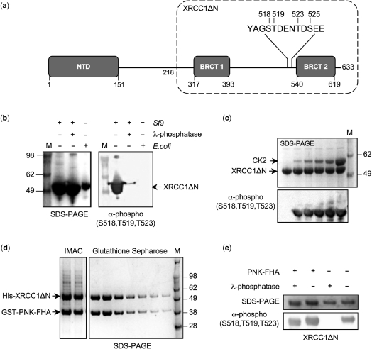
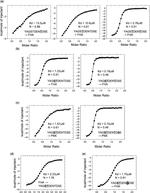
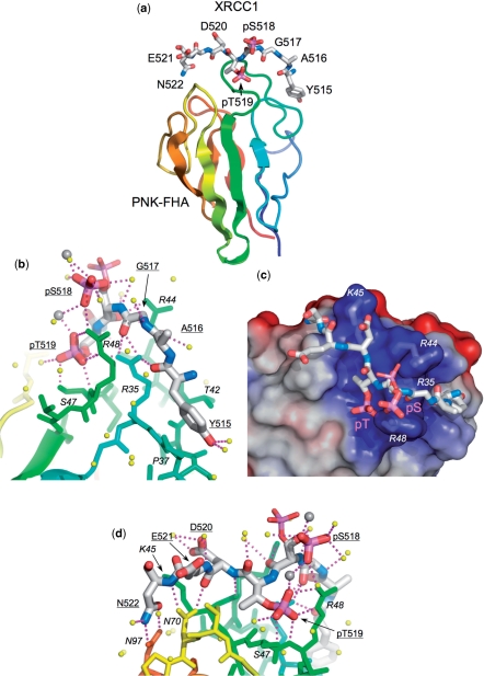
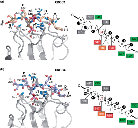
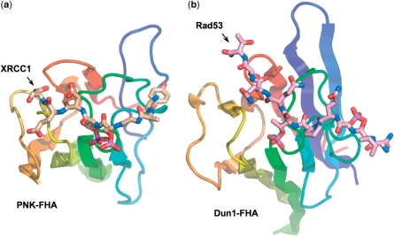
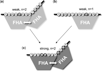
Short-patch repair of DNA single-strand breaks and gaps (SSB) is coordinated by XRCC1, a scaffold protein that recruits the DNA polymerase and DNA ligase required for filling and sealing the damaged strand. XRCC1 can also recruit end-processing enzymes, such as PNK (polynucleotide kinase 3'-phosphatase), Aprataxin and APLF (aprataxin/PNK-like factor), which ensure the availability of a free 3'-hydroxyl on one side of the gap, and a 5'-phosphate group on the other, for the polymerase and ligase reactions respectively. PNK binds to a phosphorylated segment of XRCC1 (between its two C-terminal BRCT domains) via its Forkhead-associated (FHA) domain. We show here, contrary to previous studies, that the FHA domain of PNK binds specifically, and with high affinity to a multiply phosphorylated motif in XRCC1 containing a pSer-pThr dipeptide, and forms a 2:1 PNK:XRCC1 complex. The high-resolution crystal structure of a PNK-FHA-XRCC1 phosphopeptide complex reveals the basis for this unusual bis-phosphopeptide recognition, which is probably a common feature of the known XRCC1-associating end-processing enzymes.


Figures

Figure 1.
Figure 1.
PNK–FHA protects CK2-phosphorylated XRCC1 from dephosphorylation. (a) Schematic representation of the domain structure of human XRCC1. The predicted amino-acid boundaries for each domain are indicated, with a dotted box delineating the extent of the XRCC1ΔN expression construct used in this study. The positions of the known phosphorylation sites within the linker region connecting the two C-terminal BRCT domains are also shown. (b) XRCC1ΔN expressed in insect cells (Sf9) is phosphorylated, as detected by a phospho-specific polyclonal antibody (Bethyl Laboratories, A300-059A). Incubation with λ-phosphatase readily removes the detected sites of phosphorylation. In contrast, XRCC1ΔN expressed in E.coli is not phosphorylated. One hundred micrograms of purified XRCC1ΔN protein was incubated with 50 μg of λ-phosphatase for a period of 1 h at room temperature, before being analysed by SDS–PAGE. (c) Incubation of the E.coli expressed XRCC1ΔN with recombinant maize CKII reproduces the phosphorylation observed in Sf9 cells. Sixty micrograms of purified XRCC1ΔN was incubated with increasing amounts of CKII (ranging from 0 to 95 μg), for a period of 2 h at room temperature. (d) Cell lysates from insect cells expressing His6-tagged XRCC1ΔN and E.coli expressing GST-tagged PNK–FHA were mixed, then applied to two sequential affinity columns (Talon/IMAC and Glutathione Sepharose). The SDS–PAGE analyses of the eluates from each step are shown. (e) The presence of the PNK–FHA domain protects, at least, the three phosphorylation sites detected by the polyclonal antibody in XRCC1 from the enzymatic action of λ-phosphatase. SDS–PAGE gels were stained with SimplyBlue SafeStain (Invitrogen, Paisley, UK).
Figure 2.
Figure 2.
Isothermal titration calorimetry of PNK binding to XRCC1 phospho-peptides. (a) ITC-binding curves for injection of XRCC1 peptides phosphorylated on Thr523 (left), Thr519 (middle), and on both Thr519 and Thr523 (right), into a microcalorimeter cell containing PNK–FHA (see Methods section). The mono-phosphorylated peptides show weak binding, while the doubly phosphorylated peptide binds with sub-micromolar affinity. Both peptides containing pThr518 show binding stoichiometry, (corresponding to the inflexion point of the curve) consistent with a 2:1 PNK–FHA:XRCC1 peptide complex. (b) As (a) but for peptide phosphorylated on pSer518 and pThr519 (left), and on pSer518, pThr519 and pThr523 (right). Affinities are >8-fold and >50-fold tighter, respectively, than the mono-phosphorylated pThr519 peptide. Binding stoichiometry in both cases indicates a 2:1 PNK–FHA:XRCC1 peptide complex. (c) Binding curves for injection of a pSer518 and pThr519 peptide (left) or a pSer518, pThr519, pThr523 and pSer525 peptide (right), into a cell containing full length human PNK. Affinities compare with those for the isolated FHA domain, and the binding stoichiometry remains consistent with a 2:1 PNK:XRCC1 peptide complex. (d) Inverted experiment, where PNK–FHA protein is injected into a cell containing an XRCC1 pSer518, pThr519 peptide. In this orientation of the experiment the binding stoichiometry is again consistent with formation of a 2:1 PNK:XRCC1 peptide complex, confirming that the observation is not a result of the experimental set-up. (e) Binding curves for injection of an XRRC1 peptide, phosphorylated on Ser518 and on Thr519, but with Thr523 mutated to valine. Removal of the threonine residue ablates the second binding site, so that only a single FHA domain now binds.
Figure 3.
Figure 3.
Structure of PNK–FHA–XRCC1 phosphopeptide complex. (a) Overview of the binding of the XRCC1 peptide (stick model) to residues forming the inter-strand loops in the PNK–FHA domain (cartoon rainbow coloured N-terminal:blue → C-terminal:red). This and all other molecular graphics were made with MacPyMol (DeLano Scientific). (b) Interactions of the N-terminal end of the XRCC1 peptide with PNK–FHA (see text for details). The phosphate group on pSer518 shows dual conformations in the crystal structure, (shown as thick and thin sticks), due to interactions with a calcium ion involved in formation of the crystal lattice. Ordered water molecules are shown as yellow spheres, Ca2+ ions as larger grey spheres. (c) The central part of the XRCC1 peptide consisting of the negatively charged residues pSer518, pThr519 and Asp520 binds in a very basic recess on the FHA domain surface, generated by the side chains of arginine residues 35, 44 and 48, and Lys 45. The molecular surface of PNK–FHA is shown coloured by electrostatic potential – positive:blue → negative:red. (d) Interactions of the C-terminal end of the XRCC1 peptide (see text for details). The C-terminal Asn522 of the XRCC1 peptide is anchored by a bidentate amide-amide interaction with PNK residue Asn97.
Figure 4.
Figure 4.
Comparison of PNK–FHA interactions with XRCC1 and XRCC4. PNK–FHA interacts in a similar way with the XRCC1 phosphopeptide (a), as with the XRCC4 peptide (b) apart from the additional interactions with pSer518 and Asn522 in the XRCC1 complex. That Ser518 was not phosphorylated in the XRCC4 complex is probably responsible for the different conformation observed for Arg44 in the two complexes. In both cases, the residues at −2 and −3 relative to the pThr, make no side-chain contacts with the protein that could mediate selectivity for these positions, and the specificity of FHA for the XRCC1 and XRCC4 sequences resides in the polar and electrostatic interactions with phosphorylated and acidic residues at 0, −1, +1 and +2, the hydrophobic interaction with Tyr at −4, and (for XRCC1) with the amide-side chain of the Asn at +3. In the schematics on the right, residues with a red (strictly) or orange (highly) background are conserved across FHA domains. Those with a green background are involved in hydrophobic contacts with the bound XRCC1/XRCC4 peptide, whilst those with a grey background are involved in making specific hydrogen bonds (as indicated by a line).
Figure 5.
Figure 5.
Different modes of bis-phosphopeptide-binding in PNK and Dun1. The binding mode to PNK–FHA of the XRCC1 bis-phosphopeptide (a) with adjacent pSer-pThr residues is markedly different to that recently observed for a yeast Rad53 phosphopeptide containing two non-adjacent pThr residues, to the FHA of the DNA damage checkpoint kinase Dun1 (PDB code 2JQL) (b). While the C-terminal phosphorylated residue is in a similar topological position on the FHA domain, the N-terminal pThr in Dun1 binds in the −4 subsite, which is occupied by a tyrosine in PNK–FHA complexes with XRCC1 and XRCC4 peptides.
Figure 6.
Figure 6.
Hierarchical co-operative binding of PNK–FHA domains. PNK–FHA binds with modest affinity (Kd ∼10 μM) to the XRCC1 FHA-binding region when phosphorylated on either Thr518 (a) or Thr523 (b). An FHA bound to the pThr518 site can stabilize the very weak interaction of a second FHA domain bound to the Thr523 site, even when not phosphorylated, probably by direct protein–protein contacts. However an FHA domain bound to pThr523 cannot stabilize the binding of a second FHA to the unphosphorylated Thr518 site. When both sites are fully phosphorylated (c), which is the likely result of the hierarchical phosphorylation of this region by CK2 in vivo, two FHA domains can bind with >50-fold higher affinity (Kd ∼0.1 μM), suggesting the presence of a far more stable complex than had been suggested by previous studies.

Similar articles

Cited by

References

    1. Caldecott KW, Aoufouchi S, Johnson P, Shall S. XRCC1 polypeptide interacts with DNA polymerase beta and possibly poly(ADP-ribose) polymerase, and DNA ligase III is a novel molecular ‘nick-sensor’ in vitro. Nucleic Acids Res. 1996;24:4387–4394. - PMC - PubMed
    1. Cappelli E, Taylor R, Cevasco M, Abbondandolo A, Caldecott K, Frosina G. Involvement of XRCC1 and DNA ligase III gene products in DNA base excision repair. J. Biol. Chem. 1997;272:23970–23975. - PubMed
    1. Rass U, Ahel I, West SC. Defective DNA repair and neurodegenerative disease. Cell. 2007;130:991–1004. - PubMed
    1. Jilani A, Ramotar D, Slack C, Ong C, Yang XM, Scherer SW, Lasko DD. Molecular cloning of the human gene, PNKP, encoding a polynucleotide kinase 3'-phosphatase and evidence for its role in repair of DNA strand breaks caused by oxidative damage. J. Biol. Chem. 1999;274:24176–24186. - PubMed
    1. Mani RS, Karimi-Busheri F, Cass CE, Weinfeld M. Physical properties of human polynucleotide kinase: hydrodynamic and spectroscopic studies. Biochemistry. 2001;40:12967–12973. - PubMed

Publication types

MeSH terms

Substances