

Loss of Rad51c leads to embryonic lethality and modulation of Trp53-dependent tumorigenesis in mice
- PMID: 19155299
- PMCID: PMC2754281
- DOI: 10.1158/0008-5472.CAN-08-3057
Loss of Rad51c leads to embryonic lethality and modulation of Trp53-dependent tumorigenesis in mice
Abstract
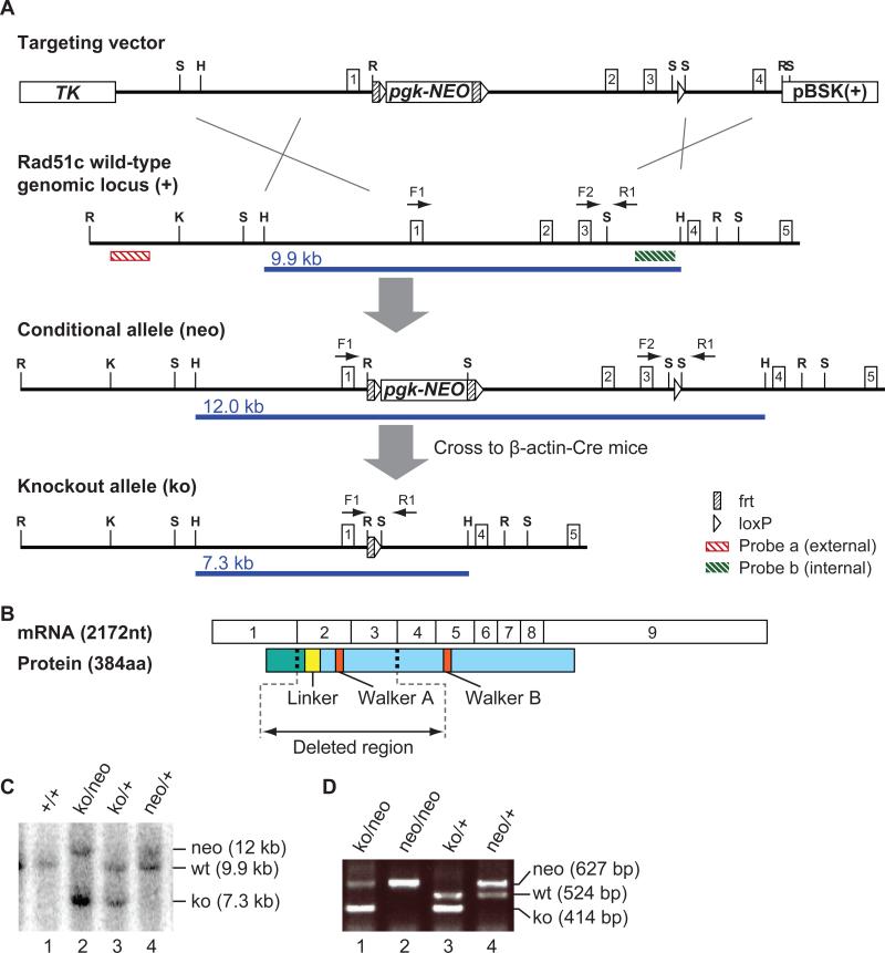
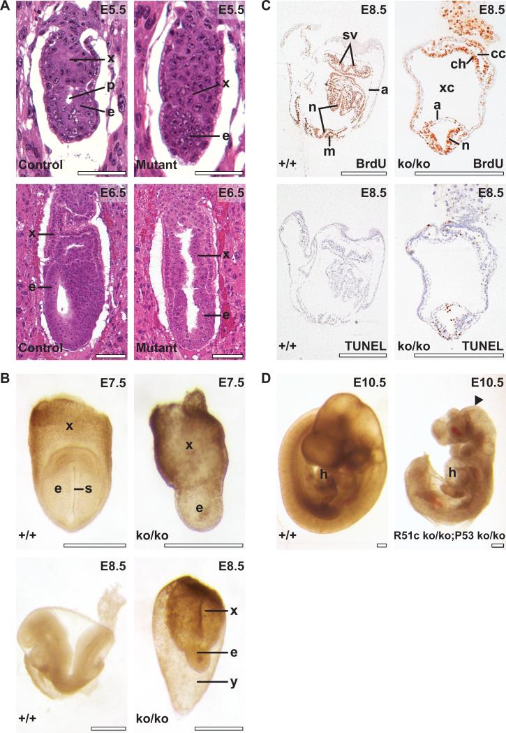
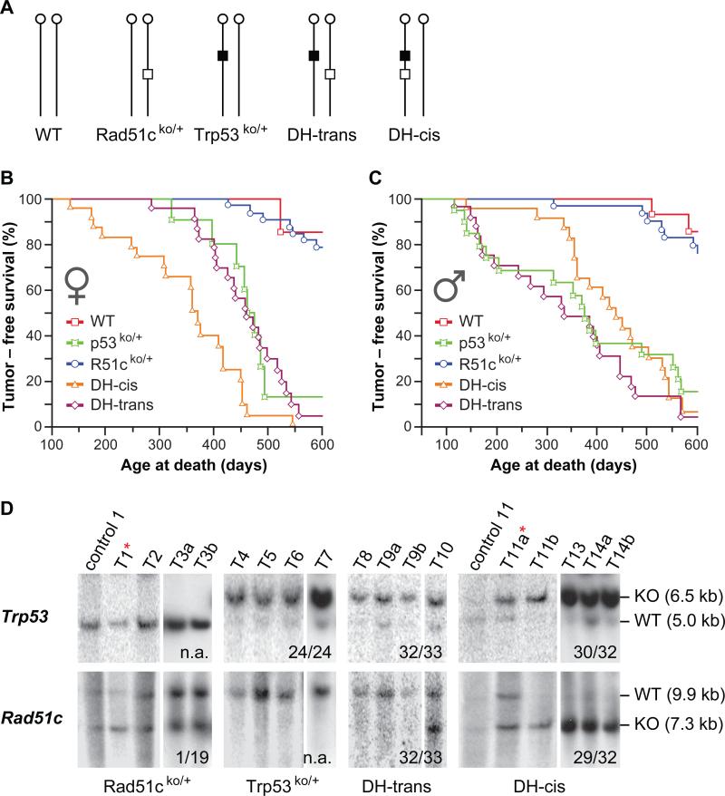
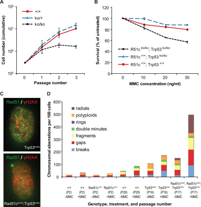
RecA/Rad51 protein family members (Rad51, Rad51b, Rad51c, Rad51d, Xrcc2, and Xrcc3) are essential for DNA repair by homologous recombination, and their role in cancers has been anticipated. Here we provide the first direct evidence for a tumor suppressor function for a member of the Rad51 family. We show that Rad51c deficiency leads to early embryonic lethality, which can be delayed on a Trp53-null background. To uncover the role of Rad51c in tumorigenesis, we have exploited the fact that Rad51c and Trp53 are both closely located on the mouse chromosome 11. We have generated double heterozygous (DH) mice carrying mutant alleles of both genes either on different (DH-trans) or on the same chromosome (DH-cis), the latter allowing for a deletion of wild-type alleles of both genes by loss of heterozygosity. DH-trans mice, in contrast to DH-cis, developed tumors with latency and spectrum similar to Trp53 heterozygous mice. Strikingly, Rad51c mutation in DH-cis mice promoted the development of tumors of specialized sebaceous glands and suppressed tumors characteristic of Trp53 mutation. In addition, DH-cis females developed tumors significantly earlier than any other group.
Figures
Similar articles
-
Loss of Rad51c accelerates tumourigenesis in sebaceous glands of Trp53-mutant mice.J Pathol. 2015 Jan;235(1):136-46. doi: 10.1002/path.4455. J Pathol. 2015. PMID: 25270124
-
Rad51c- and Trp53-double-mutant mouse model reveals common features of homologous recombination-deficient breast cancers.Oncogene. 2016 Sep 1;35(35):4601-10. doi: 10.1038/onc.2015.528. Epub 2016 Jan 25. Oncogene. 2016. PMID: 26820992
-
XRCC3 loss leads to midgestational embryonic lethality in mice.DNA Repair (Amst). 2021 Dec;108:103227. doi: 10.1016/j.dnarep.2021.103227. Epub 2021 Sep 22. DNA Repair (Amst). 2021. PMID: 34601382 Free PMC article.
-
Homologous recombination-deficient mutation cluster in tumor suppressor RAD51C identified by comprehensive analysis of cancer variants.Proc Natl Acad Sci U S A. 2022 Sep 20;119(38):e2202727119. doi: 10.1073/pnas.2202727119. Epub 2022 Sep 13. Proc Natl Acad Sci U S A. 2022. PMID: 36099300 Free PMC article.
-
RAD51 paralogs: roles in DNA damage signalling, recombinational repair and tumorigenesis.Semin Cell Dev Biol. 2011 Oct;22(8):898-905. doi: 10.1016/j.semcdb.2011.07.019. Epub 2011 Jul 28. Semin Cell Dev Biol. 2011. PMID: 21821141 Review.
Cited by
-
Impaired DNA double-strand break repair contributes to the age-associated rise of genomic instability in humans.Cell Death Differ. 2016 Nov 1;23(11):1765-1777. doi: 10.1038/cdd.2016.65. Epub 2016 Jul 8. Cell Death Differ. 2016. PMID: 27391797 Free PMC article.
-
Hypomorphic Brca2 and Rad51c double mutant mice display Fanconi anemia, cancer and polygenic replication stress.Nat Commun. 2023 Mar 11;14(1):1333. doi: 10.1038/s41467-023-36933-y. Nat Commun. 2023. PMID: 36906610 Free PMC article.
-
Influence of homologous recombinational repair on cell survival and chromosomal aberration induction during the cell cycle in gamma-irradiated CHO cells.DNA Repair (Amst). 2010 Jul 1;9(7):737-44. doi: 10.1016/j.dnarep.2010.03.009. DNA Repair (Amst). 2010. PMID: 20434408 Free PMC article.
-
A Survey of Essential Genome Stability Genes Reveals That Replication Stress Mitigation Is Critical for Peri-Implantation Embryogenesis.Front Cell Dev Biol. 2020 May 29;8:416. doi: 10.3389/fcell.2020.00416. eCollection 2020. Front Cell Dev Biol. 2020. PMID: 32548123 Free PMC article. Review.
-
Arabidopsis RAD51, RAD51C and XRCC3 proteins form a complex and facilitate RAD51 localization on chromosomes for meiotic recombination.PLoS Genet. 2017 May 31;13(5):e1006827. doi: 10.1371/journal.pgen.1006827. eCollection 2017 May. PLoS Genet. 2017. PMID: 28562599 Free PMC article.
References
-
- Shrivastav M, De Haro LP, Nickoloff JA. Regulation of DNA double-strand break repair pathway choice. Cell Res. 2008;18:134–47. - PubMed
-
- Bianco PR, Tracy RB, Kowalczykowski SC. DNA strand exchange proteins: a biochemical and physical comparison. Front Biosci. 1998;3:D570–603. - PubMed
-
- Kawabata M, Kawabata T, Nishibori M. Role of recA/RAD51 family proteins in mammals. Acta Med Okayama. 2005;59:1–9. - PubMed
Publication types
MeSH terms
Substances
Grants and funding
LinkOut - more resources
Full Text Sources
Other Literature Sources
Molecular Biology Databases
Research Materials
Miscellaneous
