. 2009 Feb 1;69(3):1230-9.
doi: 10.1158/0008-5472.CAN-08-2970. Epub 2009 Jan 20.

Senescent stromal-derived osteopontin promotes preneoplastic cell growth

Affiliations

Senescent stromal-derived osteopontin promotes preneoplastic cell growth

Ermira Pazolli et al. Cancer Res. .

Abstract

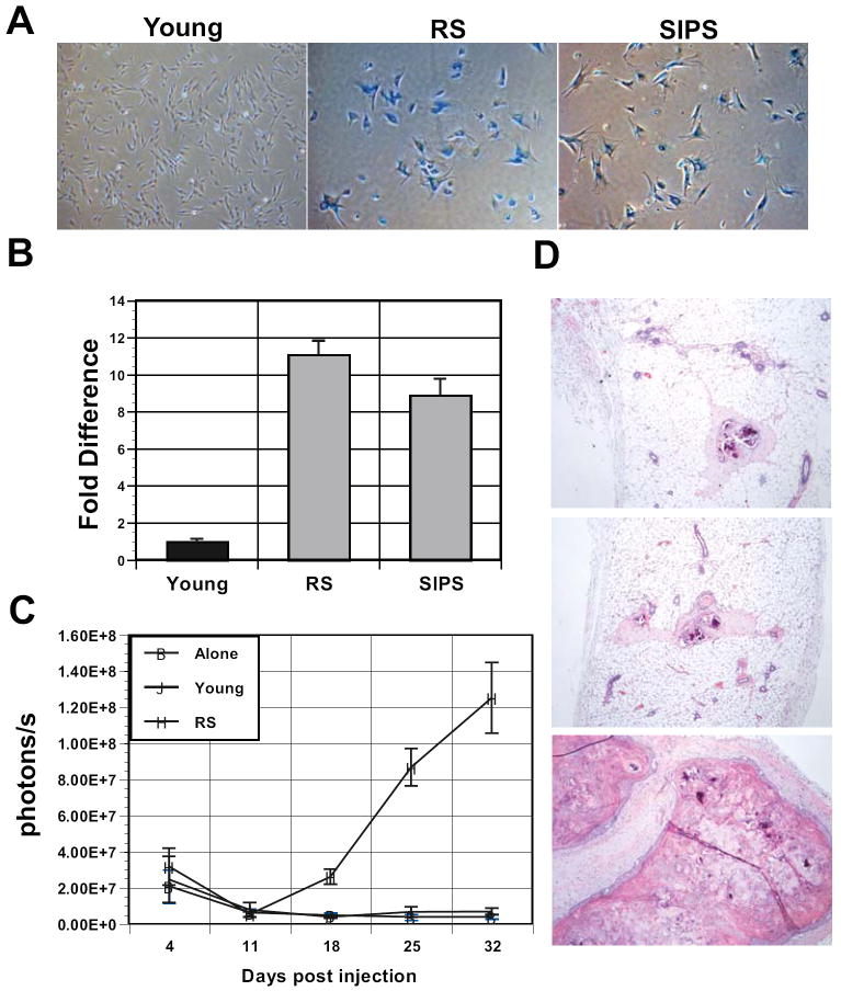
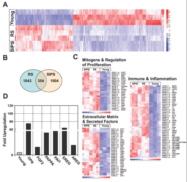
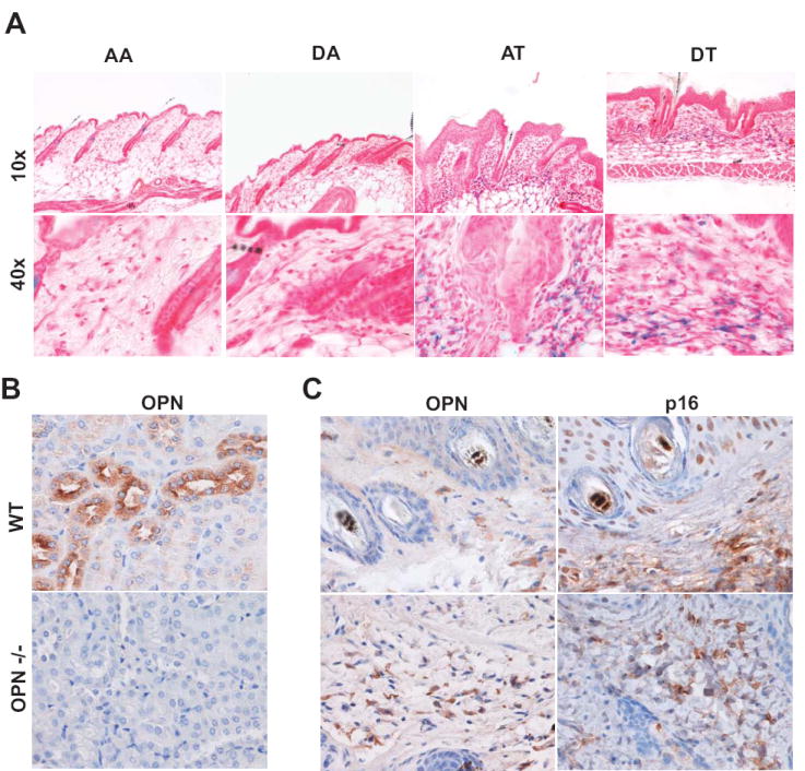
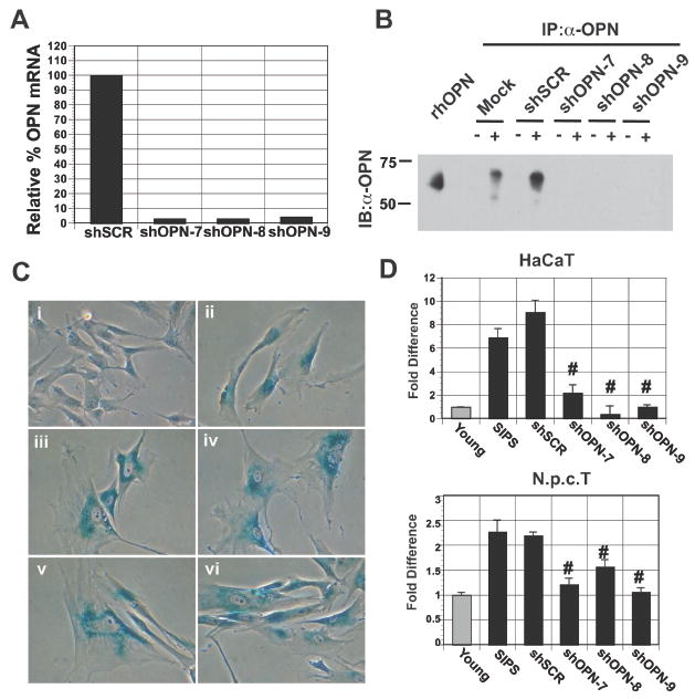
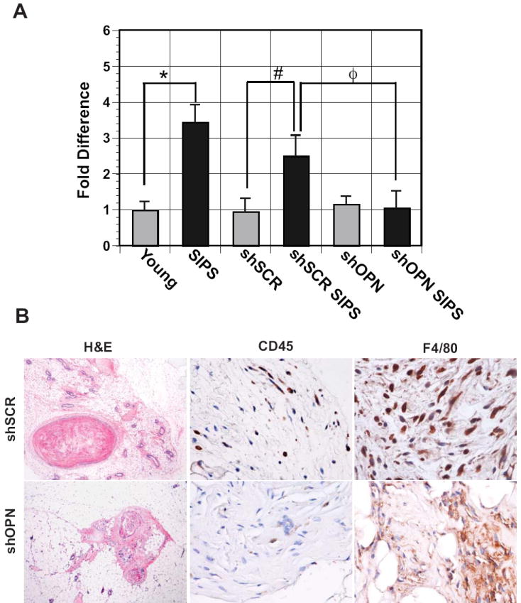
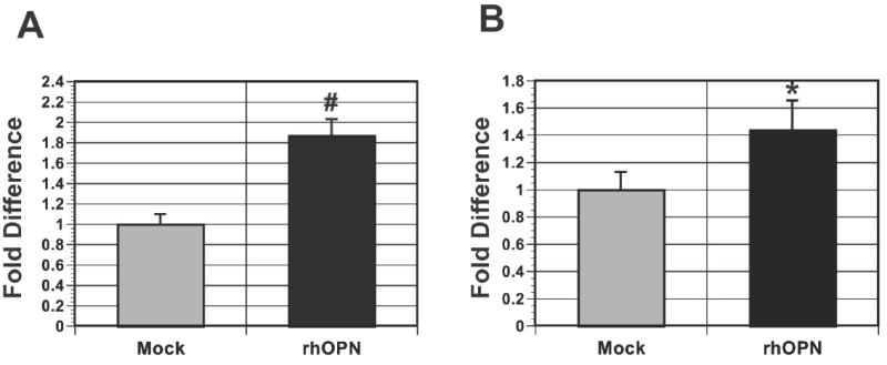
Alterations in the tissue microenvironment collaborate with cell autonomous genetic changes to contribute to neoplastic progression. The importance of the microenvironment in neoplastic progression is underscored by studies showing that fibroblasts isolated from a tumor stimulate the growth of preneoplastic and neoplastic cells in xenograft models. Similarly, senescent fibroblasts promote preneoplastic cell growth in vitro and in vivo. Because senescent cells accumulate with age, their presence is hypothesized to facilitate preneoplastic cell growth and tumor formation in older individuals. To identify senescent stromal factors directly responsible for stimulating preneoplastic cell growth, we carried out whole-genome transcriptional profiling and compared senescent fibroblasts with their younger counterparts. We identified osteopontin (OPN) as one of the most highly elevated transcripts in senescent fibroblasts. Importantly, reduction of OPN protein levels by RNA interference did not affect senescence induction in fibroblasts; however, it dramatically reduced the growth-promoting activities of senescent fibroblasts in vitro and in vivo, showing that OPN is necessary for paracrine stimulation of preneoplastic cell growth. In addition, we found that recombinant OPN was sufficient to stimulate preneoplastic cell growth. Finally, we show that OPN is expressed in senescent stroma within preneoplastic lesions that arise following 7,12-dimethylbenz(a)anthracene/12-O-tetradecanoylphorbol-13-acetate treatment of mice, suggesting that stromal-derived OPN-mediated signaling events affect neoplastic progression.


Figures

Figure 1
Figure 1. Senescent fibroblasts stimulate the growth of preneoplastic cells
A, continued passage (RS) and treatment with 100 ug/ml bleomycin (SIPS) induces robust senescence characterized by induction of senescence associated β–galactosidase (SA-βGal) staining. Magnification 20X. B, fibroblasts undergoing RS or SIPS stimulate the in vitro growth of HaCaT keratinocytes expressing click beetle red luciferase. HaCaT cells were plated on lawns of young, RS, or SIPS fibroblasts and their growth was measured by relative luciferase activity. Wells containing HaCAT cells and young fibroblasts were set to 1. Fold increase in growth over wells with young fibroblasts is plotted. p<0.05. C, HaCAT cells were injected alone or with young or senescent fibroblasts (RS) into NOD/SCID mice. For each injection, 2.5×10⁵ HaCaT cells were coinjected with 7.5×10⁵ fibroblasts. In vivo imaging of luciferase activity was used to assess HaCaT cell growth weekly. Each line represents the average photons/sec for 6 independent injection sites. Student’s t test at day 28, p < 0.05. D, Hematoxylin and eosin stains of histological sections obtained from xenografts injected with HaCaT cells alone or in the presence of young or senescent fibroblasts (top to bottom). Magnification 10X.
Figure 2
Figure 2. Analysis of the senescence transriptome reveals significant overlap between cells undergoing replicative and stress-induced premature senescence
A, microarray analysis of young BJ fibroblasts and BJ fibroblasts undergoing RS or bleomycin induced SIPS. Hierarchical clustering of consistent transcript alterations across all conditions and replicas are plotted. B, Venn diagram plots genes that significantly overlap in fibroblasts undergoing RS and SIPS versus young. C, Gene ontology analysis (GO) of upregulated classes in cells undergoing RS and SIPS compared to their younger counterparts (i.e. Immune & Inflammation, Mitogens & Regulation of Proliferation, Extracellular Matrix & Secreted Factors). D, Fold upregulation of select genes in senescent fibroblasts (SIPS) was confirmed by qRT-PCR. Expression in young fibroblasts was set as one for all genes.
Figure 3
Figure 3. Senescent stroma is present in preneoplastic lesions following DMBA-TPA treatment
A, Representative SA-βGal staining of frozen tissue sections reveal robust staining within the stromal compartment (n=3). Skin treatments are as follows: (1) AA, acetone followed by weekly acetone treatment (2) DA, DMBA followed by weekly acetone treatment; (3) AT, acetone followed by weekly TPA treatment; (4) DT, DMBA followed by weekly TPA treatments. B, Immunohistochemical analysis of osteopontin (OPN) in kidney sections from wild type (C57/B6) and OPN deficient mice. Magnification upper panel 10X and lower panel 40X. C, OPN and p16 expression in DMBA/TPA induced papillomas (upper panel) and associated skin (lower panel). Serial paraffin embedded tissue sections were stained with antibodies against OPN and the cell cycle inhibitor p16. Magnification 40X. (n=2 of 6).
Figure 4
Figure 4. Senescent-derived OPN is necessary for preneoplastic cell growth
A, Stable knockdown of OPN expression by viral-based RNAi is revealed by qRT-PCR. OPN mRNA expression in senescent fibroblasts transduced with a control hairpin (shSCR) was set to 100%. Three independent short hairpins targeting OPN (shOPN-7, -8, -9) resulted in robust knockdown of OPN mRNA. B, Western blot analysis (IB) of OPN immunoprecipitations (IP) from cultured supernatants obtained from young (-) or senescent (+) fibroblasts mock transduced, transduced with a control hairpin (shSCR), or transduced with hairpins targeting OPN (shOPN-7, -8, -9). Recombinant human OPN (rhOPN) is included as a reference. C, RNAi directed loss of OPN does not affect the induction of senescence upon bleomycin treatment. SA-βGal expression in i) young, ii) SIPS, iii) shSCR SIPS, iv) shOPN-7 SIPS, v) shOPN-8 SIPS, and vi) shOPN-9 SIPS BJ fibroblasts. Magnification 40X. D, Upper Panel: Quantification of HaCAT cell growth when plated on lawns of fibroblasts. Preneoplastic cell growth was measured by relative luciferase activity and wells where HaCAT cells were plated with young BJ fibroblasts were set to 1. OPN expression was depleted in senescent BJ fibroblasts by one of three independent hairpins (shOPN-7, -8, -9) to greater than 95% in all cases. A control hairpin (shSCR) was used as a negative control. Depletion of OPN expression reduced preneoplastic cell growth to levels observed in cells plated with young fibroblasts. #p<0.05. Lower Panel: Quantification of N.p.c.T cell growth when plated on lawns of fibroblasts. Loss of OPN expression (shOPN-7, -8, -9) resulted in reduced growth similar to that observed with HaCaT cells shown in upper panel. #p<0.05.
Figure 5
Figure 5. Loss of OPN expression in senescent fibroblasts results in reduced preneoplastic cell growth in vivo
Fibroblasts were uninfected (young and SIPS), infected with a control virus (shSCR, sSCR SIPS are young and senescent fibroblasts, respectively), or a virus that knocks down OPN expression (shOPN, shOPN SIPS are young and senescent fibroblasts, respectively) greater than 95%. Relative to SIPS fibroblasts, knockdown of OPN resulted in a significant reduction in growth of HaCaT xenografts in NOD/SCID mice (n=3). *p=0.003, #p=0.04, and Φp= 0.06. B, Immunohistochemical analysis of xenografts obtained from mice injected with HaCaT cells and senescent fibroblasts expressing a control hairpin (shSCR) (upper panel) or a hairpin targeting OPN (shOPN) (lower panel). Shown are Hematoxylin and Eosin (H&E) and staining for the immune markers CD45 and F4/80, which identify leukocytes and macrophages, respectively. Photos focus on stromal sections showing CD45 or F4/80 positive staining. Magnification for H&E, 10X and for CD45 and F4/80, 40X.
Figure 6
Figure 6. OPN is sufficient to stimulate the growth of preneoplastic cells
Cell growth was measured by relative luciferase activity and the growth of cells receiving BSA alone was set to 1. A, addition of 100 ng/ml of rhOPN was sufficient to stimulate the growth of HaCaT cells. #p<0.05. B, addition of 100 ng/ml of rhOPN was sufficient to stimulate the growth of N.p.c.T. cells. *p=0.05.

References

    1. DePinho RA. The age of cancer. Nature. 2000;408(6809):248–54. - PubMed
    1. Sakr WA, Haas GP, Cassin BF, Pontes JE, Crissman JD. The frequency of carcinoma and intraepithelial neoplasia of the prostate in young male patients. J Urol. 1993;150(2 Pt 1):379–85. - PubMed
    1. Hahn WC, Counter CM, Lundberg AS, Beijersbergen RL, Brooks MW, Weinberg RA. Creation of human tumor cells with defined genetic elements. Nature. 1999;400:464–8. - PubMed
    1. Hahn WC, Meyerson M. Telomerase activation, cellular immortalization and cancer. Ann Med. 2001;33(2):123–9. - PubMed
    1. Zhao JJ, Roberts TM, Hahn WC. Functional genetics and experimental models of human cancer. Trends Mol Med. 2004;10(7):344–50. - PubMed

Publication types

MeSH terms