. 2009 Feb 1;69(3):1080-8.
doi: 10.1158/0008-5472.CAN-08-2520. Epub 2009 Jan 20.

IFN regulatory factor 8 sensitizes soft tissue sarcoma cells to death receptor-initiated apoptosis via repression of FLICE-like protein expression

Affiliations

IFN regulatory factor 8 sensitizes soft tissue sarcoma cells to death receptor-initiated apoptosis via repression of FLICE-like protein expression

Dafeng Yang et al. Cancer Res. .

Abstract

IFN regulatory factor 8 (IRF8) has been shown to suppress tumor development at least partly through regulating apoptosis of tumor cells; however, the molecular mechanisms underlying IRF8 regulation of apoptosis are still not fully understood. Here, we showed that disrupting IRF8 function resulted in inhibition of cytochrome c release, caspase-9 and caspase-3 activation, and poly(ADP-ribose) polymerase cleavage in soft tissue sarcoma (STS) cells. Inhibition of the mitochondrion-dependent apoptosis signaling cascade is apparently due to blockage of caspase-8 and Bid activation. Analysis of signaling events upstream of caspase-8 revealed that disrupting IRF8 function dramatically increases FLIP mRNA stability, resulting in increased IRF8 protein level. Furthermore, primary myeloid cells isolated from IRF8-null mice also exhibited increased FLIP protein level, suggesting that IRF8 might be a general repressor of FLIP. Nuclear IRF8 protein was absent in 92% (55 of 60) of human STS specimens, and 99% (59 of 60) of human STS specimens exhibited FLIP expression, suggesting that the nuclear IRF8 protein level is inversely correlated with FLIP level in vivo. Silencing FLIP expression significantly increased human sarcoma cells to both FasL-induced and tumor necrosis factor-related apoptosis-inducing ligand (TRAIL)-induced apoptosis, and ectopic expression of IRF8 also significantly increased the sensitivity of these human sarcoma cells to FasL- and TRAIL-induced apoptosis. Taken together, our data suggest that IRF8 mediates FLIP expression level to regulate apoptosis and targeting IRF8 expression is a potentially effective therapeutic strategy to sensitize apoptosis-resistant human STS to apoptosis, thereby possibly overcoming chemoresistance of STS, currently a major obstacle in human STS therapy.


Figures
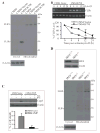
Figure 1
Figure 1. IRF8 regulates cytochrome C-mediated activation of caspases 3 and 9 in the mitochondria-dependent apoptosis pathway
A. Cytochrome C release assay. Tumor cells were treated with IFN-γ overnight, following by incubation with recombinant FasL for 4 h. Cytosol (C) and organelle-enriched mitochondrion (M) fractions were then prepared. The fractions were analyzed by Western blotting analysis for cytochrome C and Apaf1 protein levels. The blots of cytosol fractions were stripped and reprobed with anti-mouse β-actin antibody. B. Caspases 3 and 9 activation. Tumor cells were treated with IFN-γ overnight, followed by incubation with recombinant FasL for various time as indicated. Cytosolic fractions were then prepared and analyzed for activated caspase 9 and caspase 3, as well as cleaved PARP. The blots were stripped and reprobed with anti-mouse β-actin antibody.
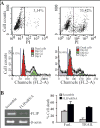
Figure 2
Figure 2. Disruption of IRF8 function inhibits caspase 8 and Bid activation
A. IP-Western blotting analysis of caspase 8 activation in the Fas DISC. Tumor cells were treated with IFN-γ overnight and then incubated with oligomerized anti-Fas mAb (the antibody was attached to Dynad beads through biotin-streptavidin) for 10 min at room temperature with rotation. The antibody-bound cells were pulled down using a magnetic stand. Bead-bound cells were lysed and the protein complex associated with the anti-Fas antibody was resolved in 4-20% SDS-polyacrulamide gels and analyzed by Western blotting using Fas- (left panel) and caspase 8-specific antibodies (right panel). The protein molecular weight markers are indicated at the left. Fas, caspase 8 and the heavy chain of the antibody (IgGH) are indicated at the right. B. Western blotting analysis of caspase 8 activation. Tumor cells were cultured in the presence of IFN-γ overnight and followed by incubation with recombinant FasL for 4 h. Cytosol fractions were prepared and analyzed by Western blotting with caspase 8-specific antibody (upper panel). The cleaved caspase 8 is indicated by an arrow. The membrane was stripped and reprobed with anti-mouse β-actin. The protein molecular weight markers are indicated at the right and caspase 8 is indicated at the left (top panel). Bottom panel: Western blotting analysis of procaspase 8 in the indicated tumor cells. Tumor cells were treated as described above. To assess the procaspase 8 levels between the tumor cell sublines and effects of IFN-γ and FasL, less cell lysate (as compared to top panel) were loaded to the gels and analyzed by Western blotting analysis as in the top panel. C. Western blotting analysis of Bid activation. Tumor cells were treated as in B. The cytosol fractions were prepared and analyzed using tBid-specific antibody.
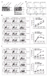
Figure 3
Figure 3. IRF8 functions as a repressor of FLIP
A. Disruption of IRF8 function increased FLIP protein level in tumor cells. Cytosol and organelle-enriched mitochondrion fractions were prepared from the indicated tumor cell line/sublines and analyzed by Western blotting analysis using a FLIP-specific antibody. The blots were stripped and reprobed with anti-mouse β-actin antibody. The protein molecular weight markers are indicated at the right and the locations of FLIPL and FLIPs are indicated at the left. B. IRF8 mediates the stability of FLIPs mRNA. Tumor cells were cultured in the presence of actinomycin D (Act D). Total RNA was isolated from the tumor cells at different time points as indicated and analyzed by RT-PCR for FLIPs mRNA level (top panel). β-actin was used as reference. The FLIPs band intensity at each time point was quantified and normalized to the β-actin band intensity at the same time point. The FLIPs mRNA level in untreated cells was set at 100% and the FLIPs mRNA level at the various time points were expressed as percent of that of untreated cells (bottom panel). C. Effects of ectopic expression of vFLIP on apoptosis. Tumor cells were stably transfected with mammalian expression vector alone (CMS4.Vector) or vector containing the vFLIP coding sequence (CMS4.vFLIP), respectively. The transfected tumor cells were then treated with recombinant IFN-γ overnight, followed by incubation with recombinant FasL for 4 h for Western blotting analysis and 24 h for apoptosis analysis. The treated tumor cells were fractionated to prepare cytosolic fractions and analyzed by Western blotting analysis for active caspase 8 protein level (top panel). The blot was stripped and reprobed with anti-mouse β-actin antibody. The treated cells were also stained with PI and analyzed for by flow cytometry for apoptotic cell death (bottom panel). D. Western blotting analysis of IRF8 and FLIP in IRF8-deficient and wt myeloid cells isolated from IRF8 null and wt littermate control mice. Total cell lysate (for IRF8 analysis, top panel) and cytosol and organelle-enriched mitochondrion fractions (for FLIP analysis, bottom panel) were prepared and analyzed by Western blotting using IRF8- and FLIP-specific antibodies, respectively. The blots were stripped and reprobed with anti-mouse β-actin antibody.
Figure 4
Figure 4. Immunohistochemical analysis of IRF8 and FLIP protein levels in human STS
Microsections of human STS specimens derived from 60 human STS patients were printed on glass slides as tissue microarray and immune-stained using IRF8-specifc antibody (A) or FLIP-specific antibody (B) as described in Material and Methods. The antibody-specific staining is shown as brown color. A. Four representative specimens with IRF8 staining: in the nuclei of tumor cells (a), cytosolic but no nuclear IRF8 staining of tumor cells (b), lymphoid aggregates in STS specimens showing strong nuclear staining (c), and IRF8 staining in the nuclei of tonsil lymphocytes (d). B. Three representative specimens with scattered low FLIP staining of tumor cells (a), strong FLIP staining of tumor cells (b), and FLIP staining is positive control tissue (c).
Figure 5
Figure 5. Silencing FLIP expression enhances FasL and TRAIL-induced apoptosis in human sarcoma cells
A. Comparison of cell death detection methods. HT1080 tumor cells were incubated in the absence (left 2 panels) or presence (right 2 panels) of FasL overnight and either directly stained with PI (top 2 panels), or fixed with 70% ethanol and then stained with PI as described in materials and methods (bottom 2 panel). The stained cells were analyzed by flow cytometry. B. Silencing FLIP expression increased the sensitivity of the tumor cells to FasL- and TRAIL-induced apoptosis. HT1080 cells were transiently transfected with scramble siRNA or FLIP-specific siRNA for approximately 24 h. The cells were analyzed for FLIP mRNA level using RT-PCR (left panel). The scramble- and FLIP siRNA-transfected cells were also treated with recombinant FasL or TRAIL protein overnight and analyzed for cell death by PI staining and flow cytometry analysis (right panel). Column, mean; bar, SD.
Figure 6
Figure 6. Ectopic expression of IRF8 sensitized human sarcoma cells to FasL- and TRAIL-induced apoptosis
A. RT-PCR analysis of IRF8 expression in non-transfected (HT1080), vector-transfected (HT1080.Vector) and IRF8-transfected (HT1080.IRF8) human sarcoma cells. β-actin was used as normalization standard (left panel). RT-PCR and Western blotting analysis of FLIP expression in HT1080, HT1080.Vector and HT1080.IRF8 human sarcoma cells (right panel). Upper panel is FLIP mRNA level and lower panel shows FLIP protein level. B. Fas-mediated apoptosis in HT1080 sarcoma cell line/sublines. Tumor cells were cultured in the presence of different concentrations of recombinant human FasL for approximately 18 h. The cells were then stained with PI and analyzed by flow cytometry. Shown are histograms of one of three representative experiments (left panel) and plot of FasL concentration against percentage of cell death (top right panel). The tumor cells were also treated with FasL (25ng/ml) and analyzed for cell death at different time points (bottom right panel). C. TRAIL-induced apoptosis in HT1080 sarcoma cell line/sublines. Tumor cells were cultured in the presence of different concentrations of recombinant human TRAIL for approximately 18 h. The cells were then stained with PI and analyzed by flow cytometry. Shown are histograms of one of three representative experiments (left panel) and plot of FasL concentration against percentage of cell death (top right panel). The tumor cells were also treated with TRAIL protein (25ng/ml) and analyzed for cell death at different time points (bottom right panel). D. Cell surface TRAIL receptors DR4 and DR5 expression levels. Tumor cells were stained with anti-DR4- and DR5-specific antibodies, respectively, and analyzed with flow cytometry. Isotype-matched IgG control staining is depicted as gray areas, and DR4- or DR5-specific staining is depicted as solid lines.

Similar articles

Cited by

References

    1. Mocellin S, Rossi CR, Brandes A, Nitti D. Adult soft tissue sarcomas: conventional therapies and molecularly targeted approaches. Cancer Treat Rev. 2006;32:9–27. - PubMed
    1. Kanno Y, Levi BZ, Tamura T, Ozato K. Immune cell-specific amplification of interferon signaling by the IRF-4/8-PU.1 complex. J Interferon Cytokine Res. 2005;25:770–9. - PubMed
    1. Levi BZ, Hashmueli S, Gleit-Kielmanowicz M, Azriel A, Meraro D. ICSBP/IRF-8 transactivation: a tale of protein-protein interaction. J Interferon Cytokine Res. 2002;22:153–60. - PubMed
    1. Holtschke T, Lohler J, Kanno Y, et al. Immunodeficiency and chronic myelogenous leukemia-like syndrome in mice with a targeted mutation of the ICSBP gene. Cell. 1996;87:307–17. - PubMed
    1. Gabriele L, Phung J, Fukumoto J, et al. Regulation of apoptosis in myeloid cells by interferon consensus sequence-binding protein. J Exp Med. 1999;190:411–21. - PMC - PubMed

Publication types

MeSH terms

Substances