. 2009 Feb 1;182(3):1325-33.
doi: 10.4049/jimmunol.182.3.1325.

MicroRNA-513 regulates B7-H1 translation and is involved in IFN-gamma-induced B7-H1 expression in cholangiocytes

Affiliations

MicroRNA-513 regulates B7-H1 translation and is involved in IFN-gamma-induced B7-H1 expression in cholangiocytes

Ai-Yu Gong et al. J Immunol. .

Abstract

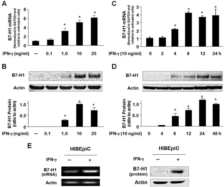
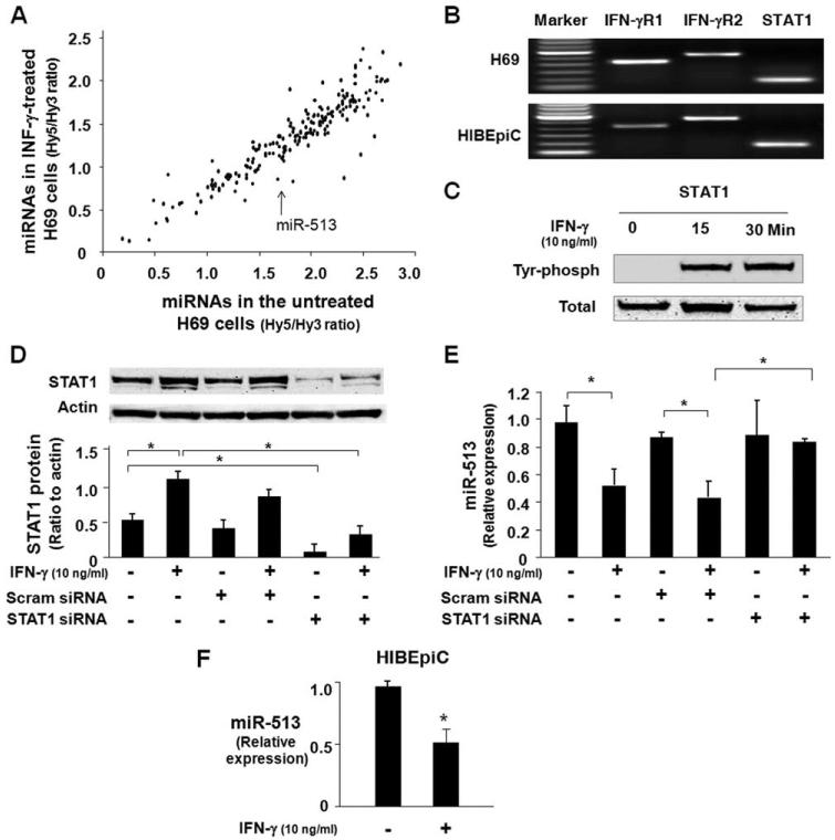
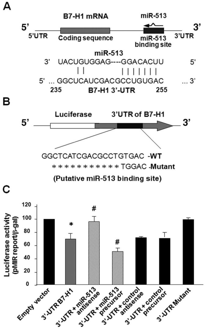
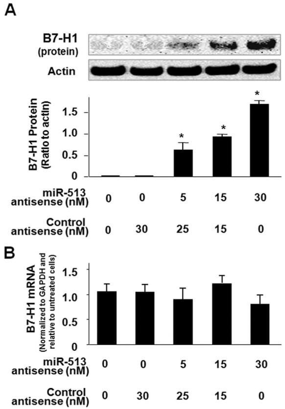
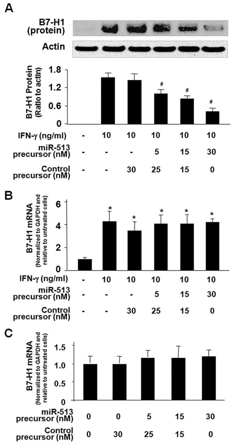
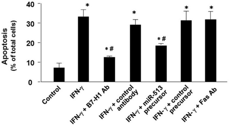
Biliary epithelial cells (cholangiocytes) respond to proinflammatory cytokines such as IFN-gamma and actively participate in the regulation of biliary inflammatory response in the liver. B7-H1 (also known as CD274 or PD-L1) is a member of the B7 costimulatory molecules and plays a critical immunoregulatory role in cell-mediated immune responses. In this study, we show that resting human cholangiocytes in culture express B7-H1 mRNA, but not B7-H1 protein. IFN-gamma induces B7-H1 protein expression and alters the microRNA (miRNA) expression profile in cholangiocytes. Of those IFN-gamma-down-regulated miRNAs, we identified microRNA-513 (miR-513) with complementarity to the 3'-untranslated region of B7-H1 mRNA. Targeting of the B7-H1 3'-untranslated region by miR-513 results in translational repression. Transfection of cholangiocytes with an antisense oligonucleotide to miR-513 induces B7-H1 protein expression. Additionally, transfection of miR-513 precursor decreases IFN-gamma-induced B7-H1 protein expression and consequently influences B7-H1-associated apoptotic cell death in cocultured Jurkat cells. Thus, miR-513 regulates B7-H1 translation and is involved in IFN-gamma-induced B7-H1 expression in human cholangiocytes, suggesting a role for miRNA-mediated gene silencing in the regulation of cholangiocyte response to IFN-gamma.


Figures

FIGURE 1
FIGURE 1
Posttranscriptional suppression of B7-H1 exists in human cholangiocytes and IFN-γ induces cholangiocyte B7-H1 protein expression. A and B, Dose-dependent expression of B7-H1 at the message (A) and protein (B) levels in H69 cells following IFN-γ stimulation. H69 cells were exposed to culture medium with various doses of IFN-γ (0, 0.1, 1.0, 10, and 25 ng/ml) for 8 h (for real-time PCR) or 24 h (for Western blotting). A representative Western blot from three independent experiments is shown in B. Actin was blotted as a loading control. Densitometric levels of B7-H1 signals were quantified and expressed as the ratio to actin. C and D, Time-dependent expression of B7-H1 expression in H69 cells induced by IFN-γ. Cells were exposed to IFN-γ (10 ng/ml) for 2-48 h followed by real-time PCR (C) and Western blotting (D) for B7-H1. E, Expression of B7-H1 in HIBEpiC cells upon IFN-γ stimulation. HIBEpiC cells were exposed to culture medium with or without IFN-γ (10 ng/ml) for 8 h (for RT-PCR) or 24 h (for Western blotting). *, p < 0.05, vs the nonstimulated control.
FIGURE 2
FIGURE 2
IFN-γ alters miRNA expression profile in cholangiocytes and decreases miR-513 expression in cholangiocytes in a STAT1-dependent manner. A, Microarray analysis of miRNA expression in H69 cells after IFN-γ stimulation for 8 h. Data were presented as the mean values of the mean Hy5/Hy3 ratios from three independent experiments. Each dot represents one miRNA. B, Expression of IFN-γ receptors and STAT1 in cholangiocytes. The mRNAs of two IFN-γ receptors (IFN-γR1 and IFN-γR2) and STAT1 were detected in both H69 cells and HIBEpiC cells by RT-PCR. C, Activation of STAT1 via tyrosine phosphorylation in cholangiocytes induced by IFN-γ. H69 cells were exposed to IFN-γ (10 ng/ml) for 15 or 30 min and tyrosine phosphorylation of STAT1 was assessed by Western blotting using Abs recognizing the tyrosine-phosphorylated (Tyr phosph) form of STAT1. Total STAT1 was also blotted as a control. D, Knockdown of STAT1 expression in H69 cells by siRNA gene silencing. H69 cells were treated with a siRNA to STAT1 for 48 h with an additional incubation for 8 h in the absence or presence of IFN-γ (10 ng/ml) followed by Western blotting for STAT1. A scrambled control siRNA (Scram siRNA) was used as the control. E, Knockdown of STAT1 blocked IFN-γ-induced decrease of miR-513 expression. H69 cells were treated with either the STAT1 siRNA or scrambled control siRNA for 48 h with an additional incubation for 8 h in the absence or presence of IFN-γ (10 ng/ml). Total RNA was isolated and the expression of miR-513 was quantified by real-time PCR. Data are representative of three independent experiments. F, IFN-γ decreases miR-513 expression in HIBEpiC cells. Cells were exposed to IFN-γ (10 ng/ml) for 8 h followed by real-time PCR for miR-513. *, p < 0.05, vs the nonstimulated control (in F) or as indicated (in D and E).
FIGURE 3
FIGURE 3
miR-513 targets a potential binding site in the B7-H1 3′-UTR resulting in translational suppression. A, The schematic of B7-H1 mRNA shows a potential binding site in the B7-H1 3′-UTR for miR-513. B, The complementary miR-513-binding site in the B7-H1 3′-UTR was inserted downstream of a luciferase reporter on the pMIR-Report plasmid. A control plasmid with the mutant 3′-UTR sequence was also generated. WT, Wild type. C, Targeting of B7-H1 3′-UTR by miR-513 results in translational suppression. H69 cells were transfected with the reporter constructs simultaneously with or without the miR-513 antisense oligonucleotide or the miR-513 precursor. After 24 h, cells were harvested and luciferase activities were measured and normalized to the control β-galactosidase level. A scrambled antisense oligonucleotide and a nonspecific precursor were used as the controls. These data are representative of three independent experiments. *, p < 0.05, vs the empty vector; #, p < 0.05, vs the B7-H1 3′-UTR reporter construct.
FIGURE 4
FIGURE 4
miR-513 antisense transfection induces B7-H1 protein expression in H69 cells. A, Transfection of miR-513 antisense induces B7-H1 protein expression. H69 cells were treated with an antisense oligonucleotide to miR-513 or a nonspecific control antisense for 72 h followed by Western blotting for B7-H1. A representative Western blot from three independent experiments and densitometric levels of B7-H1 signals are shown. *, p < 0.05, vs the nonantisense-treated control. B, miR-513 antisense transfection does not affect B7-H1 mRNA levels. H69 cells were exposed to miR-513 antisense or a nonspecific control antisense for 72 h followed by real-time PCR analysis for B7-H1 mRNA.
FIGURE 5
FIGURE 5
Transfection of miR-513 precursor reduces IFN-γ-induced B7-H1 protein expression in H69 cells. A, miR-513 precursor reduces IFN-γ-induced B7-H1 protein expression. H69 cells were transfected with miR-513 precursor or a control nonspecific precursor for 48 h and then exposed to IFN-γ (10 ng/ml) for 24 h followed by Western blotting for B7-H1. A representative Western blot from three independent experiments and densitometric levels of B7-H1 signals are shown. #, p < 0.05, vs IFN-γ-treated alone. B, miR-513 precursor transfection does not affect IFN-γ-stimulated B7-H1 mRNA expression. H69 cells were transfected with miR-513 precursor or a control nonspecific precursor for 48 h and then exposed to IFN-γ (10 ng/ml) for 24 h followed by real-time PCR analysis for B7-H1 mRNA. *, p < 0.05, vs non-IFN-γ-stimulated control. C, miR-513 precursor transfection does not affect B7-H1 mRNA expression in nonstimulated cells. H69 cells were transfected with miR-513 precursor or a control nonspecific precursor for 48 h in the absence of IFN-γ followed by B7-H1 mRNA real-time PCR analysis.
FIGURE 6
FIGURE 6
miR-513 influences B7-H1-associated apoptotic cell death in cocultured Jurkat cells. H69 cells were first exposed to IFN-γ for 24 h in the presence or absence of a neutralizing Ab to B7-H1. Some cells were transfected with miR-513 precursor or a control precursor for 48 h before exposure to IFN-γ. H69 cells were then cocultured with Jurkat cells in the presence of PMA with or without a neutralizing Ab to Fas for 24 h. Jurkat cells were separated from H69 cells and harvested for apoptosis assay by DAPI staining. These data are representative of three independent experiments. Ab; *, p < 0.05, vs Jurkat cells cocultured with non-IFN-γ-stimulated H69 cells as the control; #, p < 0.05, vs Jurkat cells cocultured with IFN-γ-stimulated H69 cells without Ab or precursor treatment.

Similar articles

Cited by

References

    1. Lazaridis KN, Strazzabosco M, LaRusso NF. The cholangiopathies: disorders of biliary epithelia. Gastroenterology. 2004;127:1565–1577. - PubMed
    1. Strazzabosco M, Fabris L, Spirli C. Pathophysiology of cholangiopathies. J. Clin. Gastroenterol. 2005;39:S90–S102. - PubMed
    1. Alpini G, McGill JM, LaRusso NF. The pathobiology of biliary epithelia. Hepatology. 2002;35:1256–1268. - PubMed
    1. Dong H, Strome S, Salomao DR, Salomao DR, Tamura H, Hirano F, Flies DB, Roche PC, Lu J, Zhu G, et al. Tumor-associated B7-H1 promotes T-cell apoptosis: a potential mechanism of immune evasion. Nat. Med. 2002;8:793–800. - PubMed
    1. Yamazaki T, Akiba H, Iwai H, Matsuda H, Aoki M, Tanno Y, Shin T, Tsuchiya H, Pardoll DM, Okumura K, et al. Expression of programmed death 1 ligands by murine T cells and APC. J. Immunol. 2002;169:5538–5545. - PubMed

Publication types

MeSH terms