Comparative Study
. 2009 Feb 1;182(3):1351-61.
doi: 10.4049/jimmunol.182.3.1351.

IL-12 enhances CTL synapse formation and induces self-reactivity

Affiliations
Comparative Study

IL-12 enhances CTL synapse formation and induces self-reactivity

Mary A Markiewicz et al. J Immunol. .

Abstract

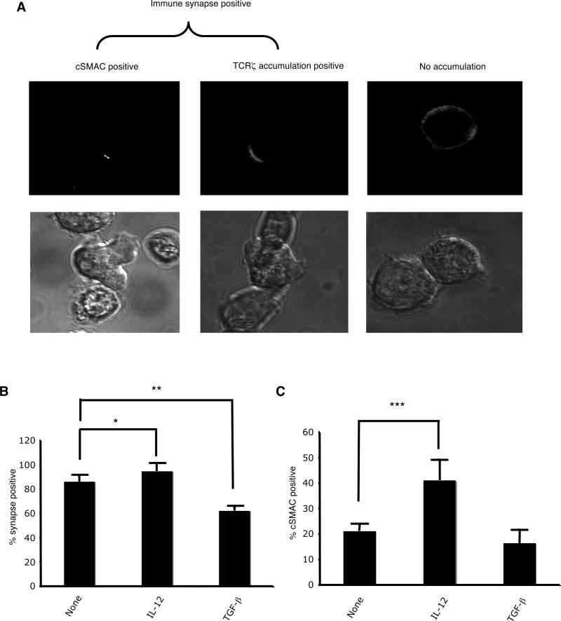
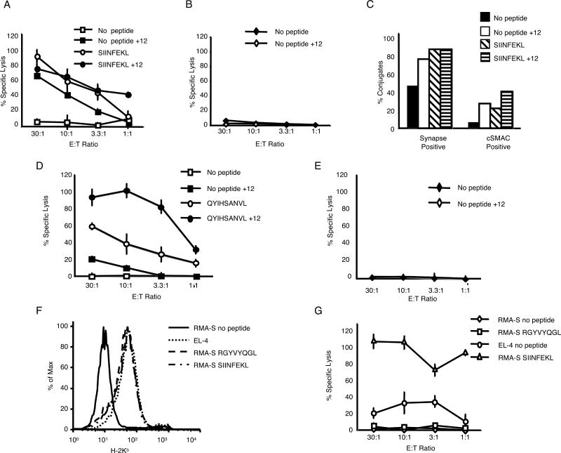
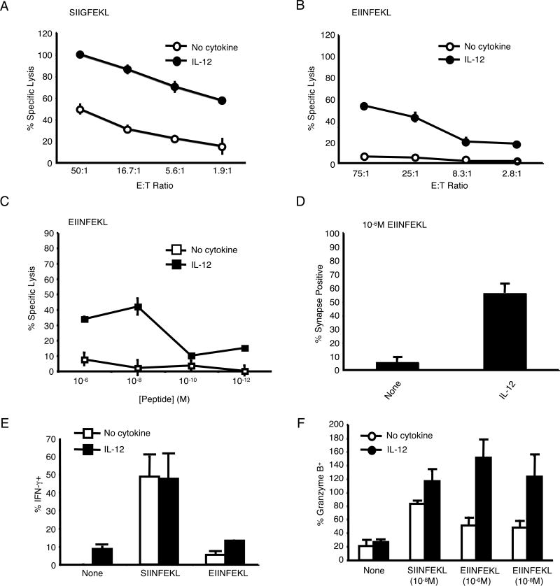
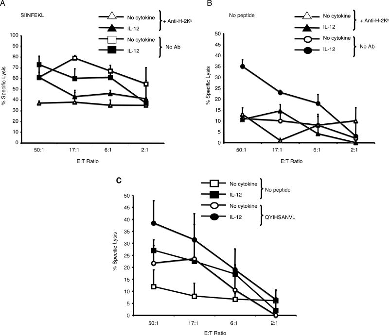
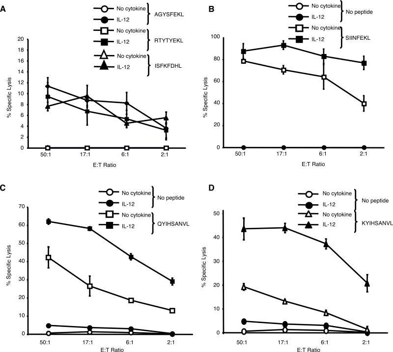
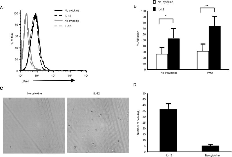
Immunological synapse formation between T cells and target cells can affect the functional outcome of TCR ligation by a given MHC-peptide complex. Although synapse formation is usually induced by TCR signaling, it is not clear whether other factors can affect the efficiency of synapse formation. Here, we tested whether cytokines could influence synapse formation between murine CTLs and target cells. We found that IL-12 enhanced synapse formation, whereas TGFbeta decreased synapse formation. The enhanced synapse formation induced by IL-12 appeared to be functional, given that IL-12-treated cells could respond to weak peptides, including self-peptides, to which the T cells were normally unresponsive. These responses correlated with expression of functionally higher avidity LFA-1 on IL-12-treated CTLs. These findings have implications for the function of IL-12 in T cell-mediated autoimmunity.


Figures

FIGURE 1
FIGURE 1
IL-12 enhances and TGF-β decreases antigen-driven synapse formation by OT-1 CTL. (A) Examples of synapse positive and cSMAC positive conjugates formed between OT-1 CTL and RMA-S cells pulsed with SIINFEKL peptide. (B) Percentage of synapse positive conjugates (+/- SD) formed between untreated, IL-12-treated, or TGF-β-treated OT-1 CTL and RMA-S/SIINFEKL (10-6M). No synapses were seen under any condition when conjugates were formed with RMA-S cells in the absence of exogenous peptide. (C) Percentage of synapse positive conjugates that were cSMAC positive formed between untreated, IL-12-treated, or TGF-β-treated OT-1 CTL and RMA-S/SIINFEKL (10-6M). These results are the average of five independent experiments. *p<0.02, **p<0.0003, ***p<0.03 in a two-sided student's T-test.
FIGURE 2
FIGURE 2
IL-12-treated OT-1 CTL lyse histocompatible target cells in a peptide-dependent manner. (A and B) Untreated and IL-12-treated OT-1 CTL were tested for cytotoxicity (+/- SD) against the histocompatible cell line EL-4 (H-2b) (+/- 10-6M SIINFEKL) and the histo-incompatible cell line P815 (H-2Dd). These results are representative of multiple independent experiments. (C) Percentage of synapse positive and cSMAC positive conjugates formed between untreated or IL-12-treated CTL and unpulsed or SIINFEKL (10-6 M)-pulsed EL-4 cells. These results are representative of two independent experiments. (D and E) Untreated and IL-12-treated DUC18 CTL were tested for cytotoxicity (+/- SD) against the histocompatible cell line P815 (H-2Dd) (+/- 10-6M QYIHSANV) and the histo-incompatible cell line EL-4 (H-2b). (F) Surface expression of H-2Kb on unpulsed EL-4, RMA-S, and RMA-S pulsed with 10-6M peptides. (G) Lysis by IL-12-treated OT-1 CTL (+/- SD) of the surface H-2b high cell line EL-4, the surface H-2b low cell line RMA-S, or RMA-S pulsed with 10-6M SIINFEKL or RGYVYQGL (control peptide). These data are representative of multiple independent experiments.
FIGURE 3
FIGURE 3
IL-12 enhances peptide sensitivity and decreases peptide-specificity of OT-1 CTL. (A) Untreated and IL-12-treated OT-1 CTL were tested for cytotoxicity against RMA-S cells pulsed with the intermediate agonist SIIGFEKL (10-6M) (+/-SD). Lysis of SIINFEKL at an E:T of 50:1 was 67% (No cytokine) and 78% (+IL-12). Lysis with control peptide at an E:T of 50:1 was 6% for both types of CTL. (B) Untreated and IL-12-treated CTL were tested for cytotoxicity against RMA-S cells pulsed with the weak agonist/antagonist EIINFEKL (10-6M) (+/- SD). Lysis of SIINFEKL at an E:T of 75:1 was 95% (No cytokine) and 100% (+IL-12). Lysis with control peptide at an E:T of 75:1 was 5% for both types of CTL. (C) Untreated and IL-12-treated OT-1 CTL were tested for cytotoxicity against RMA-S cells pulsed with various concentrations of EIINFEKL at an E:T ratio of 30:1 (+/- SD). Lysis of SIINFEKL at 10-6M was 65% (No cytokine) and 70% (+IL-12). Lysis with control peptide at 10-6M was 5% (No cytokine) and 15% (+IL-12). (D) The percentage (+/-SD of 3 independent experiments) of untreated and IL-12-treated OT-1 CTL that formed synapses with RMA-S cells pulsed with EIINFEKL (10-6M). No cSMACs were formed. (E) Percentage (+/- SD) of untreated and IL-12 treated OT-1 CTL that expressed IFN-γ as measured by intracellular staining. (F) The percentage (+/- SD) of untreated and IL-12-treated OT-1 CTL that produced granzyme B as measured by an ELISPOT assay.
FIGURE 4
FIGURE 4
IL-12-treated CTL lyse syngeneic splenocytes. (A and B) Cytotoxicity (+/- SD) of untreated and IL-12-treated OT-1 CTL against ConA-blasted C57BL/6 splenocytes. (C) Cytotoxicity (+/- SD) of untreated and IL-12-treated DUC18 CTL against ConA-blasted Balb/c splenocytes +/- QYIHSANVL (10-6M).
FIGURE 5
FIGURE 5
IL-12-treated CTL react to self-peptides. (A and B) Cytotoxicity (+/- SD) of untreated and IL-12-treated OT-1 CTL against RMA-S cells pulsed with the self-peptides AGYSFEKL, RYTYTEKL, or ISFKFDHL, or the antigenic peptide, SIINFEKL. (C and D) Cytotoxicity (+/- SD) of untreated and IL-12-treated DUC18 CTL against T2-Kd cells pulsed with the wild-type ERK2 peptide, KYIHSANVL, or the antigenic peptide, QYIHSANVL. These results are representative of two independent experiments.
FIGURE 6
FIGURE 6
LFA-1 on IL-12-treated CTL is in a higher activation state. (A) Untreated and IL-12-treated OT-1 CTL were stained with an LFA-1-specific (black lines) or control (gray lines) antibody. (B) The percentage of untreated and IL-12-treated OT-1 CTL capable of binding to ICAM-1 in a plate adhesion assay. The values shown are the average of 6 wells at each condition +/- SD. These data are representative of multiple separate experiments. *p<0.01, **p<0.003 in a two-sided student's T-test. (C and D) The number of untreated and IL-12-treated OT-1 CTL that adhered to ICAM-1 under shear stress of 1 dyn/cm2 was determined. A representative field is pictured. The values shown are the average number of cells adhered in each of 5 different fields +/- SD. These data are representative of two separate experiments.

Similar articles

Cited by

References

    1. Hemmer B, Fleckenstein BT, Vergelli M, Jung G, McFarland H, Martin R, Wiesmuller KH. Identification of high potency microbial and self ligands for a human autoreactive class II-restricted T cell clone. J Exp Med. 1997;185:1651–1659. - PMC - PubMed
    1. Hemmer B, Gran B, Zhao Y, Marques A, Pascal J, Tzou A, Kondo T, Cortese I, Bielekova B, Straus SE, McFarland HF, Houghten R, Simon R, Pinilla C, Martin R. Identification of candidate T-cell epitopes and molecular mimics in chronic Lyme disease. Nat Med. 1999;5:1375–1382. - PubMed
    1. Hemmer B, Pinilla C, Appel J, Pascal J, Houghten R, Martin R. The use of soluble synthetic peptide combinatorial libraries to determine antigen recognition of T cells. J Pept Res. 1998;52:338–345. - PubMed
    1. Mason D. A very high level of crossreactivity is an essential feature of the T-cell receptor. Immunol Today. 1998;19:395–404. - PubMed
    1. Kersh GJ, Allen PM. Essential flexibility in the T-cell recognition of antigen. Nature. 1996;380:495–498. - PubMed

Publication types

MeSH terms