

Potential contribution of IL-7 to allergen-induced eosinophilic airway inflammation in asthma
- PMID: 19155487
- PMCID: PMC2851244
- DOI: 10.4049/jimmunol.182.3.1404
Potential contribution of IL-7 to allergen-induced eosinophilic airway inflammation in asthma
Abstract
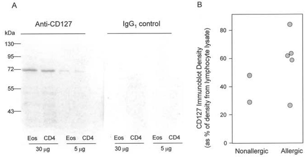
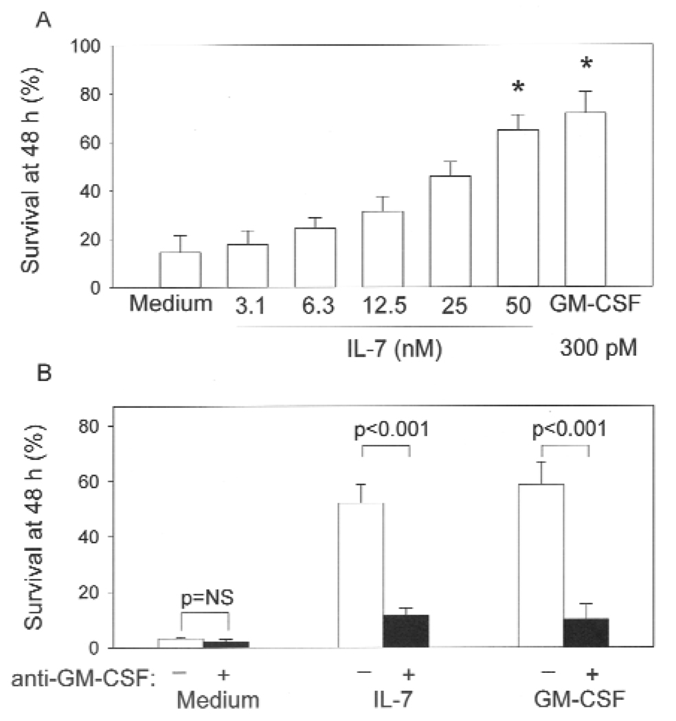
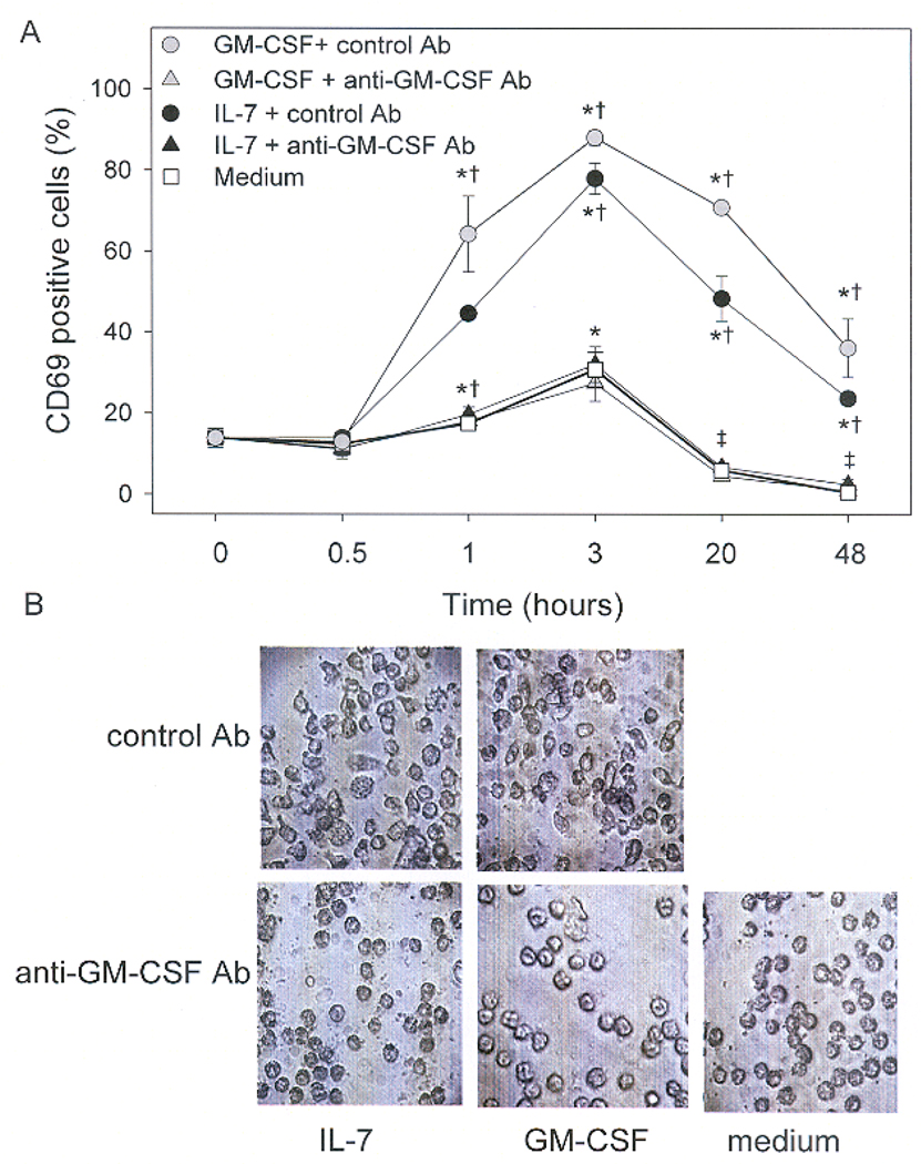
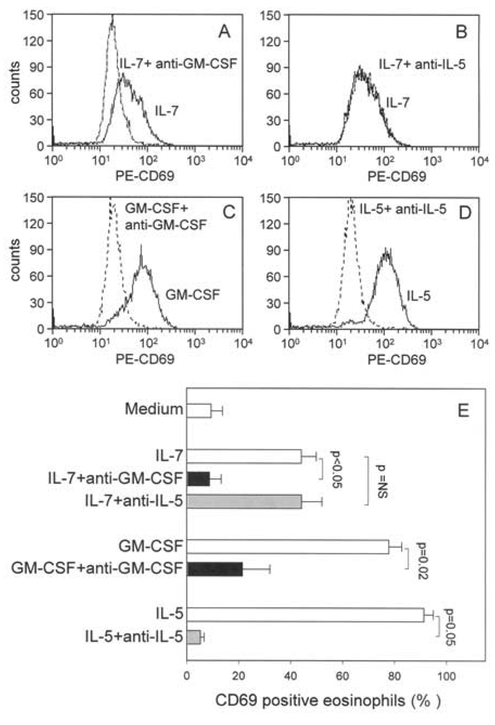
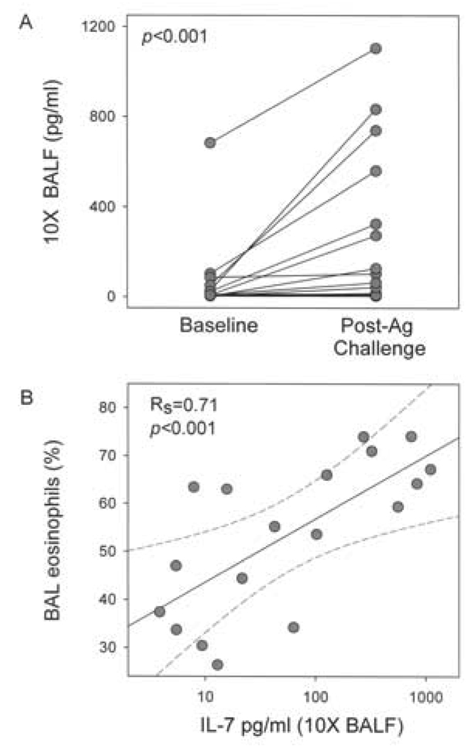
The primary function of IL-7 is to promote maturation and survival of T cells. Through microarray expression analysis, we previously observed that human blood eosinophils express mRNA for IL-7R alpha (CD127) and its common gamma chain (CD132). The purpose of this study was to determine whether eosinophils have functional IL-7 receptors and to assess the potential contribution of IL-7 to eosinophilic airway inflammation by evaluating its presence in bronchoalveolar lavage (BAL) fluid of subjects with atopic asthma before and after segmental bronchoprovocation with allergen. Immunoblot analysis revealed that CD127 is present in highly purified human blood eosinophils. Furthermore, eosinophils responded to IL-7 with phosphorylation of STAT5, up-regulation of the activation marker CD69, and prolonged survival. Neutralization of GM-CSF but not IL-5 significantly blunted these functional responses, suggesting that IL-7 mediates its effects by promoting eosinophil release of autologous GM-CSF. Notably, the suppressive effect of anti-GM-CSF on STAT5 phosphorylation occurred within 10 min of eosinophil exposure to IL-7. Thus, IL-7 likely activates eosinophil release of preformed rather than newly synthesized GM-CSF. The biological relevance of IL-7 to eosinophilia in vivo was implicated in a study of airway allergen challenge in patients with allergic asthma. IL-7 concentrations in BAL fluid increased significantly 48 h after segmental allergen challenge and were highly correlated with BAL eosinophils (r = 0.7, p < 0.001). In conclusion, the airway response to allergen is associated with the generation of IL-7, which may contribute to airway inflammation by promoting enhanced eosinophil activation and survival. Activation of eosinophils is a novel function for IL-7.
Conflict of interest statement
Disclosures
The authors have no financial conflict of interest.
Figures

Similar articles
-
CD69 surface expression on human lung eosinophils after segmental allergen provocation.Eur Respir J. 1999 Jun;13(6):1253-9. doi: 10.1183/09031936.99.13612609. Eur Respir J. 1999. PMID: 10445598
-
Effects of allergen challenge on eosinophils, eosinophil cationic protein, and granulocyte-macrophage colony-stimulating factor in mild asthma.Am J Respir Crit Care Med. 1995 Jun;151(6):1915-24. doi: 10.1164/ajrccm.151.6.7767540. Am J Respir Crit Care Med. 1995. PMID: 7767540
-
Mepolizumab Attenuates Airway Eosinophil Numbers, but Not Their Functional Phenotype, in Asthma.Am J Respir Crit Care Med. 2017 Dec 1;196(11):1385-1395. doi: 10.1164/rccm.201611-2234OC. Am J Respir Crit Care Med. 2017. PMID: 28862877 Free PMC article.
-
IL-5 and IL-5 receptor in asthma.Mem Inst Oswaldo Cruz. 1997;92 Suppl 2:75-91. doi: 10.1590/s0074-02761997000800012. Mem Inst Oswaldo Cruz. 1997. PMID: 9698919 Review.
-
Essential Mechanisms of Differential Activation of Eosinophils by IL-3 Compared to GM-CSF and IL-5.Crit Rev Immunol. 2016;36(5):429-444. doi: 10.1615/CritRevImmunol.2017020172. Crit Rev Immunol. 2016. PMID: 28605348 Free PMC article. Review.
Cited by
-
Thy1+IL-7+ lymphatic endothelial cells in iBALT provide a survival niche for memory T-helper cells in allergic airway inflammation.Proc Natl Acad Sci U S A. 2016 May 17;113(20):E2842-51. doi: 10.1073/pnas.1512600113. Epub 2016 May 2. Proc Natl Acad Sci U S A. 2016. PMID: 27140620 Free PMC article.
-
Interleukin-7 gene polymorphism rs766736182 associates with the risk of asthma in children.J Clin Lab Anal. 2019 Feb;33(2):e22675. doi: 10.1002/jcla.22675. Epub 2018 Sep 21. J Clin Lab Anal. 2019. PMID: 30239047 Free PMC article.
-
Interleukin-7 Biology and Its Effects on Immune Cells: Mediator of Generation, Differentiation, Survival, and Homeostasis.Front Immunol. 2021 Dec 2;12:747324. doi: 10.3389/fimmu.2021.747324. eCollection 2021. Front Immunol. 2021. PMID: 34925323 Free PMC article. Review.
-
Multi-trait genetic analysis of asthma and eosinophils uncovers pleiotropic loci in East Asians.Nat Commun. 2025 May 31;16(1):5081. doi: 10.1038/s41467-025-60405-0. Nat Commun. 2025. PMID: 40450010 Free PMC article.
-
Stress-induced eosinophil activation contributes to postoperative morbidity and mortality after lung resection.Sci Transl Med. 2024 Aug 21;16(761):eadl4222. doi: 10.1126/scitranslmed.adl4222. Epub 2024 Aug 21. Sci Transl Med. 2024. PMID: 39167663 Free PMC article.
References
-
- Giliani S, Mori L, de Saint BG, Le Deist F, Rodriguez-Perez C, Forino C, Mazzolari E, Dupuis S, Elhasid R, Kessel A, Galambrun C, Gil J, Fischer A, Etzioni A, Notarangelo LD. Interleukin-7 receptor alpha (IL-7Ralpha) deficiency: cellular and molecular bases. Analysis of clinical, immunological, and molecular features in 16 novel patients. Immunol. Rev. 2005;203:110–126. - PubMed
-
- Kovanen PE, Leonard WJ. Cytokines and immunodeficiency diseases: critical roles of the gamma(c)-dependent cytokines interleukins 2, 4, 7, 9, 15, and 21, and their signaling pathways. Immunol. Rev. 2004;202:67–83. - PubMed
-
- Guimond M, Fry TJ, Mackall CL. Cytokine signals in T-cell homeostasis. J Immunother. 2005;28:289–294. - PubMed
-
- Snyder KM, Mackall CL, Fry TJ. IL-7 in allogeneic transplant: clinical promise and potential pitfalls. Leuk. Lymphoma. 2006;47:1222–1228. - PubMed
-
- Nunnari G, Pomerantz RJ. IL-7 as a potential therapy for HIV-1-infected individuals. Expert. Opin. Biol. Ther. 2005;5:1421–1426. - PubMed
Publication types
MeSH terms
Substances
Grants and funding
LinkOut - more resources
Full Text Sources
Other Literature Sources
Medical
Miscellaneous
