

Increased IL-15 production is associated with higher susceptibility of memory CD4 T cells to simian immunodeficiency virus during acute infection
- PMID: 19155491
- PMCID: PMC2662754
- DOI: 10.4049/jimmunol.182.3.1439
Increased IL-15 production is associated with higher susceptibility of memory CD4 T cells to simian immunodeficiency virus during acute infection
Abstract
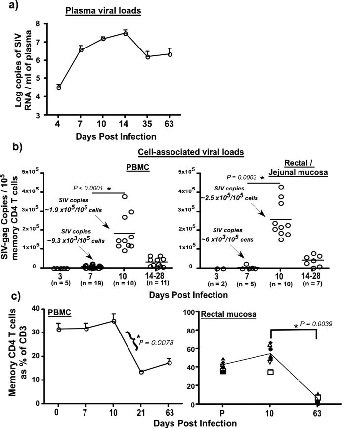
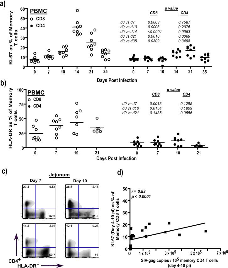
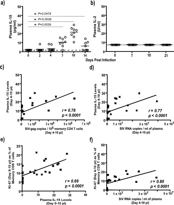
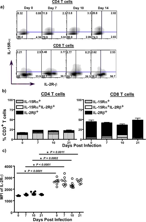
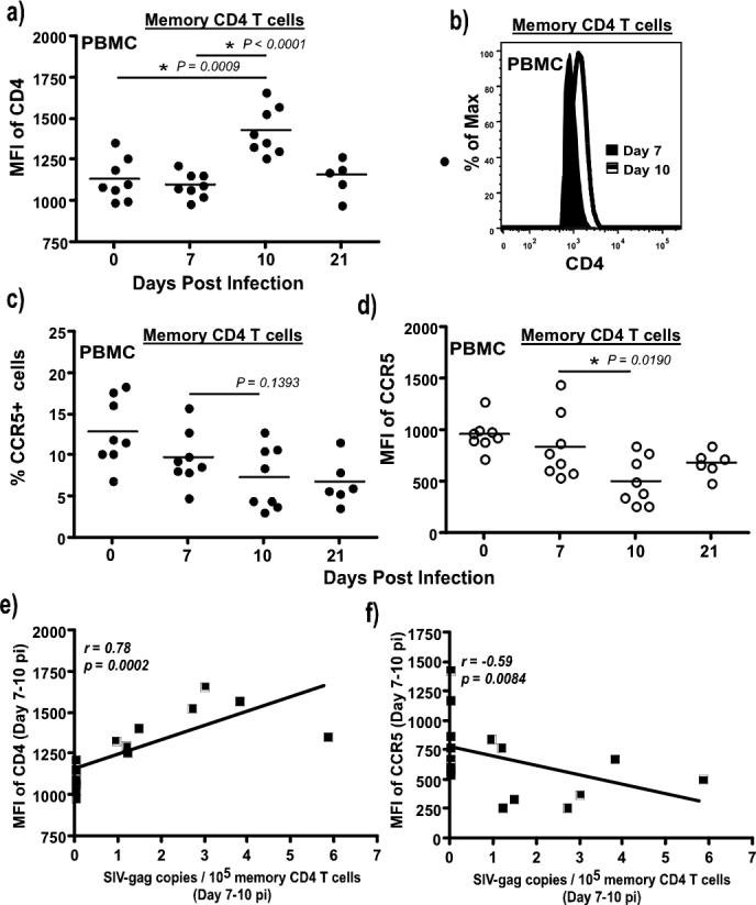
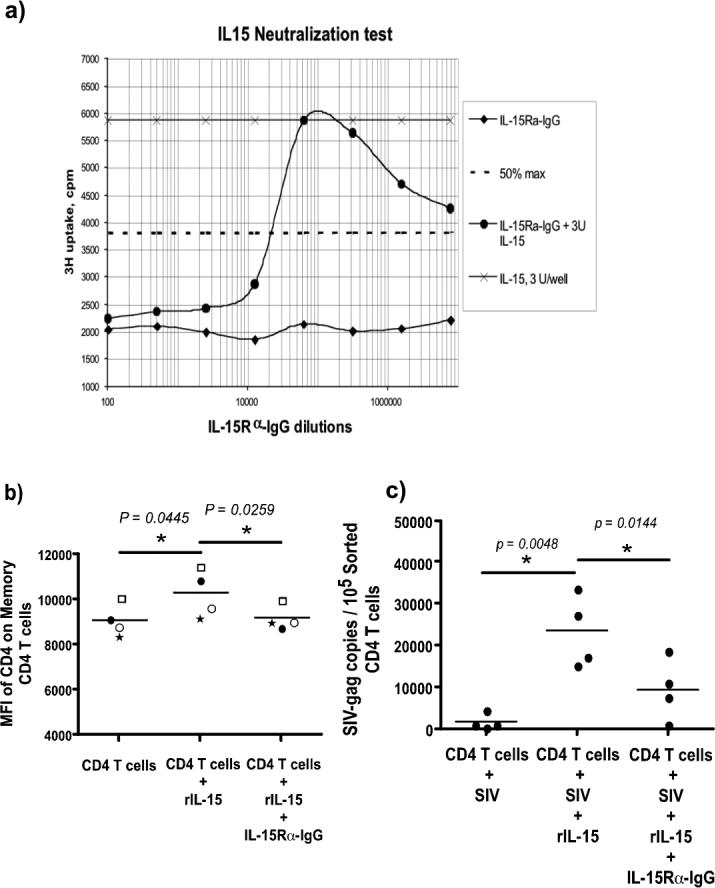
Acute SIV infection is characterized by explosive infection of memory CD4 T cells in peripheral and mucosal tissues. Interestingly, relatively few memory CD4 T cells are infected until as late as days 7-8 after challenge. However, by day 10 postinfection, most of the memory CD4 T cells are infected and carry viral DNA. The rapidity with which infection expands within 2-3 days to encompass virtually the entire memory CD4 T cell compartment suggests significant alterations in the susceptibility of memory CD4 T cells to infection during this period. The mechanism(s) underlying this increased permissiveness to infection is not known. In this study, we show that IL-15 secretion significantly correlates with the up-regulated expression of CD4 on memory CD4 T cells that is associated with increased permissiveness to SIV infection. Activation and proliferation of memory CD8, but not memory CD4 T cells, preceded the amplification of viral infection. Although memory CD4 T cells did not express normal activation markers, they displayed a significant up-regulation in the density of CD4 but not CCR5 expression between days 7 and 10 postinfection that correlated with increased plasma IL-15 levels and infection in these cells. Culture of purified CD4 T cells with IL-15 and/or SIV was associated with a significant increase in the expression of CD4 and infection of these sorted cells. Our results demonstrate that IL-15 contributes to the increased susceptibility of memory CD4 T cells to SIV during the early phase of acute SIV infection.
Figures
Similar articles
-
Differential Impact of In Vivo CD8+ T Lymphocyte Depletion in Controller versus Progressor Simian Immunodeficiency Virus-Infected Macaques.J Virol. 2015 Sep;89(17):8677-86. doi: 10.1128/JVI.00869-15. Epub 2015 Jun 10. J Virol. 2015. PMID: 26063417 Free PMC article.
-
Profound CD4+/CCR5+ T cell expansion is induced by CD8+ lymphocyte depletion but does not account for accelerated SIV pathogenesis.J Exp Med. 2009 Jul 6;206(7):1575-88. doi: 10.1084/jem.20090356. Epub 2009 Jun 22. J Exp Med. 2009. PMID: 19546246 Free PMC article.
-
Treatment with IL-7 prevents the decline of circulating CD4+ T cells during the acute phase of SIV infection in rhesus macaques.PLoS Pathog. 2012;8(4):e1002636. doi: 10.1371/journal.ppat.1002636. Epub 2012 Apr 12. PLoS Pathog. 2012. PMID: 22511868 Free PMC article.
-
CD4+ CCR5+ T-cell dynamics during simian immunodeficiency virus infection of Chinese rhesus macaques.J Virol. 2007 Dec;81(24):13865-75. doi: 10.1128/JVI.00452-07. Epub 2007 Sep 26. J Virol. 2007. PMID: 17898067 Free PMC article.
-
The Hitchhiker Guide to CD4+ T-Cell Depletion in Lentiviral Infection. A Critical Review of the Dynamics of the CD4+ T Cells in SIV and HIV Infection.Front Immunol. 2021 Jul 21;12:695674. doi: 10.3389/fimmu.2021.695674. eCollection 2021. Front Immunol. 2021. PMID: 34367156 Free PMC article.
Cited by
-
Therapeutic Potential of IL-15 and N-803 in HIV/SIV Infection.Viruses. 2021 Sep 2;13(9):1750. doi: 10.3390/v13091750. Viruses. 2021. PMID: 34578331 Free PMC article. Review.
-
Early short-term antiretroviral therapy is associated with a reduced prevalence of CD8(+)FoxP3(+) T cells in simian immunodeficiency virus-infected controller rhesus macaques.AIDS Res Hum Retroviruses. 2011 Jul;27(7):763-75. doi: 10.1089/AID.2010.0251. Epub 2011 Jan 17. AIDS Res Hum Retroviruses. 2011. PMID: 21142402 Free PMC article.
-
Functions of IL-15 in anti-viral immunity: multiplicity and variety.Cytokine. 2012 Sep;59(3):467-78. doi: 10.1016/j.cyto.2012.05.020. Epub 2012 Jun 15. Cytokine. 2012. PMID: 22704694 Free PMC article. Review.
-
IL-15 delays suppression and fails to promote immune reconstitution in virally suppressed chronically SIV-infected macaques.Blood. 2011 Sep 1;118(9):2520-9. doi: 10.1182/blood-2011-05-351155. Epub 2011 Jul 14. Blood. 2011. PMID: 21757617 Free PMC article.
-
Immunobiology of the IL-15/IL-15Rα complex as an antitumor and antiviral agent.Cytokine Growth Factor Rev. 2017 Dec;38:10-21. doi: 10.1016/j.cytogfr.2017.08.002. Epub 2017 Sep 1. Cytokine Growth Factor Rev. 2017. PMID: 28888485 Free PMC article. Review.
References
-
- Li Q, Duan L, Estes JD, Ma ZM, Rourke T, Wang Y, Reilly C, Carlis J, Miller CJ, Haase AT. Peak SIV replication in resting memory CD4+ T cells depletes gut lamina propria CD4+ T cells. Nature. 2005;434:1148–1152. - PubMed
-
- Mattapallil JJ, Douek DC, Hill B, Nishimura Y, Martin M, Roederer M. Massive infection and loss of memory CD4+ T cells in multiple tissues during acute SIV infection. Nature. 2005;434:1093–1097. - PubMed
-
- Veazey RS, DeMaria M, Chalifoux LV, Shvetz DE, Pauley DR, Knight HL, Rosenzweig M, Johnson RP, Desrosiers RC, Lackner AA. Gastrointestinal tract as a major site of CD4+ T cell depletion and viral replication in SIV infection. Science. 1998;280:427–431. - PubMed
-
- Cumont MC, Diop O, Vaslin B, Elbim C, Viollet L, Monceaux V, Lay S, Silvestri G, Le Grand R, Muller-Trutwin M, Hurtrel B, Estaquier J. Early divergence in lymphoid tissue apoptosis between pathogenic and nonpathogenic simian immunodeficiency virus infections of nonhuman primates. J Virol. 2008;82:1175–1184. - PMC - PubMed
Publication types
MeSH terms
Substances
Grants and funding
LinkOut - more resources
Full Text Sources
Research Materials
