Comparative Study
. 2009 Feb 1;182(3):1481-9.
doi: 10.4049/jimmunol.182.3.1481.

OX40-enhanced tumor rejection and effector T cell differentiation decreases with age

Affiliations
Comparative Study

OX40-enhanced tumor rejection and effector T cell differentiation decreases with age

Carl E Ruby et al. J Immunol. .

Abstract

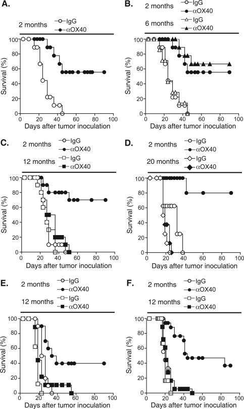
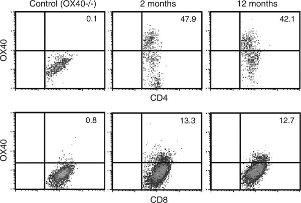
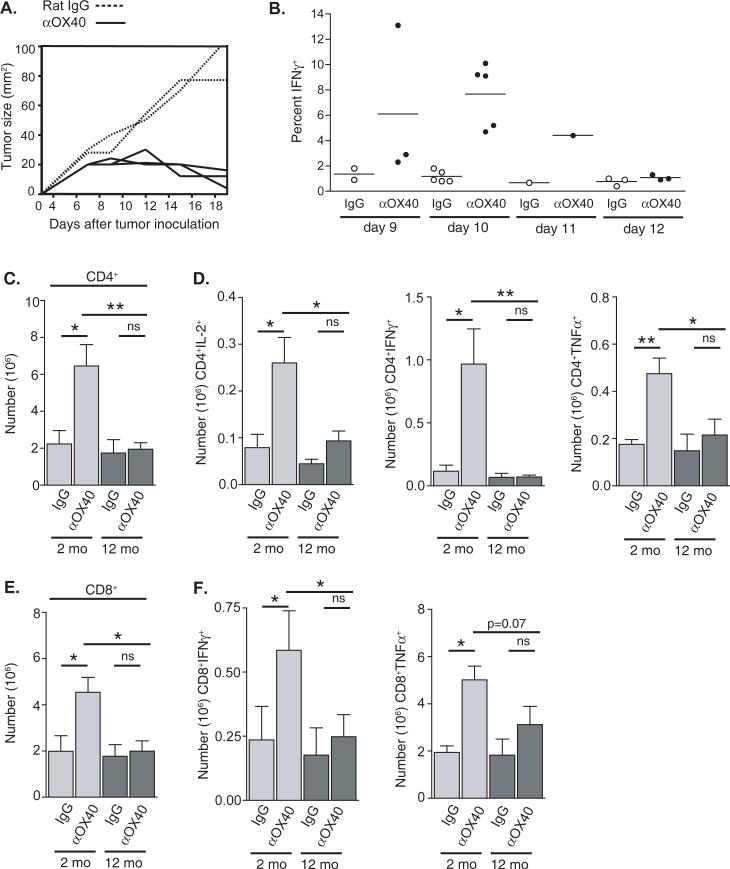
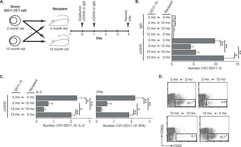
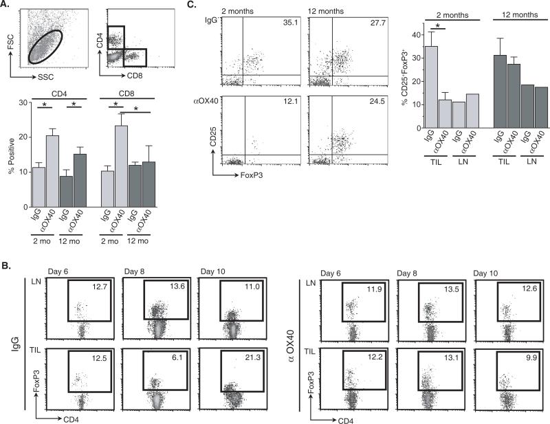
OX40 agonists have potent immunotherapeutic effects against a variety of murine tumors, yet it is unclear the role that age-related immune senescence plays on their efficacy. We found that middle-aged and elderly tumor-bearing mice (12 and 20 mo old, respectively) treated with anti-OX40 were less responsive compared with young mice 6 mo or less of age. Decreased tumor-free survival was observed in both male and female mice, and was not due to changes in the surface expression of OX40 on T cells in older animals. Enumeration of cytokine-producing effector T cells in tumor-bearing mice revealed a significant decline in these cells in the older mice treated with anti-OX40 compared with their younger counterparts. The decrease of this critical T cell population in middle-aged mice was not a result of inherent T cell deficiencies, but was revealed to be T cell extrinsic. Finally, combining IL-12, an innate cytokine, with anti-OX40 boosted levels of differentiated effector T cells in the older anti-OX40-treated mice and partially restored the defective antitumor responses in the middle-aged mice. Our data show that the anti-OX40-enhancement of tumor immunity and effector T cell numbers is decreased in middle-aged mice and was partially reversed by coadministration of the proinflammatory cytokine IL-12.


Figures

FIGURE 1
FIGURE 1
The age of the tumor-bearing host affects MCA205 tumor free survival following systemic OX40 stimulation. A-D, Female C57BL/6 mice, ages two, six, 12 and 20 months, were injected s.c. with 4-5 × 10⁵ MCA205 tumor cells. Three and seven days later 250 μg anti-OX40 or rat IgG were injected i.p. E, Male C57BL/6 mice, ages two and 12 month, were injected with 4 × 10⁵ MCA205 tumor cells. Three and seven days later 250 μg anti-OX40 or rat IgG were injected i.p. F. Female BALB/C mice, ages two and 12 month, were injected with 1 × 10⁵ CT26 tumor cells. Three and seven days later 250 μg anti-OX40 or rat IgG were injected i.p. Mice were then monitored for tumor growth.
FIGURE 2
FIGURE 2
Surface expression of OX40 on T cells infiltrating the tumors of two- and 12- month old mice. C57BL/6 mice ages two and 12 months and OX40−/− mice two months of age were challenge with 1 × 10⁶ MCA205 tumor cells. Tumors were surgically resected 12 days following challenge and the lymphocytes infiltrating the tumors were harvested. CD4 and CD8 T cells were then analyzed by FACS for the expression of OX40. Gates for OX40 staining were based on data from OX40−/− mice. The data are representative of two independent experiments.
FIGURE 3
FIGURE 3
Age-related differences in the numbers of CD4 and CD8 T cells in the tumor draining lymph nodes of anti-OX40 and rat IgG treated tumor-bearing mice ten days after MCA205 tumor challenge. Two- and 12-month old mice were challenged with 1 × 10⁶ MCA205 s.c. and then treated i.p. with 250 μg anti-OX40 or rat IgG. A. After tumor challenge two-month old mice were measured over time for tumor growth. Each curve represents an individual animal. B. Tumor-draining lymph nodes from two-month old tumor-bearing mice were harvested on various days after tumor challenge. Cells from the tumor draining lymph nodes of anti-OX40-treated mice (closed circles) or rat IgG-treated mice (open circles) were restimulated with anti-CD3 (1 μg/mL) for six hours and CD8 T cells were analyzed for intracellular IFNγ production. Each circle represents one animal. C-F. Tumor draining lymph nodes were harvested ten days after tumor challenge and CD4 C. and CD8 E. T cells enumerated. T cells were then restimulated with anti-CD3 for six hours and analyzed for cytokine production via intracellular cytokine staining. D. Number of CD4 T cell producing IL-2, IFNγ, and TNFα F. Number of CD8 T cell producing of IFNγ and TNFα.
FIGURE 4
FIGURE 4
The age of the recipient and not the adoptively transferred transgenic TCR CD4 T cell determines the extent of an OX40-enhanced antigen-specific T cell immune response. A. Schematic of the crisscross adoptive transfer of two- or 12-month old DO11.10 CD4 T cells into two- or 12-month old BALB/c recipients and immunization (500 μg ovalbumin, 50 μg anti-OX40 or rat IgG s.c.) schedule. B. Enumeration of the antigen-specific DO11.10 T cells and C. cytokine (IL-2 and IFNγ)-producing DO11.10 T cells from the draining LNs four days after challenge, D. Representative FACs plots of DO11.10 T cells (CD4+KJ-126+) with an activation phenotype (CD62LlowCD25+) from the antigen-draining LNs four days after challenge. Statistical representation is measured between age-matched adoptively transferred DO11.10 and described in the Materials and Methods.
FIGURE 5
FIGURE 5
Co-administration of exogenous IL-12 and anti-OX40 increases the acquisition of an effector CD4 T cell phenotype and accumulation of differentiated effector CD4 T cells. Two- or 12-month old DO11.10 CD4 T cells were adoptively transferred into age-matched BALB/c recipients, and immunized s.c. with ovabumin (500 μg) and anti-OX40 (50 μg) or IL-12 (100 ng). A second dose of anti-OX40 was administered the next day and IL-12 was injected s.c. daily for the next three days. Antigen-draining LNs were harvested four days after challenge and analyzed. A. Representative FACs plots showing the frequency of DO11.10 T cells (CD4+KJ-126+) producing IFNγ. B. Enumeration of IFNγ-producing DO11.10 T cells.
FIGURE 6
FIGURE 6
Age-related increase of CD4+CD25+FoxP3+ T cells within the tumors of 12-month old anti-OX40 treated animals. Two- and 12-month old mice were challenged with 1 ×10⁶ MCA205 s.c. and then treated i.p. with 250 μg anti-OX40 or rat IgG, as previously described. Tumors were resected 12 days after tumor challenge and TILs harvested (Materials and Methods). A. The frequency of CD4 and CD8 T cells from processed tumors was determined using the gate described. B. Time course of CD4+FoxP3+ T cell accumulation in the distal LNs and TILs of IgG- and anti-OX40-treated MCA205 tumor-bearing mice (two-months old). C. The frequency of CD4+ T cells expressing a CD25+FoxP3+ phenotype from TILs and pooled distal LNs from the same mice were assessed by FACs 12 days after tumor challenge.
FIGURE 7
FIGURE 7
Systemic administration of IL-12 to anti-OX40-treated 12-month old mice increased MCA205 tumor-free survival and reduces the accumulation of CD4+CD25+FoxP3+ in the tumors mice treated anti-OX40 and IL-12. Female C57BL/6 mice, two- and 12-months of age, were injected s.c. with 4-5 × 10⁵ MCA205 tumor cells. A. IL-12 (100 ng) or PBS (control) was injected i.p. daily four-to-nine days after tumor challenge. B. Three and seven days after tumor challenge 250 μg anti-OX40 was injected i.p. IL-12 (100 ng) or PBS (control) was injected i.p. daily four-to-nine days after tumor challenge. Mice were then monitored for tumor growth. C. Three and seven days after tumor challenge 250 μg anti-OX40 was injected i.p. IL-12 (100 ng) or PBS (control) was injected i.p. daily four-to-nine days after tumor challenge. Tumors were resected 12 days after tumor challenge and TILs harvested (Materials and Methods). The frequency of CD4+ T cells expressing CD25+ and FoxP3+ from the tumors and pooled contra-lateral lymph nodes from the same mice were assessed by FACs.

References

    1. Ershler WB. The influence of an aging immune system on cancer incidence and progression. J Gerontol. 1993;48:B3–7. - PubMed
    1. Yung RL. Changes in immune function with age. Rheum Dis Clin North Am. 2000;26:455–473. - PubMed
    1. Linton PJ, Haynes L, Tsui L, Zhang X, Swain S. From naive to effector--alterations with aging. Immunol Rev. 1997;160:9–18. - PubMed
    1. Effros RB, Walford RL. The immune response of aged mice to influenza: diminished T-cell proliferation, interleukin 2 production and cytotoxicity. Cell Immunol. 1983;81:298–305. - PubMed
    1. Melero I, Shuford WW, Newby SA, Aruffo A, Ledbetter JA, Hellstrom KE, Mittler RS, Chen L. Monoclonal antibodies against the 4-1BB T-cell activation molecule eradicate established tumors. Nat Med. 1997;3:682–685. - PubMed

Publication types

MeSH terms