. 2009 Feb 1;182(3):1641-7.
doi: 10.4049/jimmunol.182.3.1641.

TSLP conditions the lung immune environment for the generation of pathogenic innate and antigen-specific adaptive immune responses

Affiliations

TSLP conditions the lung immune environment for the generation of pathogenic innate and antigen-specific adaptive immune responses

Mark B Headley et al. J Immunol. .

Abstract

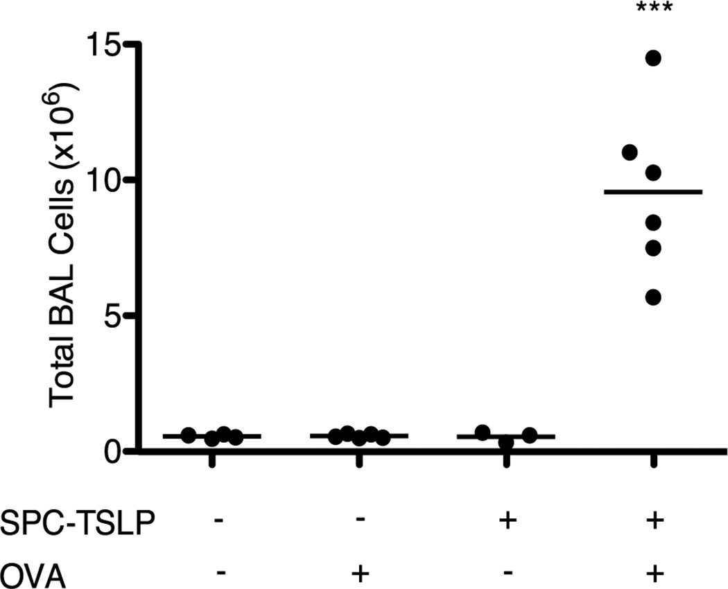
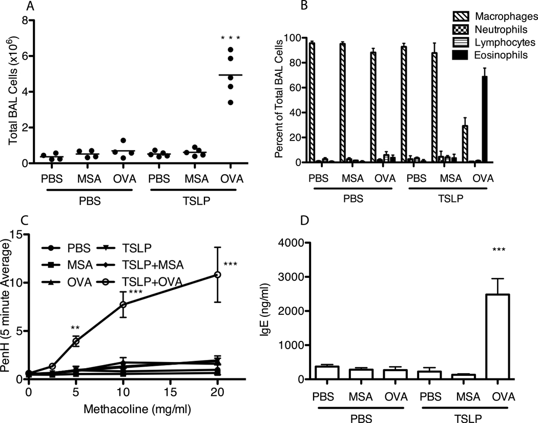
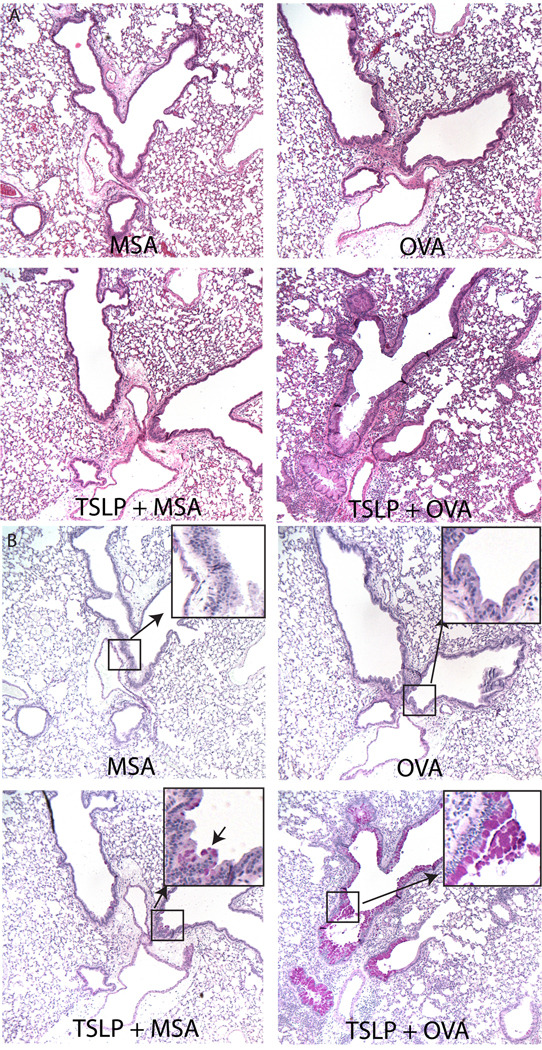
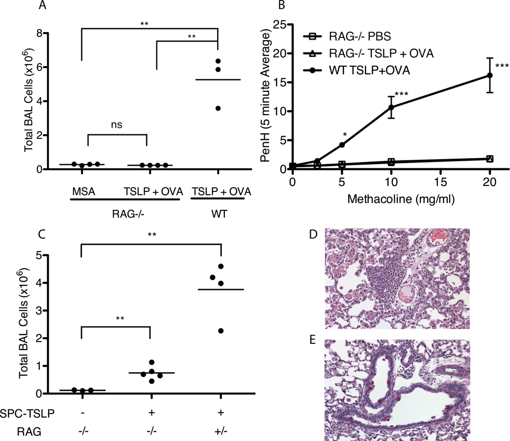
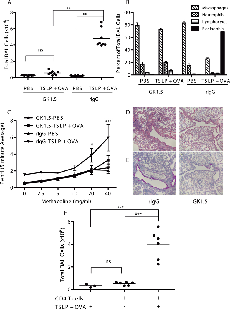
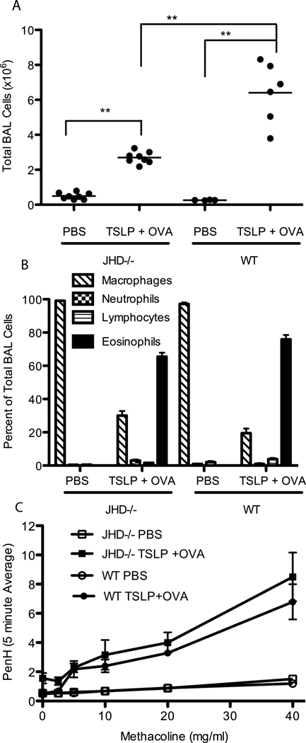
Thymic stromal lymphopoietin (TSLP) is crucial for the development of atopic diseases in humans and mice. Mice that express a lung-specific TSLP transgene (surfactant protein C promoter (SPC)-TSLP) develop a spontaneous and progressive asthma-like disease, suggesting that TSLP expression alone was sufficient for disease development. In this study, we show that, in fact, TSLP alone only causes a weak innate response that is insufficient for development of full airway inflammatory disease. Complete disease development requires both TSLP and antigenic stimulation. These data suggest that the spontaneous lung inflammation observed in SPC-TSLP mice reflects a TSLP-driven predisposition toward the development of aberrant responses against innocuous environmental Ags. This provides evidence that TSLP may act directly to induce susceptibility to the inappropriate allergic responses that characterize atopy and asthma. We additionally show that disease development requires CD4 T cells but not B cells. Further, we reveal a TSLP-driven innate response involving mucus overproduction and goblet cell metaplasia. Taken together, these data suggest a multifaceted model of TSLP-mediated airway inflammation, with an initial activation of resident innate immune cells, followed by activation of the adaptive immune system and full disease development. This study provides new insight into the unique features of the asthma pathology contributed by the innate and adaptive immune responses in response to TSLP stimulation.


Figures

Figure 1
Figure 1
SPC-TSLP mice develop airway inflammation with earlier onset in response to OVA challenge relative to non-OVA treated SPC-TSLP mice. Total BAL cell counts from SPC-TSLP mice treated i.n. with OVA or PBS every other day for 14 days beginning at 4–6 weeks of age. Mice were analyzed between 6 and 8 weeks of age. Statistic are one-way ANOVA with Tukey post-test, ***=p≤0.001 when compared to all other groups.
Figure 2
Figure 2
Acute intranasal TSLP induces airway inflammation only in the presence of foreign antigen. WT Balb/c mice were treated i.n. every other day for 14 days with PBS or 500ng TSLP with or without 25 mg MSA or OVA. (A) Total number of cells isolated from BAL. (B) Cells isolated from Bal were cytospun onto glass specimen slides and stained with modified wright-giemsa stain to different between macrophages, neutrophils, lymphocytes, and eosinophils. Values shown are mean ± SEM with n≥4 for each group. (C) PenH as a measure of AHR on Day 14 in response to increasing concentrations of methacoline. (D) Serum from whole blood collected from animals at sacrifice was assayed for total IgE by ELISA. Values shown represent mean ± SEM with n=3 per group, *** indicates that labeled group is significant against all other groups with p≤0.001.
Figure 3
Figure 3
Disease phenotype in the lung of intranasal TSLP-treated mice varies depending on the presence of antigen. Paraffin sections of lungs excised from mice treated with MSA, OVA, TSLP+MSA, or TSLP+OVA every other day for 14 days were stained with (A) H&E or (B) PAS. Pink staining cells under PAS represent mucous producing goblet cells. Magnified inset highlights rare pink-staining cells in airway of TSLP+MSA treated animals not present in MSA or OVA alone treated animals.
Figure 4
Figure 4
TSLP-mediated airway inflammatory disease requires an intact lymphocyte compartment for full disease development. (A) Total number of cells isolated from BAL taken from Rag2−/− or WT Balb/c mice that were treated every other day for 14 days with MSA or TSLP+OVA. Statistics are from one-way ANOVA with a Tukey post-test, ** = significant with p≤0.01 (B) AHR, represented as the PenH value measured over increasing doses of methacoline. Points are mean±SEM. Statistics are two-way repeated measures ANOVA with Bon Ferroni post-tests, *** = p≤0.001 and *=p≤0.05 with n≥3 for each group. (C) Total BAL Cell counts from SPC-TSLP X Rag−/− or SPC-TSLP X Rag +/− (heterozygous control) mice sacrificed between 4 – 6 months of age. Statistics are from two-tailed student T-tests comparing groups, *** = p≤0.001 and **=p≤0.01 (D&E) Representative paraffin sections of lung excised from SPC-TSLP X Rag−/− mice and stained with H&E (D) or PAS (E) showing presence of rare pockets of lung infiltrating cells and pink staining goblet cells.
Figure 5
Figure 5
CD4 T cells are required for TSLP-mediated airway inflammation. (A) Total BAL cell counts from WT Balb/c mice treated i.p. with rIgG or anti-CD4 antibody (GK1.5), to acutely deplete CD4 T cells, over the time course of i.n. treatment with PBS or TSLP+OVA.(B)Differential cell counts performed on wright-giemsa stained BAL cells. (C) Assessment of AHR, points are mean±SEM. Statistics are two-way repeated measures ANOVA with Bon Ferroni post-tests, *** = p≤0.001 and *=p≤0.05 with n=4 for each group. (D,E) Paraffin sections from lungs excised from TSLP+OVA treated mice that received either rIgG or GK1.5 and stained with H&E (D) or PAS (E). (F) Total BAL cell counts from rag2−/− mice that were adoptively transferred with 5 × 10⁶ CD4 T Cells one day prior to initiating PBS or TSLP+OVA treatment every other day for 14 days. (A&F) Statistics are one-way ANOVA with Tukey post-test ***=p≤0.001 and **=p≤0.01.
Figure 6
Figure 6
B cells aren’t required for TSLP induced airway inflammation but serve to amplify the response. JHD−/− (B cell deficient) or WT Balb/c mice were treated i.n. with PBS or TSLP+OVA every other day for 14 days. (A) Total BAL cell counts, (B) differential cell counts performed on BAL cells (C) assessment of AHR as measured by PenH. (A) Statistics are one-way ANOVA with Tukey post-test ***=p≤0.001 and **=p≤0.01. (C) Points are mean±SEM, both JHD−/− and WT treated with TSLP+OVA are significantly different (p≤0.001) than either the JHD−/− or WT PBS treated groups but not from each other by two-way repeated measures ANOVA with Bon Ferroni post tests.

Similar articles

Cited by

References

    1. Cohn L, Elias JA, Chupp GL. Asthma: mechanisms of disease persistence and progression. Annual review of immunology. 2004;22:789–815. - PubMed
    1. Elias JA, Zhu Z, Chupp G, Homer RJ. Airway remodeling in asthma. J Clin Invest. 1999;104:1001–1006. - PMC - PubMed
    1. Burrows B, Martinez FD, Halonen M, Barbee RA, Cline MG. Association of asthma with serum IgE levels and skin-test reactivity to allergens. N Engl J Med. 1989;320:271–277. - PubMed
    1. Brightling CE, Bradding P. The re-emergence of the mast cell as a pivotal cell in asthma pathogenesis. Current allergy and asthma reports. 2005;5:130–135. - PubMed
    1. Hammad H, Lambrecht BN. Dendritic cells and epithelial cells: linking innate and adaptive immunity in asthma. Nature reviews. 2008;8:193–204. - PubMed

Publication types

MeSH terms