. 2009;4(1):e4235.
doi: 10.1371/journal.pone.0004235. Epub 2009 Jan 21.

Neuroblastoma cell lines contain pluripotent tumor initiating cells that are susceptible to a targeted oncolytic virus

Affiliations

Neuroblastoma cell lines contain pluripotent tumor initiating cells that are susceptible to a targeted oncolytic virus

Yonatan Y Mahller et al. PLoS One. 2009.

Abstract

Background: Although disease remission can frequently be achieved for patients with neuroblastoma, relapse is common. The cancer stem cell theory suggests that rare tumorigenic cells, resistant to conventional therapy, are responsible for relapse. If true for neuroblastoma, improved cure rates may only be achieved via identification and therapeutic targeting of the neuroblastoma tumor initiating cell. Based on cues from normal stem cells, evidence for tumor populating progenitor cells has been found in a variety of cancers.

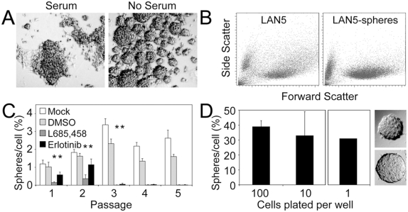
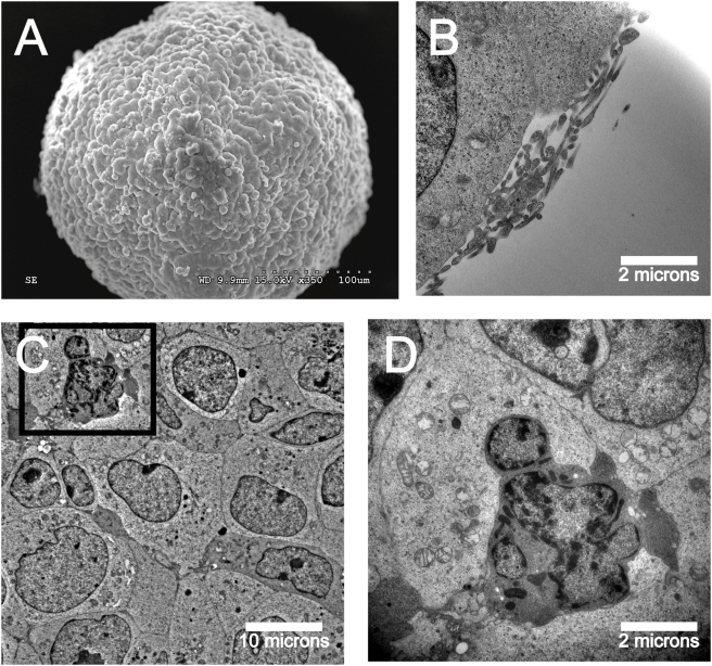
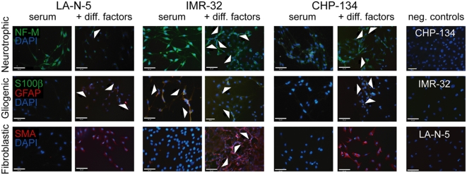
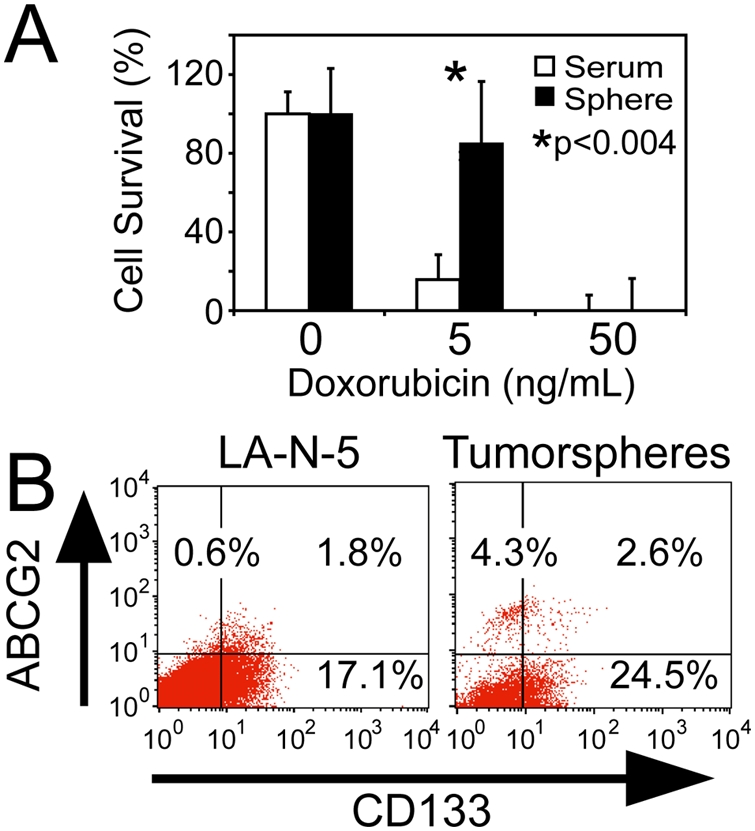
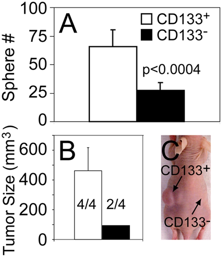
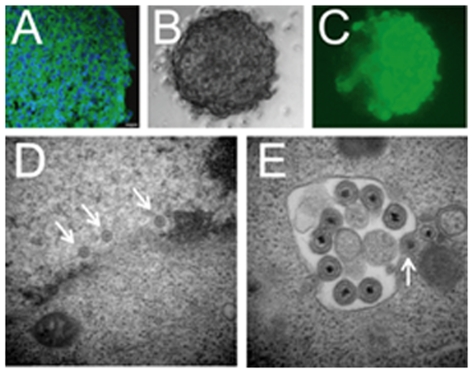
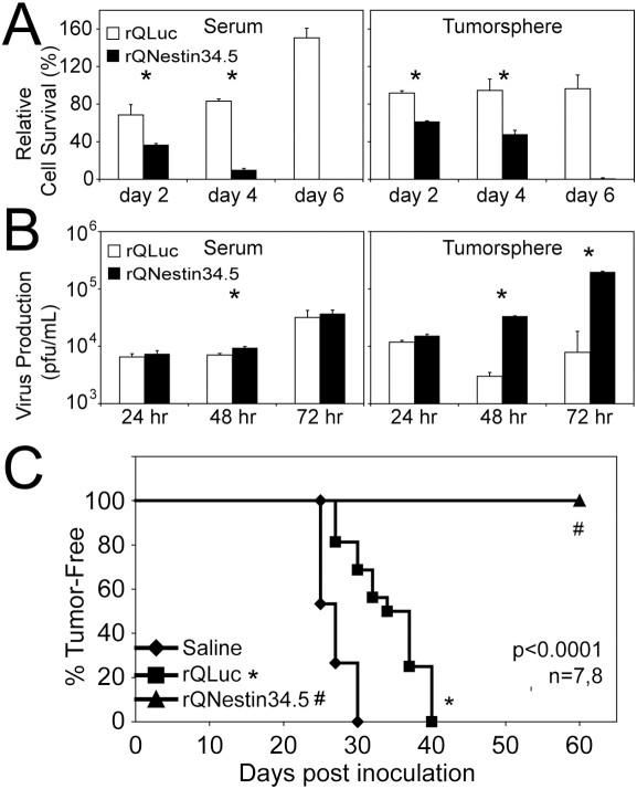
Methodology/principal findings: Four of eight human neuroblastoma cell lines formed tumorspheres in neural stem cell media, and all contained some cells that expressed neurogenic stem cell markers including CD133, ABCG2, and nestin. Three lines tested could be induced into multi-lineage differentiation. LA-N-5 spheres were further studied and showed a verapamil-sensitive side population, relative resistance to doxorubicin, and CD133+ cells showed increased sphere formation and tumorigenicity. Oncolytic viruses, engineered to be clinically safe by genetic mutation, are emerging as next generation anticancer therapeutics. Because oncolytic viruses circumvent typical drug-resistance mechanisms, they may represent an effective therapy for chemotherapy-resistant tumor initiating cells. A Nestin-targeted oncolytic herpes simplex virus efficiently replicated within and killed neuroblastoma tumor initiating cells preventing their ability to form tumors in athymic nude mice.

Conclusions/significance: These results suggest that human neuroblastoma contains tumor initiating cells that may be effectively targeted by an oncolytic virus.


Conflict of interest statement

Competing Interests: The authors have declared that no competing interests exist.

Figures

Figure 1
Figure 1. Neuroblastoma cells form tumorspheres in serum-free media.
(A) LA-N-5 neuroblastoma cells plated in serum supplemented media show adherent growth while those plated in serum-free media (with EGF and bFGF) formed non-adherent tumorspheres. (B) Dissociated bulk and tumorsphere cells were subjected to FACS analysis. Tumorsphere cells showed increased uniformity in complexity (low side scatter) compared to bulk cultured cells. (C) Tumorsphere formation over serial passage (plating at supra-clonal density) in neurosphere media alone or supplemented with a γ-secretase inhibitor (L685,458, 1 mM) or an EGFR inhibitor (erlotinib, 10 µM). The inhibitors were dissolved in DMSO, which was used as a negative control. (D) Plating of dissociated LA-N-5 tumorsphere cells at 100 or 10 cells per well showed consistent sphere-forming efficiency regardless of plating density; 30% of wells plated with a single cell contained a single sphere. Pictures show examples of clonally derived spheres.
Figure 2
Figure 2. Ultrastructural characterization of neuroblastoma tumorspheres shows similarities to normal neurospheres.
(A) Scanning EM of neuroblastoma tumorsphere shows a smooth and uniform surface. (B) Microvilli-like structures shown on the tumorsphere surface by transmission EM in cross-section. (C) Transmission EM shows tight packing of tumor cells in the tumorsphere core. (D) Apoptotic tumorsphere cell shows nuclear blebbing, chromatin fragmentation and mitochondrial swelling.
Figure 3
Figure 3. Clonally-derived neuroblastoma tumorsphere cells show multi-lineage differentiation.
Clonally-derived tumorspheres from three different neuroblastoma cell lines were dissociated and cells were plated on poly-lysine and laminin coated chamber slides. Culture conditions were media containing serum alone or serum with neurotrophic factors (top row), gliogenic factors (middle row) or fibroblastic factors (bottom row) (except negative controls, which were serum alone without factors). Slides were stained with neurofilament-M (NF-M, green, top row), GFAP (red, middle row) S100β (green, middle row), or smooth muscle actin (red, bottom row); each were also co-stained with DAPI (blue). Negative control cultures were incubated without primary antibody and with secondary anti-mouse TRITC or anti-rabbit FITC. Arrows in the top and bottom rows indicate spindle-like cell extensions consistent with either neuronal or fibroblastic differentiation, while arrows in the middle row indicate positively stained cells. On close inspection of the GFAP/S100β stains, IMR-32 cells under serum conditions and CHP-134 cells supplemented with factors show co-staining with a mixture of green/red signals. Scale bars = 65 microns.
Figure 4
Figure 4. Neuroblastoma tumorsphere-derived cells are doxorubicin resistant and express ABCG2 and CD133.
(A) LA-N-5 bulk and tumorsphere-derived cells were assessed for doxorubicin sensitivity by MTT assay at day 7. (B) Analysis for ABCG2 and CD133 expression in neuroblastoma cells grown as bulk culture or as tumorspheres.
Figure 5
Figure 5. Side population analysis reveals asymmetric cell division of neuroblastoma cells.
(A) Side population analysis using Hoechst 33342, +/−verapamil, of bulk LA-N-5 cells (LAN5) and LA-N-5 cells grown in doxorubicin (LAN5-doxR). (B) Analysis of ABCG2 and CD133 expression in LAN5 and LAN5-doxR cells. (C) Sphere-forming efficiency of sorted SP and non-SP (NSP) cells from LAN5 and LAN5-doxR cultures. (D) Sorted SP and NSP cells, from cultures of LAN5 and LAN5-doxR, were plated in serum containing media for 2 weeks and re-evaluated by side population, +/−verapamil. SP-derived cultures show regeneration of SP and NSP cells, while NSP-derived cultures showed only regeneration of NSP cells, not SP cells.
Figure 6
Figure 6. CD133 expressing neuroblastoma cells show increased sphere and tumor formation.
Cells sorted for CD133 expression were assayed for (A) tumorsphere formation and (B) tumorigenicity following flank implantation of 5,000 cells in immunodeficient mice. Quantification of tumor volume and frequency were performed at 21 days post-inoculation. Numerators indicate number of flanks with tumors and denominators total number of flanks injected. (C) Image of mouse injected with CD133 expressing cells on the left flank and CD133 null cells on the right flank.
Figure 7
Figure 7. Neuroblastoma tumorspheres express nestin and are efficiently infected by oHSV.
(A) Immunohistochemistry on tumorsphere cryosection for nestin (green) and DAPI co-stain (blue). Scale bar = 10 microns. (B, C) A neuroblastoma tumorsphere infected with rQNestin34.5 was imaged at 48 hours post-infection by (B) phase-contrast and (C) fluorescent microscopy for GFP. (D, E) rQNestin34.5-infected neuroblastoma tumorsphere at 48 hours post-infection evaluated by transmission electron microscopy showing viral nucleocapsids in the nucleus (arrows), and mature HSV particles in the cytoplasm in the process of acquiring their envelopes (arrows).
Figure 8
Figure 8. Neuroblastoma tumorsphere and tumor initiating cells are sensitive to a nestin-targeted oncolytic HSV.
(A) Cytotoxicity assay on LA-N-5 bulk culture and tumorsphere-derived cells infected with rQNestin34.5 or the control virus, rQLuc. (B) Virus replication assay on LA-N-5 bulk culture and tumorsphere cells infected with rQNestin34.5 or rQLuc. (C) LA-N-5 cells were infected with oHSV ex vivo, followed by implantation into immunodeficient mice. Animals were followed over time for tumor development.

References

    1. Olshan A, Bunin G. Epidemiology of Neuroblastoma. In: Brodeur G, Sawada T, Tsuchida Y, Voute P, editors. Neuroblastoma. Amsterdam: Elsevier Science; 2000. pp. 33–39.
    1. Brodeur G, Maris J. Neuroblastoma. In: Pizzo P, Poplack D, editors. Principles and Practice of Pediatric Oncology. Philadelphia: Lippincott Williams & Wilkins; 2001. pp. 896–937.
    1. George RE, Li S, Medeiros-Nancarrow C, Neuberg D, Marcus K, et al. High-risk neuroblastoma treated with tandem autologous peripheral-blood stem cell-supported transplantation: long-term survival update. J Clin Oncol. 2006;24:2891–2896. - PubMed
    1. Matthay K, Villablanca J, Seeger R, Stram D, Harris R, et al. Treatment of high-risk neuroblastoma with intensive chemotherapy, radiotherapy, autologous bone marrow transplantation, and 13-cis-retinoic acid. Children's Cancer Group. New England Journal of Medicine. 1999;341:1165–1173. - PubMed
    1. Luo L, Han ZC. Leukemia stem cells. Int J Hematol. 2006;84:123–127. - PubMed

Publication types

MeSH terms