

Differentiation of rabbit bone marrow mesenchymal stem cells into corneal epithelial cells in vivo and ex vivo
- PMID: 19156227
- PMCID: PMC2627808
Differentiation of rabbit bone marrow mesenchymal stem cells into corneal epithelial cells in vivo and ex vivo
Abstract
Purpose: To examine whether bone marrow mesenchymal stem cells (MSCs) could be differentiated into corneal epithelial cells in vivo and ex vivo.
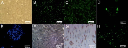
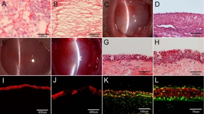
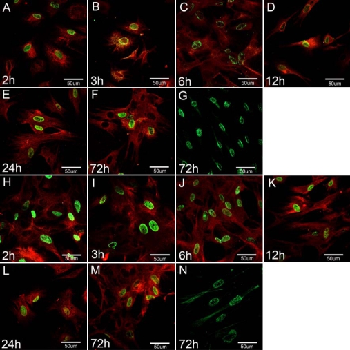
Methods: In vivo, BrdU labeled rabbit MSCs (Rb-MSCs) were suspended in the fibrin gels and transplanted onto the surface of the damaged rabbit corneas. Histology and molecular phenotype were studied on postoperative day 28. In vitro, labeled Rb-MSCs were cultured for three days in two different systems: (1) Group A: Rb-MSCs were co-cultured with rabbit limbal stem cells (Rb-LSCs) by the Transwell culture system. A suspension of Rb-LSCs was added to the upper membrane surface, and the inserts were positioned in the culture wells, which were incubated with Rb-MSCs; (2) Group B: Supernatant medium that had first been used to culture Rb-LSCs and then filtered with a 0.45 mum filter was used to culture Rb-MSCs. For both groups, immunofluorescence and flow cytometric analysis were used to examine the expression of cytokeratin 3 (CK3) in differentiated Rb-MSCs.
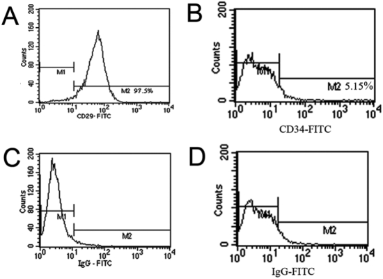
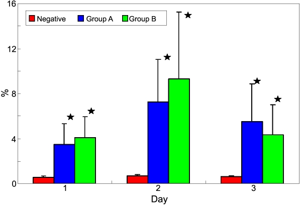
Results: In vivo, the data showed that following transplantation of Rb-MSCs, the rabbit's damaged corneal surface was successfully reconstructed and that some Rb-MSCs participated in the healing of the injured corneal epithelium and expressed CK3. In vitro, the data showed that Rb-MSCs rapidly differentiated into cells with a morphological and molecular phenotype of corneal epithelial-like cells. For both groups, the differentiated Rb-MSCs were positive for corneal epithelial-specific marker CK3. In Group A, flow cytometry analysis showed that at day one, only 3.46+/-1.9% of cells expressed CK3. This increased to 7.24+/-3.80% at day two and decreased slightly (5.50+/-3.33%) at day three. The proportion of CK3 in Group B was 4.09+/-1.84% at day one, rising to 9.31+/-5.92% after 24 h, but falling (4.37+/-2.61%) at day three. The mean differences are significant between each group and the negative control, but was not significant between Group A and Group B.
Conclusions: MSCs could differentiate into corneal epithelial-like cells in vivo and ex vivo.
Figures
References
-
- Tuli R, Seghatoleslami MR, Tuli S, Wang ML, Hozack WJ, Manner PA, Danielson KG, Tuan RS. A simple, high-yield method for obtaining multipotential mesenchymal progenitor cells from trabecular bone. Mol Biotechnol. 2003;23:37–49. - PubMed
-
- Jiang Y, Vaessen B, Lenvik T, Blackstad M, Reyes M, Verfaillie CM. Multipotent progenitor cells can be isolated from postnatal murine bone marrow, muscle, and brain. Exp Hematol. 2002;30:896–904. - PubMed
-
- Prockop DJ. Marrow stromal cells as stem cells for nonhematopoietic tissues. Science. 1997;276:71–4. - PubMed
-
- Pittenger MF, Mackay AM, Beck SC, Jaiswal RK, Douglas R, Mosca JD. Multilinenge Potential of Adult Human Mesenchymal Stem Cells. Science. 1999;284:143–7. - PubMed
Publication types
MeSH terms
Substances
LinkOut - more resources
Full Text Sources
Other Literature Sources
Medical