. 2009 Sep;39(1-2):49-58.
doi: 10.1007/s12031-008-9174-3. Epub 2009 Jan 21.

The Cdk5/p35 kinases modulate leptin-induced STAT3 signaling

Affiliations

The Cdk5/p35 kinases modulate leptin-induced STAT3 signaling

Yi He et al. J Mol Neurosci. 2009 Sep.

Abstract

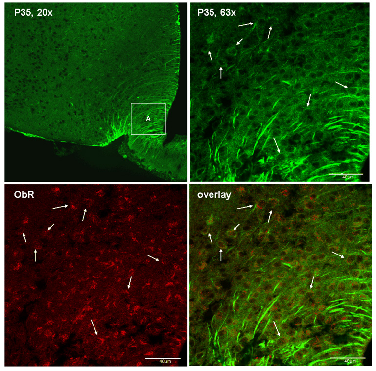
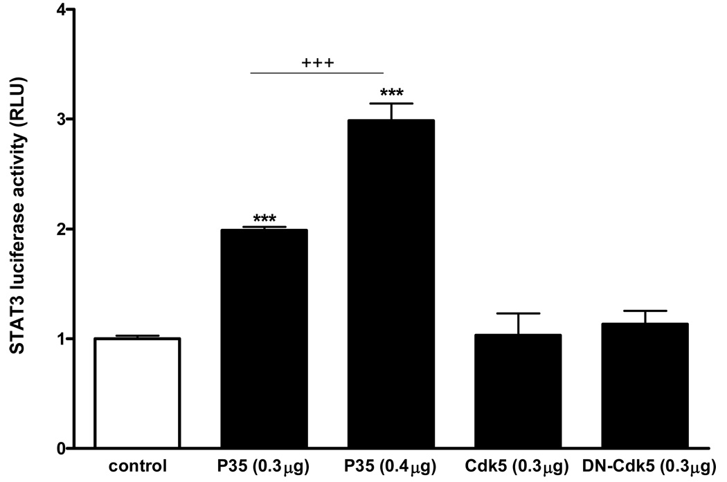
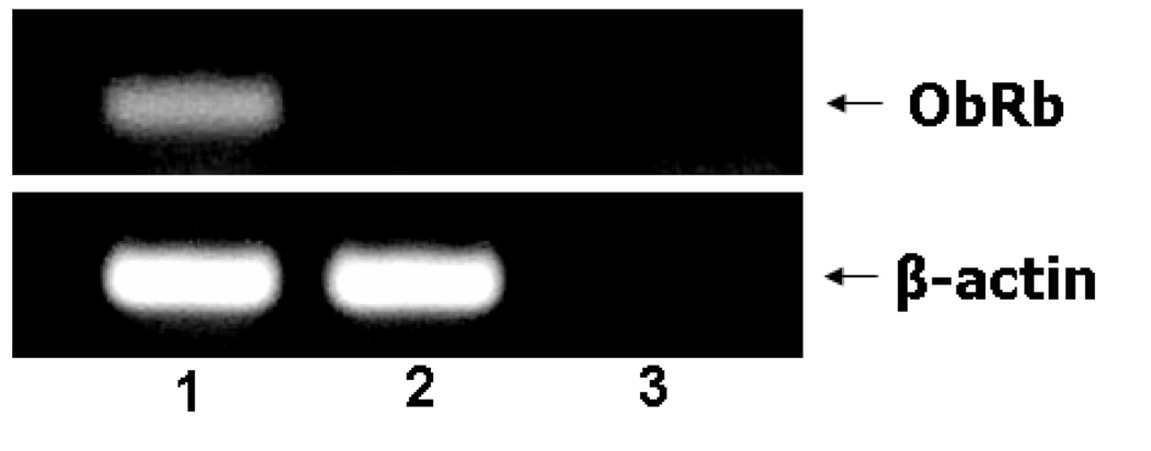
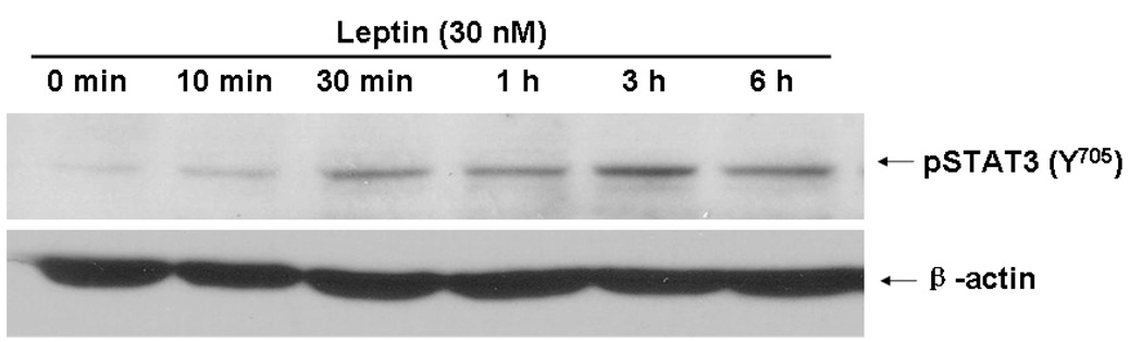
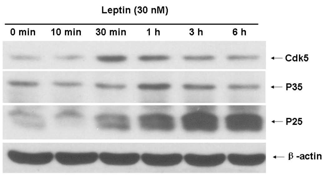
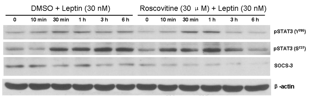
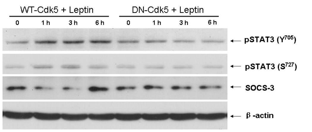
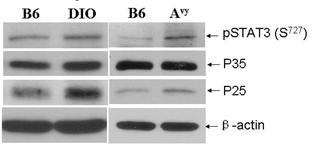
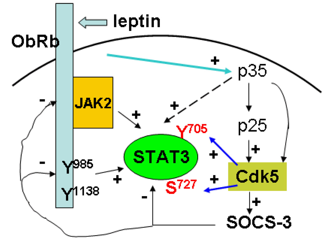
Cyclin-dependent kinase (Cdk) 5 is ubiquitously expressed in the brain and plays an essential role in central nervous system development and synaptic plasticity. The p35 kinase is a neuronal specific activator of Cdk5. Here, we show for the first time that Cdk5 activation modulates leptin signaling. P35 and its metabolite p25 were colocalized with the leptin receptor ObR in selective neurons in the hypothalamus. Overexpression of p35 alone was sufficient to induce the transcriptional activation of signal transducer and activator of transcription 3 (STAT3) in a cellular model. In retinoic acid-differentiated SH-SY5Y neuronal cells where ObRb was induced, leptin increased the expression of Cdk5, p35, and p25 kinases. The time course of induction coincided with that of phosphorylated (p)-STAT3. When Cdk5 activity was inhibited, either by roscovitine or overexpression of dominant negative Cdk5, there was a reduction of pSTAT3 activation. The results show that the activation of Cdk5 by p35 sustained leptin-induced pSTAT3 at 3-6 h. Thus, p35 is a novel modulator of leptin-induced STAT3 signaling.


Figures

Fig. 1
Fig. 1
Expression of p35 in ObR (+) neurons, shown by confocal microscopy. Top left: p35/p25 immunoreactivity in the arcuate nucleus of the hypothalamus, present in neuronal cytoplasma and in tanycytes. Top right: enlarged area A from the top left panel. Bottom left: ObR immunoreactivity in the same area A. Bottom right: confocal overlay of p35 (green) and ObR (red) immunoreactivity. Arrows indicate co-localized cells.
Fig. 2
Fig. 2
Overexpression of p35 alone, in the absence of leptin or other ligands, was sufficient to induce STAT3 transcriptional activity in HEK293 cells shown by luciferase reporter assays. Both 0.3 µg/ml and 0.4 µg/ml of p35 caused significant increases of STAT3 luciferase activity in comparison with the empty vector control (***: p < 0.005). This contrasts with the lack of effect of Cdk5 or DN-Cdk5 in the absence of ligands. The higher dose of p35 caused a greater increase than the lower dose (+++: p < 0.005 when the 0.3 and 0.4 µg groups were compared).
Fig. 3
Fig. 3. The cellular model of differentiated SH-SY5Y cells
3A. Induction of ObRb mRNA in SH-SY5Y cells and the effects of leptin on STAT3 activation. ObRb mRNA was present in retinoic acid-differentiated cells (lane 1), but not in non-differentiated cells (lane 2) or the no-template negative control (lane 3). 3B. Retinoic acid-induced differentiation of SH-SY5Y cells resulted in subcellular redistribution of ObRb. Immunocytochemistry showed that ObRb was present in the cytoplasm of the undifferentiated cells (middle panel), but at the cell surface of differentiated cells (right panel). The specificity of the staining was shown by the lack of signals in the negative control with secondary antibody only (left panel). 3C. Leptin treatment (30 nM) induced pSTAT3 throughout the study period (10 min – 6 h), with a peak at 3 h. This contrasts with the lack of changes of the β-actin signal.
Fig. 4
Fig. 4. Effect of leptin treatment (30 nM) on Cdk5 in SH-SY5Y cells
4A. Western blotting showed that leptin induced Cdk5, p35, and p25 by 30 min, with peaks at 30 min, 1 h, and 3 h, respectively. The housekeeping gene β-actin was unchanged. 4B. Immunocytochemistry showed that leptin treatment altered the subcellular distribution of p35/p25 from the cytoplasmic cluster at the basal state to a more diffuse pattern at 1 and 6 h.
Fig. 5
Fig. 5. The Cdk5 inhibitor roscovitine modulates leptin-induced pSTAT3 in SH-SY5Y cells
Leptin (30 nM) increased pSTAT3 (both Y705 and S727) between 30 min – 6 h of treatment; the increase at the later time points (3 and 6 h) was dampened by co-treatment with roscovitine, which also shifted pSTAT3-S727 activation to an earlier time. The induction of SOCS-3 was greatly diminished by roscovitine, which dampened SOCS-3 signal between 10 min and 6 h.
Fig. 6
Fig. 6
Overexpression of Cdk5 (wildtype, WT-Cdk5) in SH-SY5Y cells increased leptin-induced pSTAT3 production at 1, 3, and 6 h, whereas DN-Cdk5 (dominant negative without kinase activity) suppressed it at all time points. This was seen for both Y705 and S727, and did not affect the housekeeping gene β-actin. By contrast, WT-Cdk5 reduced SOCS-3 at 1 and 3 h but increased it at 6 h.
Fig. 7
Fig. 7
Activities of STAT3 and Cdk5 in the hypothalamus of obese mice. The hypothalamus of a 5-month old DIO mouse and a 3-month old Avy mouse both had higher levels of pSTAT3 (S727) and p25 than their respective controls. The increase of p35 was apparent in DIO but not in the Avy mouse. The house-keeping gene β-actin remained unchanged.
Fig. 8
Fig. 8
Diagram of the induction and consequences of activation of Cdk5. (1) Leptin signaling through ObRb induces activation of p35, p25, and Cdk5. (2) Cdk5 potentiates the activity of pSTAT3 by enhancing phosphorylation at both Y705 and S727 sites. (3) Cdk5 also increases SOCS-3 activity which eventually facilitates termination of STAT3 activation.

References

    1. Banks WA, Kastin AJ, Huang W, Jaspan JB, Maness LM. Leptin enters the brain by a saturable system independent of insulin. Peptides. 1996;17:305–311. - PubMed
    1. Baumann K, Mandelkow EM, Biernat J, Piwnica-Worms H, Mandelkow E. Abnormal Alzheimer-like phosphorylation of tau-protein by cyclin-dependent kinases cdk2 and cdk5. FEBS Lett. 1993;336:417–424. - PubMed
    1. Benomar Y, Roy AF, Aubourg A, Djiane J, Taouis M. Cross down-regulation of leptin and insulin receptor expression and signalling in a human neuronal cell line. Biochem.J. 2005;388:929–939. - PMC - PubMed
    1. Bjorbak C, Lavery HJ, Bates SH, Olson RK, Davis SM, Flier JS, Myers MG., Jr SOCS3 mediates feedback inhibition of the leptin receptor via Tyr985. J Biol Chem. 2000;275:40649–40657. - PubMed
    1. Cheung ZH, Fu AK, Ip NY. Synaptic roles of Cdk5: implications in higher cognitive functions and neurodegenerative diseases. Neuron. 2006;50:13–18. - PubMed

Publication types

MeSH terms