Review
. 2009 Feb;39(1):37-49.
doi: 10.1007/s12035-009-8050-7. Epub 2009 Jan 22.

Activity-dependent dendritic release of BDNF and biological consequences

Affiliations
Free PMC article
Review

Activity-dependent dendritic release of BDNF and biological consequences

Nicola Kuczewski et al. Mol Neurobiol. 2009 Feb.
Free PMC article

Abstract

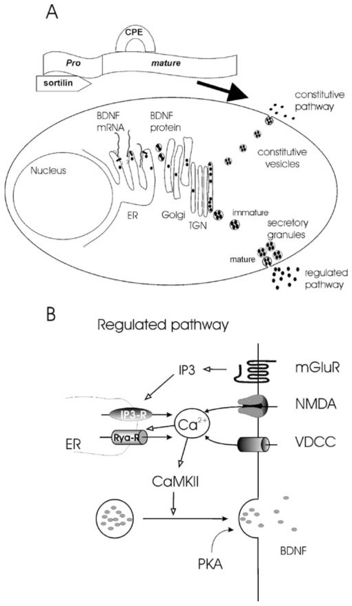
Network construction and reorganization is modulated by the level and pattern of synaptic activity generated in the nervous system. During the past decades, neurotrophins, and in particular brain-derived neurotrophic factor (BDNF), have emerged as attractive candidates for linking synaptic activity and brain plasticity. Thus, neurotrophin expression and secretion are under the control of activity-dependent mechanisms and, besides their classical role in supporting neuronal survival neurotrophins, modulate nearly all key steps of network construction from neuronal migration to experience-dependent refinement of local connections. In this paper, we provide an overview of recent findings showing that BDNF can serve as a target-derived messenger for activity-dependent synaptic plasticity and development at the single cell level.


Figures

Fig. 1
Fig. 1
Different approaches to study BDNF secretion. a Released BDNF can be detected and quantified in the medium using immunochemical essays (ELISA or Western blot). b Release of BDNF can be visualized in living BDNF-GFP expressing neurons. Scale bar, 25 μm. c BDNF released from the neuron binds to TrkB receptors at the surface of the cell-based fluorescent indicator. The activated kinase domain of the TrkB receptor phosphorylates a chimeric fluorescent protein, leading to a conformational change allowing fluorescence resonance energy transfer (FRET). BDNF is therefore detected with fluorescence readout (adapted from [40])
Fig. 2
Fig. 2
Synthesis, storage, and release of BDNF. a The pre-pro-BDNF is synthesized and sequestered in the endoplasmic reticulum (ER). Pro-BDNF then transits the Golgi apparatus and accumulates in membrane stacks of the trans-Golgi network. The trans-Golgi network can be thought of as a major protein sorting station inside the cell. At this point, two types of secretory pathways exist. Secretion via the constitutive pathway does not rely on extracellular signals or any triggering events. In the regulated pathway, fusion of the secretory granules with the plasma membrane is triggered by an intracellular rise in Ca2+. The binding of the pro form with sortiline and the mature form with carboxipeptidase E is required to address the pro-BDNF in the regulated pathway (modified from Fig. 2 in [1]; with permission. b Scheme of the signaling involved in postsynaptic regulated BDNF secretion. BDNF secretion required a postsynaptic rise in intracellular Ca2+ concentration. This Ca2+ rise can result from an influx through voltage-dependent Ca2+ channels (VDCCs) or NMDA receptors upon membrane depolarization or from activation of internal Ca2+ stores (IP3-R) following the activation of metabotropic glutamatergic receptors (mGluRs). The initial Ca2+ rise can be amplified by Ca2+-induced Ca2+ secretion via ryanodine receptors (Rya-Rs). Ca2+ activates CaMKII leading to the fusion of the secretory granules. Basal levels of PKA activation “gate” BDNF secretion
Fig. 3
Fig. 3
Action potential induced BDNF secretion. Merged images showing intracellular BDNF-GFP fluorescence (green) and secreted BDNF-GFP detected using an anti-GFP antibody (red) under non-permeabilizing conditions for antibody detection. BDNF-GFP secretion was induced by the firing activity produced by the K+ channel antagonist 4-amminopyridine (4AP) in the presence of glutamatergic antagonists (left). BDNF-GFP secretion was impaired by TTX (right). Lower panel: cell attached recordings showing the firing activity in the two conditions. The experiments were performed on primary hippocampal cell cultures transfected at 11 days in vitro (DIV) and stimulated at 13 DIV. Scale bar, 25 μm
Fig. 4
Fig. 4
BDNF secretion depends on the neuronal output rather than synaptic inputs. Suprathreshold excitatory postsynaptic potentials (EPSPs) trigger action potentials that back-propagate (b-APs) from the soma to the dendrites. Center, b-APs-induced BDNF secretion and synaptic strengthening. Left, this process can be reinforced by conditions that facilitate the back propagation of APs such as the action of neuromodulators. Right, when the same suprathreshold glutamatergic activity is generated in a context that prevents APs back-propagation, such as a concomitant activation of dendritic inhibition (GABA), BDNF secretion and synaptic strengthening will not occur. Modified from Comunicative & Intergrative Biology 1(2):153

References

    1. Lessmann V, Gottmann K, Malcangio M. Neurotrophin secretion: current facts and future prospects. Prog Neurobiol. 2003;69:341–374. - PubMed
    1. Lu B, Pang PT, Woo NH. The yin and yang of neurotrophin action. Nat Rev Neurosci. 2005;6:603–614. - PubMed
    1. Huang EJ, Reichardt LF. Neurotrophins: roles in neuronal development and function. Annu Rev Neurosci. 2001;24:677–736. - PMC - PubMed
    1. Thoenen H. Neurotrophins and neuronal plasticity. Science. 1995;270:593–598. - PubMed
    1. Ernfors P, Merlio JP, Persson H. Cells expressing mRNA for neurotrophins and their receptors during embryonic rat development. Eur J Neurosci. 1992;4:1140–1158. - PubMed

Publication types

MeSH terms

Substances