. 2009 Feb;42(2):89-103.
doi: 10.1080/08916930802629554.

Lupus-prone MRL/faslpr/lpr mice display increased AID expression and extensive DNA lesions, comprising deletions and insertions, in the immunoglobulin locus: concurrent upregulation of somatic hypermutation and class switch DNA recombination

Affiliations

Lupus-prone MRL/faslpr/lpr mice display increased AID expression and extensive DNA lesions, comprising deletions and insertions, in the immunoglobulin locus: concurrent upregulation of somatic hypermutation and class switch DNA recombination

Hong Zan et al. Autoimmunity. 2009 Feb.

Abstract

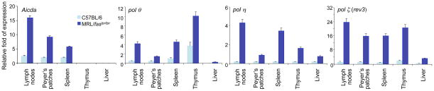
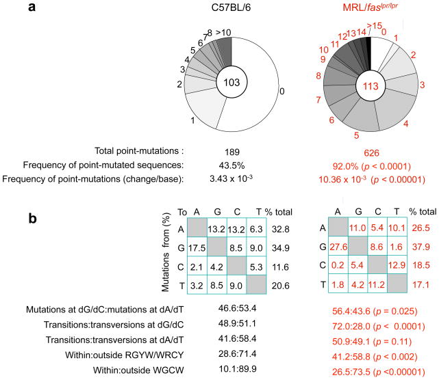
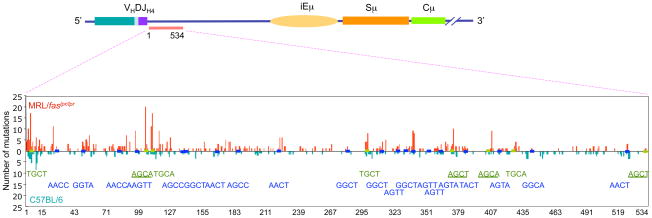
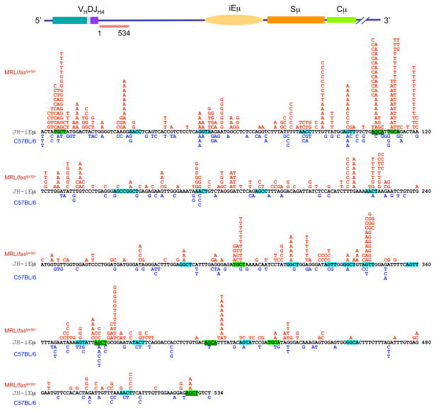
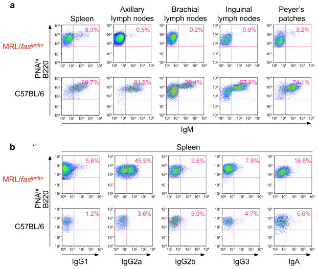
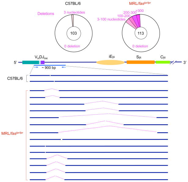
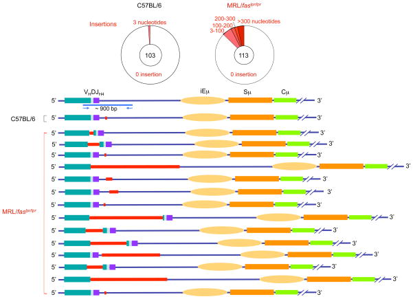
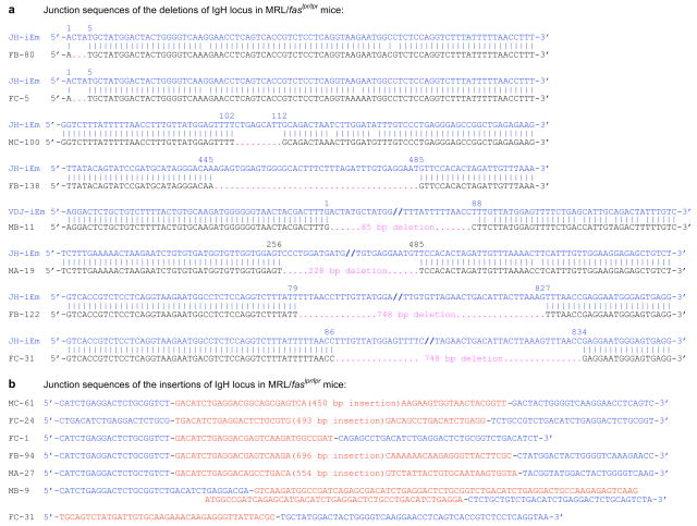
Systemic lupus erythematosus (SLE) is an autoimmune disease characterized by the production of an array of pathogenic autoantibodies, including high-affinity anti-dsDNA IgG antibodies. These autoantibodies are mutated and class-switched, mainly to IgG, indicating that immunoglobulin (Ig) gene somatic hypermutation (SHM) and class switch DNA recombination (CSR) are important in their generation. Lupus-prone MRL/fas(lpr/lpr) mice develop a systemic autoimmune syndrome that shares many features with human SLE. We found that Ig genes were heavily mutated in MRL/fas(lpr/lpr) mice and contained long stretches of DNA deletions and insertions. The spectrum of mutations in MRL/fas(lpr/lpr) B cells was significantly altered, including increased dG/dC transitions, increased targeting of the RGYW/WRCY mutational hotspot and the WGCW AID-targeting hotspot. We also showed that MRL/fas(lpr/lpr) greatly upregulated CSR, particularly to IgG2a and IgA in B cells of the spleen, lymph nodes and Peyer's patches. In MRL/fas(lpr/lpr) mice, the significant upregulation of SHM and CSR was associated with increased expression of activation-induced cytidine deaminase (AID), which mediates DNA lesion, the first step in SHM and CSR, and translesion DNA synthesis (TLS) polymerase (pol) theta, pol eta and pol zeta, which are involved in DNA synthesis/repair process associated with SHM and, possibly, CSR. Thus, in lupus-prone MRL/fas(lpr/lpr) mice, SHM and CSR are upregulated, as a result of enhanced AID expression and, therefore, DNA lesions, and dysregulated DNA repair factors, including TLS polymerases, which are involved in the repair process of AID-mediated DNA lesions.


Figures

Figure 1
Figure 1
Aicda, polθ, pol η,and pol ζ (rev3) are preferentially expressed in lymph nodes, Peyer’s patches and spleen of lupus-prone MRL/faslpr/lpr mice. Total RNA was prepared from spleen, lymph nodes, Peyer’s patches, thymus and liver of non-immunized 8-week old MRL/faslpr/lpr or age-matched non-autoimmune C57BL/6 mice. The levels of Aicda, polθ, pol η, and pol ζ (rev3) transcripts were analyzed by real-time qRT-PCR using SYBR green and normalized to gapdh expression. Data are mean values ± SD from 3 independent experiments.
Figure 2
Figure 2
Mutation frequency is greatly increased and mutation spectrum is significantly altered in lupus-prone MRL/faslpr/lpr mice. (a) Pie charts depict the proportions of sequences that carry 1, 2, 3, etc. point-mutations over the 534 bp intronic JH4-iEμ DNA in GC (PNAhi) B cells from Peyer’s patches of three 11-week old MRL/faslpr/lpr and C57BL/6 mice. The numbers of sequences analyzed are at the center of the pies. (b) Compilations, with the numbers indicating percentages of all mutations scored in the pool of all point-mutations from MRL/faslpr/lpr and C57BL/6 mice. Below the compilations, the ratio of mutations at dG/dC to those at dA/dT, the ratio of transition:transversion substitutions at both dC/dG and dA/dT, and the ratio of mutations within and outside RGYW/WRCY mutational hotspots and WGCW AID hotspots are indicated. The significance of differences in the mutation frequency/spectrum between MRL/faslpr/lpr and C57BL/6mice was analyzed with the χ2 test. p < 0.05 were considered statistically significant.
Figure 3
Figure 3
SHM is upregulated and preferentially targets the WGCW AID hotspot in lupus-prone mice. The 534 bp intronic JH4-iEμ DNA of Peyer’s patch PNAhi B cells of 11-week-old lupus-prone MRL/faslpr/lpr and age-matched non-autoimmune C57BL/6 mice were sequence analyzed. Mutations in MRL/faslpr/lpr mouse DNA are shown in red, mutations in C57BL/6 mouse DNA are shown in blue. WGCW AID hotspots are shown in green. RGYW/WRCY mutational hotspots are shown in blue. AGCT and AGCA motifs, which are iterations of both WGCW and RGYW are underlined.
Figure 4
Figure 4
SHM is significantly upregulated in the lupus-prone mice. The 534 bp intronic JH4-iEμ DNA of Peyer’s patch B cells of 11-week-old lupus-prone MRL/faslpr/lpr and non-autoimmune C57BL/6 mice were sequence analyzed. Mutations in MRL/faslpr/lpr mouse DNA are shown in red, mutations in C57BL/6 mouse DNA are shown in blue. WGCW AID hotspots are highlighted green. RGYW/WRCY mutational hotspots are highlighted blue. AGCT and AGCA motifs, which are iterations of both WGCW and RGYW are underlined.
Figure 5
Figure 5
CSR is significantly upregulated in lupus-prone MRL/faslpr/lpr mice. (a) Cells from spleen, lymph nodes or Peyer’s patches of MRL/faslpr/lpr or non-autoimmune C57BL/6 mice were stained with PE-labeled anti-B220 mAb, and FITC-labeled anti-IgM mAb, the IgM- B220+ cells are considered as switched B cells; (b) Cells from spleen, lymph nodes or Peyer’s patches of MRL/faslpr/lpr or non-autoimmune C57BL/6 mice were stained with PE-labeled anti-B220 mAb, and FITC-labeled anti-IgA mAb; (c) Spleen cells from MRL/faslpr/lpr or non-autoimmune C57BL/6 mice were stained with PE-labeled anti-B220 mAb, and FITC-labeled anti-IgG1, anti-IgG2a, anti-IgG2b, or anti-IgG3 mAb. Stained cells were then analyzed by FACS.
Figure 6
Figure 6
Significant increased CSR to all isotypes in GC B cells of lupus-prone mice. (a) GC (PNAhi) B cells from spleen, lymph nodes or Peyer’s patches of lupus-prone MRL/faslpr/lpr or non-autoimmune C57BL/6 mice were stained with PE-labeled anti-B220 mAb, FITC-labeled anti-IgM mAb and then analyzed for surface fluorescence by FACS. (b) GC (PNAhi) B cells from the spleens of MRL/faslpr/lpr or C57BL/6 mice were stained with PE-labeled anti-B220 mAb, FITC-labeled anti-IgM, anti-IgG1, anti-IgG2a, anti-IgG2b, anti-IgG3 or anti-IgA mAb, and then analyzed for surface fluorescence by FACS.
Figure 7
Figure 7
The Igh locus in the B cells of lupus-prone MRL/faslpr/lpr mice contains a high frequency of deletions. Pie charts depict the proportions of sequences that contain different numbers of deletions (pink) over the about 900 bp intronic VJ558DJH4-iEμ DNA in GC (PNAhi) B cells from Peyer’s patches of three 11-week-old MRL/faslpr/lpr and three 11-week-old C57BL/6 mice. Depicted are locations and sizes of deletions.
Figure 8
Figure 8
The Igh locus in the B cells of lupus-prone MRL/faslpr/lpr mice contains a high frequency of insertions. Pie charts depict the proportions of sequences that contain different numbers of insertions (red) over the about 900-bp VJ558DJH4-iEμ DNA in GC (PNAhi) B cells from Peyer’s patches of three 11-week old MRL/faslpr/lpr and three 11-week-old C57BL/6 mice. Depicted are locations and sizes of insertions.
Figure 9
Figure 9
The junction sequences of deletions and deletions in the Igh locus of MRL/faslpr/lpr mice. VJ558DJH4-iEμ DNA of GC (PNAhi) B cells from Peyer’s patches of MRL/faslpr/lpr or C57BL/6 mice were amplified and sequenced. Each sequence is compared with germline Igh sequence. (a) The deleted nucleotides are indicated by pink dots. The numbers on top of each aligned sequence indicate upstream and downstream breakpoints of recombined sequences. (b) The inserted sequences are in red and underlined. The upstream and downstream VJ558DJH-iEμ sequences linked to the inserted DNA are in blue.

References

    1. Shlomchik MJ, Madaio MP. The role of antibodies and B cells in the pathogenesis of lupus nephritis. Springer Semin Immunopathol. 2003;24:363–375. - PubMed
    1. Diamond B. Autoimmunity. Immunol Rev. 2005;204:5–8. - PubMed
    1. Shlomchik MJ, Aucoin AH, Pisetsky DS, Weigert MG. Structure and function of anti-DNA autoantibodies derived from a single autoimmune mouse. Proc Natl Acad Sci USA. 1987;84:9150–9154. - PMC - PubMed
    1. Shlomchik M, Mascelli M, Shan H, Radic MZ, Pisetsky D, Marshak-Rothstein A, Weigert M. Anti-DNA antibodies from autoimmune mice arise by clonal expansion and somatic mutation. J Exp Med. 1990;171:265–292. - PMC - PubMed
    1. van Es JH, Gmelig Meyling FH, van de Akker WR, Aanstoot H, Derksen RH, Logtenberg T. Somatic mutations in the variable regions of a human IgG anti-double-stranded DNA autoantibody suggest a role for antigen in the induction of systemic lupus erythematosus. J Exp Med. 1991;173:461–470. - PMC - PubMed

Publication types

MeSH terms

Substances