Review
. 2009 Nov 15;87(15):3267-76.
doi: 10.1002/jnr.21992.

Myelin-associated glycoprotein and its axonal receptors

Affiliations
Review

Myelin-associated glycoprotein and its axonal receptors

Ronald L Schnaar et al. J Neurosci Res. .

Abstract

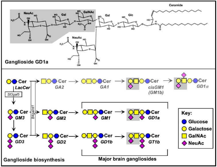
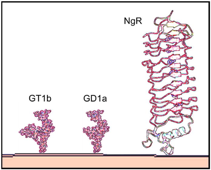
Myelin-associated glycoprotein (MAG) is expressed on the innermost myelin membrane wrap, directly apposed to the axon surface. Although it is not required for myelination, MAG enhances long-term axon-myelin stability, helps to structure nodes of Ranvier, and regulates the axon cytoskeleton. In addition to its role in axon-myelin stabilization, MAG inhibits axon regeneration after injury; MAG and a discrete set of other molecules on residual myelin membranes at injury sites actively signal axons to halt elongation. Both the stabilizing and the axon outgrowth inhibitory effects of MAG are mediated by complementary MAG receptors on the axon surface. Two MAG receptor families have been described, sialoglycans (specifically gangliosides GD1a and GT1b) and Nogo receptors (NgRs). Controversies remain about which receptor(s) mediates which of MAG's biological effects. Here we review the findings and challenges in associating MAG's biological effects with specific receptors.


Figures

Fig. 1
Fig. 1
Ganglioside structure and metabolism. Top: The structure of GD1a is shown with the MAG-binding determinant (NeuAc α2-3 Gal β1-4 GalNAc) shaded. Bottom: Biosynthetic pathways to the major brain gangliosides. The MAG-binding determinant is shaded, and the two glycosyltransferases discussed in the text, St3gal5 and B4galnt1 are shown. St3gal5-null mice lack all major brain gangliosides, but instead synthesize an equivalent amount of the rare gangliosides cisGM1 and GD1α, both of which carry the MAG-binding determinant.
Fig. 2
Fig. 2
The structures of MAG receptors GT1b, GD1a and NgR1. Only the portions of the molecules that extend from the membrane into the extracellular space are shown. Each MAG receptor is anchored to the cell membrane by a lipid moiety that is not shown (ceramide for GT1b/GD1a and phosphatidylinositol for NgR1). The relative sizes are accurate based on NMR structures of gangliosides (Sonnino et al., 2007) and crystallographic analyses of NgR (He et al., 2003). The gangliosides extend 2.4 nm from the cell surface, with their MAG-binding determinants at their outermost extent, whereas NgR is 8 nm in length with centrally located MAG-binding residues (Barton et al., 2003; Lauren et al., 2007).
Fig. 3
Fig. 3
Dual MAG receptor model. MAG is envisioned as interacting independently with gangliosides (GD1a or GT1b, MAG-binding determinant highlighted) and with NgR’s (NgR1 or NgR2). In each pathway, MAG engagement results in transmembrane signals. For example, NgR1 interacts with transmembrane signaling molecules p75NTR and Lingo-1 to transduce a signal that results in RhoA activation and inhibition of axon outgrowth. Gangliosides may signal via some of the same components, or via yet undefined components as shown, to activate RhoA, modulate axon and node of Ranvier structures, and stabilize axons. The proximal ganglioside transmembrane signal transducing molecules responsible for MAG’s different biological effects may be shared (as shown) or independent. Additional modulating factors (cAMP, calcium, PKC) and potential pathway crosstalk are not shown (see Yiu and He, 2003).

Similar articles

Cited by

References

    1. Arquint M, Roder J, Chia L-S, Down J, Wilkinson D, Bayley H, Braun P, Dunn R. Molecular cloning and primary structure of myelin-associated glycoprotein. Proc Natl Acad Sci U S A. 1987;84:600–604. - PMC - PubMed
    1. Atwal JK, Pinkston-Gosse J, Syken J, Stawicki S, Wu Y, Shatz C, Tessier-Lavigne M. PirB is a functional receptor for myelin inhibitors of axonal regeneration. Science. 2008;322:967–970. - PubMed
    1. Barton WA, Liu BP, Tzvetkova D, Jeffrey PD, Fournier AE, Sah D, Cate R, Strittmatter SM, Nikolov DB. Structure and axon outgrowth inhibitor binding of the Nogo-66 receptor and related proteins. EMBO J. 2003;22:3291–3302. - PMC - PubMed
    1. Bartsch S, Montag D, Schachner M, Bartsch U. Increased number of unmyelinated axons in optic nerves of adult mice deficient in the myelin-associated glycoprotein (MAG) Brain Res. 1997;762:231–234. - PubMed
    1. Bartsch U, Kirchhoff F, Schachner M. Immunohistological localization of the adhesion molecules L1, N-CAM, and MAG in the developing and adult optic nerve of mice. J Comp Neurol. 1989;284:451–462. - PubMed

Publication types

MeSH terms